Effects of equilibration media and co-culture on vitrification of sheep
blastocysts derived in vitro
*Hatem ATALLA
1, Mithat EVECEN
2, Serhat PABUCCUOĞLU
2, Sema BİRLER
21Faculty of Agriculture and Veterinary Medicine, An Najah National University, Nablus, Palestine. 2Istanbul Univetsity Cerrahpaşa Faculty of Veterinary Science, Department of Reproduction and Artificial Insemination; Istanbul, Turkey.
Summary: The effects of two vitrification protocols on the survival of sheep blastocysts were examined in embryos produced in vitro with sheep oviduct epithelial cells co-culture (CC) or without co-culture (C). Oocytes collected from slaughtered ewes were matured for 24 h, fertilized with fresh ram semen for 20 h and cultured in SOF medium for up to 9 days in vitro. For vitrification, blastocyst stage embryos were assigned to two equilibration groups (20% ethylen glycol (EG) or 10% glycerol (G) for the first equilibration), and as the second equilibration they were kept in 20% ethylen glycol plus 10% glycerol for 5 minutes. After 30 sec in vitrification solution (25% ethylen glycol + 25% glycerol), they were immediately immersed into liquid nitrogen. After thawing procedure, embryos were transferred into 0.25 M sucrose for 5 min, washed in Hepes buffered synthetic oviduct fluid (HSOF) and cultured in SOF medium for 24 h. Cleavage rates were 75.2% in C and 74.2% in CC groups, and blastocyst rates were 14.4% in C and 17.1% in CC groups. After in vitro culture of vitrified-thawed blastocysts, survival rates were 62.1, 38.4, 30.2, and 39.3% in C-EG, CC-EG, C-G and CC-G groups, respectively. This study shows that vitrification of sheep embryos using ethylene glycol instead of glycerol as a first equilibration cryoprotectant could give reasonable survival rates and that co-culture of embryos with sheep oviduct epithelial cell has no beneficial effect on vitrification of embryos.
Keywords: Co-culture, equilibration medium, in vitro fertilization, sheep vitrification.
İn vitro üretilen koyun blastosistlerinin vitrifikasyonu üzerine, ekilibrasyon medyumu ve ko-kültürün
etkileri
Özet: İki vitrifikasyon protokolünün, koyun ovidukt epitel hücreleriyle ko-kültürlü (CC) ve ko- kültürsüz (C) ortamda in vitro üretilmiş olan koyun blastosistlerinin yaşayabilirliği üzerindeki etkileri araştırıldı. Oositler, TCM 199 medyumunda 24 saat süreyle olgunlaştırıldı, 20 saat fertilize edildi ve 9 gün süreyle sentetik ovidukt sıvısı (SOF) medyumunda kültüre edildi. Embriyolar ilk olarak, %20 etilen glikol (EG) veya % 10 gliserol (G) den oluşan iki farklı medyumda ekilibrasyona bırakıldılar. İkinci ekilibrasyon amacıyla % 20 etilen glikol + % 10 gliserol içinde ve 5 dakika süreyle tutuldular. Ardından, % 25 etilen glikol + % 25 gliserol’den oluşan vitrifikasyon solüsyonu içerisinde 30 saniye tutulan embriyolar, derhal sıvı azot içerisine batırılarak donduruldu. Çözdürme işleminin ardından embriyolar, 0.25 M sukroz içine 5 dakika bekletildi, Hepes tamponlu sentetik ovidukt sıvısı (HSOF) içinde yıkandı ve 24 saat süreyle SOF içerisinde in vitro kültüre bırakıldı. Yarıklanma oranları C grubunda %75.2, CC grubunda %74.2, blastosist oranları C'de %14.4, CC grubunda ise %17.1 oldu. Dondurulmuş-çözdürülmüş blastosistlerin in vitro kültüründen sonra canlılık oranları C-EG, CC-EG, C-G ve CC-G gruplarında sırasıyla; %62.1, %38.4, %30.2 ve %39.3 oldu. Bu çalışmada, koyun embriyolarının vitrifikasyonunda; ilk ekilibrasyon medyumu olarak gliserol yerine etilen glikol kullanılmasının faydalı olduğu buna karşılık, koyun ovidukt epiteli hücreleri ile ko-kültürün etkili olmadığı saptandı.
Anahtar sözcükler: Ekilibrasyon medyumu, in vitro fertilizasyon, ko-kültür, koyun, vitrifikasyon.
Introduction
Reproductive biotechnology has been developed very rapidly in the last decades. Because of the need of a vast number of oocytes and embryos for reproductive technologies, there are numerous studies on in vitro fertilization and cryopreservation techniques. In order to
* A part of this study was presented in the 15th Annual Conference of the European Society for Domestic Animal Reproduction (ESDAR) in 2011, 15-17 September in Antalya-TURKEY and published in Reprod Dom Anim 46 (Suppl. 3), 78–161, 2011.
improve the success of in vitro fertilization, different media and culture conditions have been used (1, 3, 18, 31). Somatic cells or cells’ extracts have been used in culture systems to prevent in vitro blocks and accomplish desirable developmental rates (8, 25, 29). Co-culture with oviduct epithelial cells has been tried for ensuring oviduct
milieu in in vitro culture environment. Although some problems such as increase in the incidence of large offspring syndrome were attributed to co-culture and serum usage during in vitro culture, some experiments showed that co-culture of embryos with somatic cells may beneficially influence the development of embryos to blastocyst stage thanks to its well-defined embryotrophic effects in mouse (2), sheep (4, 7), cattle (9), goat (30) by secreting embryotrophic substances and also by removing embryotoxic substances.
Different cryopreservation regimens for preservation of embryos have been used (26, 30). Slow freezing and vitrification techniques are equally used for cryopreservation of embryos, but vitrification method is generally chosen for oocytes (5, 21, 28). Types and concentration (11, 16) of cryoprotectants, exposure time of embryos to cryoprotectants (16, 17), source of embryos as in vivo or in vitro (5, 14) developmental stage of embryos (21), and media types (16, 27) are the factors that influence the survival rate of embryos after cryopreservation and thawing. In vitro produced embryos are more sensitive to freezing conditions than in vivo embryos (6, 13, 20). Blastocyst stage embryos have more freezing tolerance than morula stage embryos in sheep (1, 12).
In this study, the effects of co-culture with oviduct epithelial cells and equilibration medium differences on the survival after vitrification of in vitro produced sheep embryos were investigated.
Materials and Methods
All chemicals were purchased from Sigma unless otherwise specified.
Ethics Statement: The study was performed in
accordance with guidelines for animal research from Istanbul University Ethics Committee on Animal Research (2017-425006).
In vitro fertilization of oocytes: Oocytes were
obtained and fertilized in vitro as reported previously (10). Briefly, ovaries obtained from a local slaughterhouse were brought to laboratory in Dulbecco’s PBS at 30-35 C. Cumulus-oocyte complexes were recovered by slicing sheep ovaries and matured for 24 h in medium 199 supplemented with 10% FCS, pyruvate and hormones (FSH and LH) (H-199+) at 38.5 C, in a humidified atmosphere of 5% CO2. Oviduct epithelial cells were
obtained by flushing oviducts by 20 ml H-199+ medium for each oviduct and after three washings cultured in H-199+ medium without pyruvate and hormones at 38.5 C, in a humidified atmosphere of 5% CO2 until using for
co-culture with presumptive embryos.
Pooled fresh ram semen collected from at least 3 kivircik rams were used for in vitro fertilization of
matured oocytes. Semen was prepared by percoll-gradient method and added to fertilization medium (Bicarbonate supplemented Synthetic Oviduct Fluid: BSOF) as 8x10⁵
-1x10⁶ spermatozoon per ml for 20 h in a humidified
atmosphere of 5% CO2 at 38.5C. Presumptive embryos
were divided into 2 groups and cultured in SOF medium at 38.5C, in a humidified atmosphere of 5% CO2, 5% O2
and 90% N2 for 9 days with (CC) or without (C) co-culture
with sheep oviduct epithelial cells. On day 4, 1.5 mM glucose was added to culture medium and oocytes not cleaved were discarded.
Toxicity tests: For toxicity tests of cryoprotectants,
all procedures in all groups were repeated without putting straws into liquid nitrogen. Embryos were then cultured in vitro in SOF medium supplemented with 1.5 mM glucose for 24 h at 38.5C, in a humidified atmosphere of 5% CO2,
5% O2 and 90% N2.
Vitrification: Embryos reached to blastocyst stage
were vitrified in two groups:
First Group (EG)
First equilibration solution= 20% ethylen glycol Second equilibration solution= 20% ethylen glycol + 10% glycerol
Vitrification solution= 25% ethylen glycol + 25% glycerol
Second Group (G)
First equilibration solution= 10% glycerol
Second equilibration solution= 20% ethylen glycol + 10% glycerol
Vitrification solution= 25% ethylen glycol + 25% glycerol
All solutions were at room temperature throughout the classical vitrification procedure. Embryos were exposed for 5 min to first equilibration medium, 5 min to second equilibration medium and were plugged into liquid nitrogen after maximum 30 sec in vitrification solution.
Survival control: Thawing procedure was the same
for two vitrification groups. Straws were put in a water bath at 37°C for 15 to 20 seconds and ingredients of the straw were poured into a petri dish. After 5 min embryos were put in 0.25 M sucrose for another 5 min. Embryos were then washed with HSOF medium and cultured in SOF medium supplemented for 1.5 mM glucose for 24 h at 38.5C, in a humidified atmosphere of 5% CO2, 5% O2
and 90% N2.
Statistics: The data were analyzed using SPSS
(Version 10.0) for Windows (MS). Developmental rates of embryos cultured in vitro in two groups, survival rates of embryos after vitrification in two groups, and vitrification medium toxicity tests of embryos were assessed by using Chi- square test (Tables 1, 3 and 4). In order to test the normality of data, Shapiro-Wilk test was
applied and since the data was non-parametric, the results of development time of embryos to the blastocyst stage between two groups were assessed by using Mann Whitney- U test (Table 2).
Results
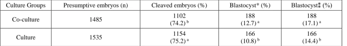
After in vitro fertilization of 3159 oocytes, 3020 presumptive embryos (C Group n= 1535; CC Group n= 1485) were cultured in vitro. Development results of embryos are shown in Table 1. After in vitro culture of embryos, 166 (14.4%) and 188 (17.1%) were developed to the blastocyst stage in C and CC groups, respectively (p<0.001). Development time of embryos to the blastocyst
stage were faster in CC group when compared to C group (6.9 ± 0.7 versus7.2 ± 0.7 day; Table 2) (p<0.005). After vitrification and thawing, survival rates of blastocyst stage embryos are shown in Table 3. According to post-thawing development rates of embryos, the group in which embryos cultured without co-culture and vitrified in EG as the first equilibration solution was the best group survived (62.1%) after vitrification (p<0.01).
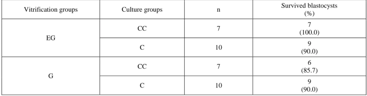
Although the cryoprotectants we used (EG and G) already embryo tested; in this study we aimed to confirm their toxicity. The toxic effects of the cryoprotectants were found similar (p>0.05) and the survival rates of embryos ranged between 85 to 100% (Table 4).
Table 1. Development results of embryos cultured in vitro. Tablo 1. İn vitro kültüre edilen embriyoların sonuçları
Culture Groups Presumptive embryos (n) Cleaved embryos (%) Blastocyst* (%) Blastocyst⁑ (%)
Co-culture 1485 1102 (74.2) b 188 (12.7) a 188 (17.1) a Culture 1535 1154 (75.2) a 166 (10.8) b 166 (14.4) b a, b: Within columns, values with different superscript are different (Chi-Square test: p<0.001).
a, b: Aynı kolonlarda farklı harf taşıyan değerler önemlidir (Ki-kare testi: p<0.001). (Blastocyst/Presumptive Oocyte)
(Blastocyst/Cleaved Embryo)
Table 2. Development time of embryos to the blastocyst stage. Tablo 2. Embriyoların blastosiste gelişim süreleri
Culture groups
Blastocyst numbers at in vitro culture day (%)
Mean day Total
Day 6 Day 7 Day 8 Day 9
Co-culture 70 (37.2) 74 (39.4) 31 (16.5) 13 (6.9) 6.9 ± 0.7 a 188 Culture 39 (23.5) 68 (41.0) 50 (30.1) 9 (5.4) 7.2 ± 0.7 b 166
a, b: Within columns, values with different superscript are different (Mann-Withney U test: P<0.005). a, b: Aynı kolonlarda farklı harf taşıyan değerler önemlidir (Mann-Withney U test: P<0.005).
Table 3. Survival rates of embryos after vitrification and thawing.
Tablo 3. Vitrifikasyon ve çözdürme sonrasında embriyoların sağ kalım oranları. Vitrification
groups
Culture groups
Cultured blastocysts after thawing (n)
Survived blastocyst number after thawing for 24h (%) EG CC 73 28 (38.4)b C 66 41 (62.1)a G CC 84 33 (39.3)b C 63 19 (30.2)b a, b: Within columns, values with different superscript are different (Chi-Square test: P<0.01).
Table 4. Results of toxicity tests after 24h of in vitro culture. Tablo 4. İn vitro kültürün 24. saatindeki toksisite test sonuçları
Vitrification groups Culture groups n Survived blastocysts
(%) EG CC 7 7 (100.0) C 10 9 (90.0) G CC 7 6 (85.7) C 10 9 (90.0)
Discussion and Conclusion
Co-culture with oviduct epithelial cells enhanced the development of ovine embryos to blastocyst stage in this study (p<0.001). The results are consistent with the study of Ledda et al. (12) who used two different media for in vitro culture of in vivo fertilized 1-2 cell ovine embryos and showed 71 vs 46% and 46 vs 13% using with or without cells in (Chatot Ziomek Bavister) CZB and (Tissue Culture Medium) TCM 199 medium groups, respectively.
Shirazi and Motaghi (23) reported that neither the presence of oviductal epithelial cells nor their conditioned medium could improve the developmental potential of ovine embryos during IVF. Pereira and Marques (19), have explained that adding somatic cells into culture media has negative effect on blastocyst development due to the raising of ammonium concentration. In a group that they used culture for 2 days and continued without co-culture, 52% of blastocyst rate was obtained. This result demonstrated that short co-culture period did not have negative effect on blastocyst development. It was also reported that somatic cells may have disadvantages such as contamination and embryonic abnormality risks and it is time-consuming procedure to prepare somatic cells (15). Despite those previous negative results; in this study not only the blastocyst rate was high, but also the development to blastocyst stage was faster in co-culture group. Mean day for embryos reaching toblastocyst stage was 6,9 ± 0,7 and 7,2 ± 0,7 in co-culture and culture groups, respectively (p<0.005). Similar to the present study, Van Wagtendonkdeleeuw et al. (27) demonstrated in cattle that development rate was faster by using co-culture with (Buffalo rat liver) BRL cells, and Donnay et al. (6) revealed that co-culture with cumulus cells accelerated the development to blastocyst stage (7.30.1 vs. 8.10.1 day) rather than BRL cells.
The present study demonstrated that using EG instead of G as the first equilibration solution improved survival rate (62.1% vs. 30.2%; p<0.01) of embryos cultured without oviduct epithelial cells and that
co-culture with sheep oviduct epithelial cells has negative effect on survival rates after vitrification of embryos (p<0.01). Songsasen et al. (24) investigated the rate and time for entering of different cryoprotectants into cell and expressed that the rates of different cryoprotectants were as EG>PG>DMSO≈G causing 76.9%, 62.5% and 55.6% survival rates, respectively.
Yang et al. (30) stated that survival of vitrified cattle embryos is affected by cryoprotectant types and exposure procedures. As consistent with present study, Ali and Shelton (2) concluded that EG was the best cryoprotectant choice in sheep. Martinez and Matkovic (14) also showed that EG was better choice than G for vitrification of sheep morulae (59.4 vs 29.7%), but they did not found a difference in overall pregnancy rate (51.3 vs 40.2%). Martinez et al. (15) reported that ovine embryos cryopreserved in 3 steps using ethylene glycol vs glycerol showed a higher hatching rate (40.0 vs 20.0%). Schiewe et al. (22) and De Paz et al. (5) compared vitrification with slow freezing method for cryopreservation of in vivo produced embryos and showed higher survival rates in slow freezing. Survival rates after vitrification of in vitro produced embryos in the present study were higher than those previous studies. Although high cryoprotectant concentrations used in vitrification method may have toxic effects on embryos (24); in this study, the toxic effects of cryoprotectants were found at both low and similar rates (p>0.05).
In conclusion, the results of this study show that using co-culture with oviduct epithelial cells for in vitro production of sheep embryos may have positive effects but time and effort consuming and also have no positive effects on survival after cryopreservation. Ethylen glycol as a first equilibration solution may increase survival rates of in vitro produced sheep embryos after vitrification.
Acknowledgement
This study was supported by the Research Fund of Istanbul University. Project No: T-863/17072000 and Project No: UDP/17290. The authors would like to thank
to Prof. Dr. Bülent EKİZ for his helps with the statistics of this study.
References
1. Alçay S, Üstüner B, Nur Z (2016): Effects of low molecular weight cryoprotectants on the post-thaw ram sperm quality and fertilizing ability. Small Ruminant Research, 136, 59-64.
2. Ali J, Shelton J (1993): Successful vitrification of day-6 sheep embryos. Journal of J Reprod Fert, 99, 65-70. 3. Bavister BD (1998): Role of oviductal secretions in
embryonic growth in vivo and in vitro. Theriogenology, 29, 143-154.
4. Birler S, Pabuccuoğlu S, Alkan S, et al. (2001): Effects of different maturation and culture media on IVF of sheep oocytes. Pakistan Vet J, 100-6 ,102.
5. De Paz P, Sanchez A, Fernandez J, et al. (1994): Sheep embryo cryopreservation by vitrification and conventional freezing. Theriogenology, 42, 327-338.
6. Donnay I, Van Langendonckt A, Auquier P, et al. (1997): Effects of co-culture and embryo number on the in vitro development of bovine embryos. Theriogenology, 47, 1549-1561.
7. Gandolfi F, Moor R (1987): Stimulation of early embryonic development in the sheep by co-culture with oviduct epithelial cells. J Reprod Fert, 81,23-28.
8. Garcia-Garcia R, Gonzalez-Bulnes A, Dominguez V, et al. (2006): Survival of frozen-thawed sheep embryos cryopreserved at cleavage stages. Cryobiology, 52, 108-113.
9. Gupta MK, Lee HT (2010): Cryopreservation of oocytes and embryos by vitrification. Korean J Reprod Med, 37, 267-291.
10. Heidari B, Shirazi A, Naderi M, et al. (2013): Effect of various co-culture systems on embryo development in ovine. Czech J Anim Sci, 58, 443-452.
11. Jang H, Jung Y, Cheong H, et al. (2008): Effects of cell status of bovine oviduct epithelial cell (BOEC) on the development of bovine IVM/IVF embryos and gene expression in the BOEC used or not used for the embryo culture. Asian-Aust J Anim Sci, 21, 980-987.
12. Ledda S, Bogliolo L, Leoni G, et al. (1995): Two culture systems showing a biphasic effect on ovine embryo development from the 1-2 cell stage to hatched blastocysts. Reprod Nutr Dev, 35, 629-637.
13. Manokaran S, Veerapandian C, Balasubramanian S (2012): In vitro fertilization of sheep oocytes matured in two different media. Indian J Small Ruminants, 18, 44-46. 14. Martinez A, Matkovic M (1998): Cryopreservation of
ovine embryos: slow freezing and vitrification. Theriogenology, 49, 1039-1049.
15. Martínez A, Valcárcel A, Furnus C, et al. (2006): Cryopreservation of in vitro-produced ovine embryos. Small Ruminant Research, 63, 288-296.
16. Nedambale T, Dinnyes A, Groen W, et al. (2004): Comparison on in vitro fertilized bovine embryos cultured in KSOM or SOF and cryopreserved by slow freezing or vitrification. Theriogenology, 62, 437-449.
17. Niemann H (1991): Cryopreservation of ova and embryos from livestock: current status and research needs. Theriogenology, 35, 109-124.
18. Paramio MT, Izquierdo D (2014): Current status of in vitro embryo production in sheep and goats. Reprod Dom Anim, 49, 37- 48.
19. Pereira R, Marques C (2008): Animal oocyte and embryo cryopreservation. Cell and Tissue Banking, 9, 267-277. 20. Riha J, Machatkova M, Pavlok A (2002): Viability of
fresh and frozen transferred IVP bovine embryos. Czech J Anim Sci, 47, 261-267.
21. Sakkas D, Trounson A, Kola I (1989): In vivo cleavage rates and viability obtained for early cleavage mouse embryos in co-culture with oviduct cells. Reprod Fer Dev, 1, 127-136.
22. Schiewe M, Rall W, Stuart L, et al. (1991): Analysis of cryoprotectant, cooling rate and in situ dilution using conventional freezing or vitrification for cryopreserving sheep embryos. Theriogenology, 36, 279-293.
23. Shirazi A, Motaghi E (2013): The in vitro fertilization of ovine oocytes in the presence of oviductal cells and its effect on the expression of zygote arrest 1 (zar1) and subsequent embryonic development. J Reprod Infert, 14, 8-16
24. Songsasen N, Buckrell B, Plante C, et al. (1995): In vitro and in vivo survival of cryopreserved sheep embryos. Cryobiology, 32, 78-91.
25. Tominaga K (2004): Cryopreservation and sexing of in vivo-and in vitro-produced bovine embryos for their practical use. Journal of Reproduction and Development, 50, 29-38.
26. Vajta G (2000): Vitrification of the oocytes and embryos of domestic animals. Anim Reprod, 60, 357-364.
27. Van Wagtendonk-de Leeuw A, Mullaart E, De Roos A, et al. (2000): Effects of different reproduction techniques: AI, MOET or IVP, on health and welfare of bovine offspring. Theriogenology, 53, 575-597.
28. Villamil PR, Lozano D, Oviedo J, et al. (2012): Developmental rates of in vivo and in vitro produced bovine embryos cryopreserved in ethylene glycol based solutions by slow freezing or solid surface vitrification. Anim Reprod, 9, 86-92.
29. Yadav P, Saini A, Kumar A, et al. (1998): Effect of oviductal cell co-culture on cleavage and development of goat IVF embryos. Anim Reprod, 51, 301-306.
30. Yang N, Lu K, Gordon I, et al. (1992): Vitrification of bovine blastocysts produced in vitro. Theriogenology, 37, 326 (abstract).
31. Youngs CR (2011): Cryopreservation of preimplantation embryos of cattle, sheep, and goats. J Vis Exp, 54, 2764. Geliş tarihi : 30.01.2018 / Kabul tarihi : 24.04.2018 Address for correspondence:
Mithat EVECEN
Istanbul University, Faculty of Veterinary Medicine, Department of Reproduction and Artificial Insemination, İstanbul, Turkey