ÇUKUROVA ÜNİVERSİTESİ TIP FAKÜLTESİ DOI: 10.17826/cumj.665994
Yazışma Adresi/Address for Correspondence: Dr. Nursel Dikmen, Hatay Mustaf Kemal Üniversitesi, Tayfur Ata Sökmen Tıp Fakültesi, Göğüs Hastalıkları Anabilim Dalı, Hatay, Turkey E-mail: [email protected] Geliş tarihi/Received: 03.02.2020 Kabul tarihi/Accepted: 16.07.2020 Çevrimiçi yayın/Published online: 30.12.2020
ARAŞTIRMA / RESEARCH
Evaluation of right heart functions by echocardiography and tissue
Doppler imaging echocardiography in obese and non-obese patients
with obstructive sleep apnea
Obez ve nonobez obstrüktif uyku apne sendromlu hastalarda sağ kalp
fonksiyonlarının ekokardiyografi ve Doppler ekokardiyografi ile değerlendirilmesi
Nursel Dikmen1 , Sebahat Genç2 , Adnan Burak Akçay3
1Hatay Mustafa Kemal Üniversitesi, Tayfur Ata Sökmen Tıp Fakültesi, Göğüs Hastalıkları Anabilim Dalı, Hatay, Turkey 2Muğla Sıtkı Koçman Üniversitesi, Göğüs Hastalıkları Anabilim Dalı, Muğla, Turkey
3T.C Sağlık Bakanlığı Ankara Şehir Hastanesi, kardiyoloji Anabilim Dalı, Ankara, Turkey
Cukurova Medical Journal 2020;45(4):1291-1301
Abstract Öz
Purpose: The aim of our study was to compare the right
ventricular systolic and diastolic functions and pulmonary artery pressure (PAP) in obese and non-obese patients with obstructive sleep apnea syndrome (OSAS) by tissue Doppler imaging (TDI) echocardiography.
Materials and Methods: This study was conducted with
69 patients, 34 obese and 35 non-obese, diagnosed moderate or severe OSAS by an overnight polysomnographic sleep study. In all patients, LV (left ventricle) and RV (right ventricle) size, left atial (LA) and RA (right atrial) dimensions, LV and RV systolic and diastolic functions and systolic PAPs were measured by M-mode, two-dimensional analysis, color flow Doppler and TDI.
Results: RV diastolic dysfunction was detected in both
groups; this impairment was significantly higher in the obese group (lateral tricuspid late diastolic myocardial annular zone velocity A'a: 0.13 ± 0.03 in non-obese patients and 0.11 ± 0.04 in obese patients). The mean systolic PAP was significantly higher in obese patients (31.2±5.6, 27.1±5.8, respectively)
Conclusion: Obstructive Sleep Apnea Syndrome
increases cardiovascular morbidity and mortality. In our study,left ventricul and right ventricul diastolic dysfunction was determined by tissue Doppler imaging in patients with moderate and severe Obstructive Sleep Apnea Syndrome. Obesity contributes to this impairment regardless of Obstructive Sleep Apnea Syndrome .
Amaç: Çalışmamızın amacı, obez ve nonobez obstrüktif
uyku apne sendromu (OUAS) hastalarında; doku doppler (DD) ekokardiyografi ile sağ ventrikül sistolik ve diyastolik fonksiyonlarının ve pulmoner arter basınçlarını (PAB) karşılaştırmaktı.
Gereç ve Yöntem: Çalışmamıza Uyku Laboratuarında
polisomnografik inceleme yapılmış ve orta ya da ağır OUAS tanısı konmuş 35 nonobez ve 34 obez olmak üzere 69 hasta alındı. Tüm hastalarda M-mode, iki boyutlu inceleme, renkli akım Doppler ve Doku Dopler yardımıyla sol ventrikül ve sağ ventrikül boyutları, sol atrium (LA) ve sağ atrium (RA) boyutları, sol ventrikül ve sağ ventrikül sistolik ve diyastolik fonksiyonları ve sistolik PAB’ ları ölçüldü.
Bulgular: Her iki grupta da RV diyastolik
fonksiyonlarında bozulma saptanmış olup, obez grupta anlamlı olarak daha fazla bozulma saptanmıştır (lateral triküspit annuler bölge geç diyastolik miyokardial hız A’a: nonobez hastalarda 0.13 ±0.03 ve obez hastalarda 0.11±0.04). Obez grupta ortalama sistolik PAB anlamlı olarak daha yüksek bulunmuştur (Sırasıyla 31.2±5.6 ve 27.1±5.8).
Sonuç: Obstrüktif Uyku Apne Sendromu kardiyovasküler
morbidite ve mortaliteyi arttırır. Çalışmamızda orta ve ağır OUAS hastalarında doku doppler incelemesi ile SV ve RV diyastolik fonksiyonlarında bozulma saptanmıştır. Obezite OUAS‟tan bağımsız olarak bu bozukluğa katkıda bulunmaktadır.
Keywords: Obesity, obstructive sleep apnea syndrome,
1292
INTRODUCTION
Obstructive sleep apnea syndrome (OSAS) is a disease characterized by repetitive partial or complete airway obstruction that develops during sleep. Cardiovascular complications are commonly seen in the patients with OSAS, causing increases in morbidity and mortality 1,2. OSAS is closely
associated with obesity; However it may also exist in the non-obese patients. The relation of obesity with OSAS has not completely been clarified with the explanations of causes and effects; however it is well-known that obesity shows parallelism with the severity of OSAS3.
Repetitive hypopnea and apnea episodes in OSAS result in hypoxemia, increased sympathetic activity, increases in pulmonary and systemic arterial pressures and changes in heart rate that are reasons for high cardiac risk. Episodes of repetitive transient nocturnal hypoxemia may cause increase in pulmonary artery pressure, right ventricular hypertrophy, cardiac arrhythmias, polycythemia, and even sudden deaths during sleep1,4. Another
important fact is that, the main risk factors (obesity, hypertension, etc.) for cardiovascular disease frequently exist simultaneously in the OSAS patients. Significant relations have been determined between OSAS, and systemic and pulmonary hypertension, cardiac failure, coronary heart disease, arrhythmias, sudden death and cerebrovascular diseases1,5. There
are various studies indicating left ventricular function disorders in OSAS. However, the relation between OSAS and right ventricular function disorder is a matter of debate, since ratios changing from 0% to 70% have been reported in various studies6.
Various echocardiographic methods are used in the determination of cardiac and hemodynamic changes that develop in OSAS. Cardiac complications developing secondary to the disease in early phases are difficult to determine by the conventional methods. In the early period; transthoracic echocardiography and tissue Doppler echocardiography, a technique with high sensitivity, are applied for the evaluation of the effects of disease on cardiac parameters; these methods can serve as useful guides for the diagnosis, follow-up and treatment of the disease. Thus early diagnosis and treatment of the patients that carry cardiac risks, can affect cardiovascular morbidity and mortality in a positive manner in the long term6.
The aim of the present study is to investigate and compare the right ventricular systolic and diastolic functions, right atrial dimensions and pulmonary artery pressures by means of conventional echocardiography and tissue Doppler echocardiography, in obese and non-obese patients with moderate and severe sleep apnea syndrome.
MATERIALS AND METHODS
This study included patients who were hospitalized for polysomnographic evaluation at the Mustafa Kemal University Sleep Clinic. The polysomnographic data of all patients were evaluated simultaneously by two physicians with a sleep certificate at the same time. Patients with moderate and severe OSAS in the test results were invited to the study, 126 patients were invited to the study. 57 patients with exclusion criteria were excluded from the study. Patients (n = 69) investigated by polysomnographic study, and diagnosed moderate (AHI = 15-30/h) or severe (AHI >30/h) OSAS in the Sleep Laboratory, were included in the study. Our study included patients over 18 years of age who had moderate to severe Osas in their polysomnography, who did not have additional respiratory, metabolic and cardiac disease, who approved the use of the data. These patients were not included in the study because the distinction between the underlying societal and cardiac diseases and whether the pathology was related to OSAS or to the underlying disease would not be clear. For this reason, Patients with severe chronic obstructive lung disease (COLD), hypertension (HT), cardiac failure, renal failure, cardiac valve disease, lung disease causing respiratory failure, atrial fibrillation (AF), cardiomyopathy (CMP), cerebrovascular disease, and hypothyroidism were excluded from the study.
Procedure
Confirmation of the Local Ethics Committee was obtained. Our study was approved by the Ethics Committee of Mustafa Kemal University Faculty of Medicine with the date and number of 2011/04. The purpose of our study was explained to the participants in detail, and informed by Helsinki rules, and an informed consent form was obtained. The patients were informed about what procedures would be applied, and they completed informed consent forms. Each patient was interviewed after the
1293
polysmonography test, demographic data of the patients, height and weight measurements were looked at. Similar numbers of patients were included in both groups. Patients were questioned with regards to age, gender, associated additional diseases, existing symptoms and risk factors, smoking habits and alcohol use. Age, gender, height and weight values of the patients were noted, and body mass indices were calculated (BMI=Weight/Height²) (kg/m²). Patients were separated to obese (BMI≥30) and non-obese (BMI<30) groups, considering the BMI.
Mallampati scores, total cholesterol, triglyceride, LDL (low-density lipoprotein) and HDL (high-density lipoprotein) levels, thyroid function tests and respiratory function tests were noted. Mallampati scores were evaluated by the same physician. Patients were divided into 4 groups according to their oropharyngeal appearance by performing oral examination, and looking at uvula and tonsil size. Class I: Uvula, soft palate, tonsil bed, front and back piasas are easily seen; Class II: Uvula and soft palate are seen; Class III: Soft palate and uvula base are visible; Class IV: Uvula was completely closed by the root of the tongue and the pharyngeal wall was not seen and the mallampathy scores of the patients were recorded. Radiographic examination of the lung was performed to exclude a possible lung disease. Electrocardiograms (ECG) were evaluated for an additional possible cardiac disease.
Epworth Sleepiness Scale (ESS) was completed for each patient. The Epworth sleepiness scale is a test used to demonstrate daytime sleepiness. It consists of 8 questions in total. Each question is filled by the patient himself/herself to give 0-3 points. In this survey, the possibility of falling asleep in certain situations is questioned on an ordinary day when the patient is not overly tired. Total scores of the patients are recorded. If the total score is 10 or more, it indicates the presence of excessive daytime sleepiness. Validity and reliability of the daytime sleepiness has been demonstrated in domestic and international studies.
Polysomnography
PSG was performed using Compumedics E Series 44 channel polysomnograph, Profusion PSG3 Software (Abbotsford, VIC, Australia). EEG, EOG, sub-mental EMG, ECG, finger pulse oximetry, thoracic and abdominal movements, body position, and bilateral anterior tibial EMG were recorded. An
oronasal thermistor and nasal cannulas were used to detect apnea and hypopnea.
All signals were digitized and stored on a personal computer. Scoring was performed according to the recommendations of Rechtschaffen and Kales and the AASM rules7,8.
By investigating polysomnographic analyses of the patients; AHI assessments, OSAS levels, sleep efficiency, percent values of sleep stages, the lowest saturation, the mean saturation, the mean desaturation, obstructive, mixed or central types of apnea (via AHI), arousal index, and the lowest, the highest and the mean pulses were recorded.
Echocardiographic evaluation
Echocardiographic evaluations were evaluated by a single cardiologist after patients rest for at least 30 minutes. For echocardiographic examination 2.5 MHz, Vivid 7 Digital ultrasound device was used (Horten, Norway, GE); 2-dimensional, M-mode, pulsed- wave Doppler and pulsed wave tissue Doppler echocardiographic parameters were obtained. Echocardiographic evaluation was made via parasternal and apical images, by telling patients to lie down at the left lateral position. Cases with technical insufficiency were not included in the study. Measurements and records were made at the end of normal inspirium and expirium. Parameters like left atrial dimensions, regional wall motion abnormalities and left ventricular hypertrophy (LVH) were evaluated via 2-dimensional B- mode images. With the ECG records, the LV dimensions, interventricular septum and LV posterior wall measurements were obtained by cutting perpendicularly the long axis at the tip of the mitral leaflets, in the M-mode. Right ventricle dimensions were measured in parasternal long axis view in M-mode, cutting the right ventricle perpendicularly at the tip of the tricuspid valve. Right ventricular free wall thickness ≤ 5mm was considered as right ventricular hypertrophy (RVH). The right atrium size was analyzed from apical 4-chamber view.
Left ventricular and right ventricular wall motions were analyzed at the left semi lateral position, from the standard left parasternal and apical views. Transmitral and tricuspid valve flow velocities were recorded using pulse-wave Doppler technique, from the apical window by using 1 to 3mm sampling volume between the open leaflet tips that lie passively throughout the diastole. By using pulse-wave
1294
Doppler and via the mitral and tricuspid valves, peak flow velocity in early diastole, the E wave, and the late end- diastolic A wave produced by atrial contraction, were measured.
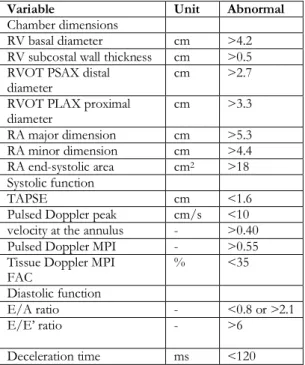
Table 1: Summary of reference limits for recommended measures of right heart structure and function9
Variable Unit Abnormal
Chamber dimensions
RV basal diameter cm >4.2 RV subcostal wall thickness cm >0.5 RVOT PSAX distal
diameter cm >2.7
RVOT PLAX proximal
diameter cm >3.3 RA major dimension cm >5.3 RA minor dimension cm >4.4 RA end-systolic area cm2 >18 Systolic function TAPSE cm <1.6
Pulsed Doppler peak cm/s <10 velocity at the annulus - >0.40 Pulsed Doppler MPI - >0.55 Tissue Doppler MPI
FAC % <35
Diastolic function
E/A ratio - <0.8 or >2.1
E/E’ ratio - >6
Deceleration time ms <120 FAC, Fractional area change; MPI, myocardial performance index; PLAX, parasternal long-axis; PSAX, parasternal short-axis; RA, right atrium; RV, right ventricle; RVD, right ventricular diameter; RVOT, right ventricular outflow tract; TAPSE, tricuspid annular plane systolic excursion.
Pulsed-wave tissue Doppler images were obtained at transducer frequency of 3.5-4.0 MHz, color Doppler screening ratio of 100-140 Hz, and Nyquist limit of 15-20 cm/s. These measurements were obtained from apical 4-chamber view, and by using 5 mm sampling volumes in basal septal and basal lateral tricuspid annular regions. Systolic (Sm: basal septal region, Sa: lateral tricuspid annulus longitudinal systolic velocity), early diastolic (Em: basal septal region, Ea: lateral tricuspid annular region), and late diastolic (Am: basal septal region, Aa: lateral tricuspid annular region) myocardial velocities, and velocities from tricuspid valve annular region, and isovolumetric contraction time (IVCT), contraction time (CT) and isovolumetric relaxation time (IVRT) were measured. Additionally, tricuspid annular plane systolic excursion (TAPSE), which indicates the RV systolic function, was also evaluated. This parameter was analyzed from the right ventricle free wall, by
placing M-mode on the lateral tricuspid annulus (Table 1).
Statistical analysis
Data analysis was performed using ‘Statistical Package for Social Sciences (SPSS) 13 for Windows’ (SPSS Inc., Chicago, Illinois) statistical program. Groups and descriptive data were compared by Chi-square test. Pearson correlation analysis was used in comparison of two continuous variables. When parametric test assumptions were provided, the means of two groups were compared by using the independent-samples-t-test. A p value <0.05 was accepted to be significant. Average - standard deviation from central spreading measures was used for continuous variables. Other distributions were expressed in rates and analyzed by chi-square tests. It was decided to perform parametric tests using the Shapiro Wilk test, one of the normality tests for continuous variables. One Way ANOVA and Student-t tests were used from the parametric tests. Of the nonparametric tests, Mann Whitney U and Kruskal Wallis tests were used. Descriptive statistics were used for the demographic features specified in the study.
RESULTS
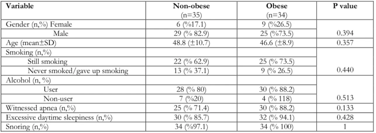
A total of 69 patients, 15 of whom were females, were included in the study. Ages of the cases varied from 28 to 67. Patients were separated to obese and non-obese groups, by considering the body mass indices (BMI) (kg/m²). These groups involved 35 non-obese (6 females, 29 males) and 34 obese (9 females, 25 males) cases. The mean ages of the non-obese and obese patients were found to be 48.8±10.7 years and 46.6±8.9 years, respectively. Tobacco and alcohol use did not differ between the groups (p<0.05) (Table 2). OSAS symptoms like snoring, witnessed apnea and excessive daytime sleepiness questioned before the polysomnographic evaluation, were similar in both groups (Table 2). When the patients were separated to moderate and severe OSAS, demographic data were found to be similar, whereas systolic blood pressure levels were significantly higher in the severe OSAS group (p<0.001).
AHI was determined to be significantly higher in the obese patients; a total of 53 (76%) patients were in the severe OSAS group, and 30 cases (88.2%) from the obese group and 23 cases (35%) from the
non-1295
obese group contributed to this total number of severe OSAS patients. In the obese group; the lowest saturation during sleep and the mean saturation
during sleep were significantly lower; the lowest and the mean saturations during sleep were found to decrease more as the AHI increased.
Table 2. Properties of the obese and non-obese patients
Variable Non-obese (n=35) Obese (n=34) P value Gender (n,%) Female 6 (%17.1) 9 (%26.5) 0.394 Male 29 (% 82.9) 25 (%73.5) Age (mean±SD) 48.8 (±10.7) 46.6 (±8.9) 0.357 Smoking (n,%) 0.440 Still smoking 22 (% 62.9) 25 (% 73.5)
Never smoked/gave up smoking 13 (% 37.1) 9 (% 26.5) Alcohol (n, %)
0.513
User 28 (% 80) 30 (% 88.2)
Non-user 7 (%20) 4 (% 118)
Witnessed apnea (n,%) 25 (% 71.4) 30 (% 88.2) 0.133
Excessive daytime sleepiness (n,%) 30 (% 85.7) 32 (% 94.1) 0.428
Snoring (n,%) 34 (%97.1) 34 (% 100) 1
However, the mean desaturation did not differ significantly between the groups (p=0.234). Minimum oxygen saturation decreased as AHI increased; these two parameters were inversely and significantly correlated with each other (r= -0.450, p=0.013). When patients were separated to moderate and severe OSAS cases; the lowest saturation (86±7 and 74±12 respectively, p=0.020) and the mean saturation (94±1 and 89±6, respectively; p=0.003) were indicated to be significantly lower in the severe OSAS group, but the mean desaturation values were significantly higher (3±1 and 9±9 respectively, p<0.001). When arousal indexes were evaluated in two patient groups; arousal index correlated negatively and significantly with the lowest saturation and the mean saturation (p<0.001); as AHI increased, arousal index also increased significantly, and positively.
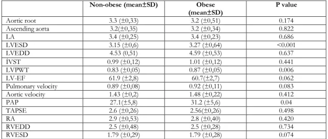
When the lowest, the highest, and the mean heart rates determined during sleep were compared between the two groups, the highest and the mean heart rate values were found to be higher in the obese cases, but this difference between the groups was not statisticaly significant. When transthoracic echocardiography was evaluated; right atrial (RA) size, right ventricular end-systolic diameter (RVESD), right ventricular end-diastolic diameter (RVEDD), ejection fraction (EF), and left atrial (LA) and left ventricular end-diastolic diameters were found to be similar in both groups (Table 3). PAP in the obese patient group was significantly higher when compared to the non-obese cases
(31.2±5.6 mmHg and 27.1±5.8 mmHg, respectively; p=0.04) (Table 3).
In both patient groups, right and left heart functions were primarily evaluated by conventional Doppler; E/A ratio was determined, which was superior to other parameters especially in indicating diastolic dysfunction. In a total of 22 patients, E/A was determined to be <0.8 for the right heart (normal range 0.8-2.1); 10 of these patients (28.6%) were from the non-obese, and 12 (35.3%) were from the obese group. In 32 patients, E/A was found to be <1 for the left heart (normal value >1); 18 of these patients (51.4%) were from non-obese group, and 14 were (41.2%) from the obese patient group (Table 4). However the mean E/A values did not differ significantly between the two groups (E/A for the right heart: in obese group 1.11±0.28, in non-obese group 1.25±0.55, p=0.225; E/A for the left heart: in obese group 1.08±0.33, in non-obese group 1±0.29, p=0.257).
When the right ventricular tissue Doppler parameters were examined; isovolumetric relaxation time (IVRT), one of the parameters that shows diastolic dysfunction (normal value 43-53, mean 48) was determined to be below the mean level in 6 (17.6%) of the obese patients, and in three cases (8.6%) of the non-obese group; however no significant difference was found between the groups (p=0.306). A’a (lateral tricuspid annular region late diastolic myocardial velocity normal value:12-14 cm/s) was found to be significantly lower in the obese cases (Table 5).
1296
Table 3. Evaluation of transthoracic echocardiography results in the obese and non-obese OSAS patients. Non-obese (mean±SD) Obese
(mean±SD) P value Aortic root 3.3 (±0,33) 3.2 (±0,51) 0.174 Ascending aorta 3.2(±0,35) 3.2 (±0,34) 0.822 LA 3.4 (±0,25) 3.4 (±0,23) 0.686 LVESD 3.15 (±0,6) 3.27 (±0,64) <0.001 LVEDD 4.53 (0,51) 4.59 (±0,53) 0.637 İVST 0.99 (±0,12) 1.01 (±0,12) 0.441 LVPWT 0.83 (±0,05) 0.87 (±0,05) 0.006 LV-EF 61.9 (±2,8) 60.7(±2,7) 0.062 Pulmonary velocity 0.89 (±0,08) 0.92 (±0,11) 0.083 Aortic velocity 1.43 (±0,2) 1.48 (±0,22) 0.412 PAP 27.1(±5,8) 31.2 (±5,6) 0.04 TAPSE 2.6 (±0,26) 2.56(±0,26) 0.498 RA 2.9 (±0,53) 2.8 (±0,40) 0.420 RVEDD 2.5 (±0,48) 2.5 (±0,28) 0.734 RVESD 1.79 (±0,29) 1.79 (±0,28) 0.074
LA; Left atrium, LVESD; Left ventricular end-systolic diameter, LVEDD; Left ventricular end-diastolic diameter, İVST; interventricular septum thickness, LVPWT; Left ventricular posterior wall thickness, LV-EF; Left ventricle ejection fraction PAP; pulmonary artery pressure TAPSE; tricuspid annular plane systolic excursion, RA; Right atrium size, RVEDD; Right ventricular end-diastolic diameter, RVESD: Right ventricular end-systolic diameter.
Table 4. Comparison of conventional echocardiography results in the obese and non-obese OSAS patients. Non-obese (mean±SD) Obese (mean±SD) P value
Right V.PWE 0.52 (±0.13) 0.51 (±0.11) 0774 Right V.PWA 0.45 (±0.14) 0.47 (±0.11) 0.623 Right E/A 1.25 (±0.55) 1.11 (±0.28) 0.225 Left V.PWE 0.70 (±0.15) 0.69 (±0.18) 0.024 Left V.PWA 0.68(±0.15) 0.66 (±0.14) 0.569 Left E/A 1 (±0.29) 1.08(±0.33) 0.257
Table 5. Comparison of tissue Doppler echocardiographic results of right heart functions in obese and non-obese OSAS patients.
Non-obese (mean±SD) Obese (mean±SD) P value
Sa 0.13 (±0.02) 0.12 (±0.02) 0.282 E'a 0.11(±0.03) 0.10(±0.03) 0.148 A'a 0.13 (±0.03) 0.11 (±0.04) 0.020 IVRT 69.7(±17.1) 64.5 (±14.4) 0.175 IVCT 71.4(±18.1) 71.8 (±15.2) 0.933 CT 265.5(±29.5) 260.9(±34.4) 0.551 E/E’ 4.9 (±2.19) 5.5 (±2.2) 0.092
Sa: lateral tricuspid annulus longitudinal systolic velocity E’a: lateral tricuspid annular region early diastolic myocardial velocity A’a: lateral tricuspid annular region late diastolic myocardial velocity, İVCT: isovolumetric contraction time, CT: Contraction time, İVRT: isovolumetric relaxation time
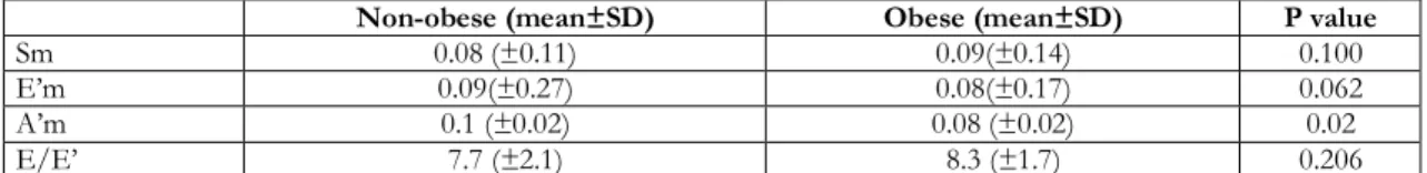
When the left ventricular tissue Doppler parameters were examined, late diastolic myocardial velocity (A), one of the parameters indicating left ventricular diastolic function impairment, was found to be
significantly lower in the obese patient group when compared to non-obese cases. Other parameters were found to be similar in both groups (Table 6).
1297
Table 6.Comparison of tissue Doppler echocardiographic results of left heart functions in obese and non-obese OSAS patients
Non-obese (mean±SD) Obese (mean±SD) P value
Sm 0.08 (±0.11) 0.09(±0.14) 0.100
E’m 0.09(±0.27) 0.08(±0.17) 0.062
A’m 0.1 (±0.02) 0.08 (±0.02) 0.02
E/E’ 7.7 (±2.1) 8.3 (±1.7) 0.206
Sm: basal septal region systolic peak flow velocity, E’m: basal septal region early diastolic and A’m: basal septal region late diastolic myocardial velocity
DISCUSSION
Obstructive sleep apnea (OSA) is a syndrome that affects 1% to 5% of the population, and it increases cardiovascular morbidity and mortality1,2. OSAS
frequency generally increases with age. In a study, prevalence of the patients with AHI value ≥15 increased by a direct proportion until 60 years of age, and the prevalence remained constant at about 20% above 60 years6. In our study, no significant statistical
difference between age groups, moderate and severe OSAS.
It is known that there is a strong relation between BMI and OSAS10. It was indicated in a study that
increases in weight by 10%, increases AHI by 32%, and causes a six-fold increase in the risk of moderate to severe OSAS development11. Being similar with
the literature, AHI increased significantly in our study, by the increase of BMI.
The primary cardiovascular diseases associated with OSAS are HT, ischemic heart disease, cardiac arrhythmias, left heart failure, PHT, right heart failure, polycythemia, stroke and sudden death. The frequency of this syndrome has been determined to be 50% in HT patients, 25% in the patients with chronic heart failure, 30% in those with acute coronary syndrome, and 60% in the cases that developed stroke12.
Negative effects of OSAS on the LV functions have been demonstrated in various studies. Impairment of LV functions caused by OSAS is considered due to increase in the intrathoracic negative pressure, intermittent hypoxia and short awakening periods13,14. In a study Hanlon and his colleagues conducted on 61 children who were obese and had hypertension, Children with OSAS were found to be more likely to have left ventricular hypertrophy (LVH) (85.7% vs. 59.4%, p = 0.047).(OSAS was associated with 4.11 times greater odds of LVH (95%
CI 1.15, 14.65; P = 0.030), remaining significant after adjustment for age, sex, race, and BMI z-score (after adjustment for hypertension, P = 0.051),The higher the intensity of OSAS, the greater the risk of LVH was determined15. Acute pulmonary hemodynamic changes during apneas have also been well-defined 16; however, it is not well-known yet whether OSAS is an independent risk factor for the PHT17.
There are not a large number of studies investigating the relation between OSAS and RV morphology, and the results of studies are variable. In studies evaluating RV diameters and hypertrophy; it was reported that OSAS does not lead to a change in RV end-diastolic and end-systolic diameters, but causes an increase in RV free wall thickness. In a study by Berman et al, RV free wall thickness was observed to increase in 71% of the cases diagnosed as OSAS6. In a study by Hanly et al., RV and LV diameters did not differ significantly between the cases of snoring without apnea or the patients with OSAS18. Guidry et al. have compared two groups, one possessing a mean RDI value of 42, and the other 5. These groups did not differ significantly with regard to RA and RV diameters, and RV systolic function; however, RV anterior free wall thickness was found to increase significantly in the group with high RDI value19. In a meta-analysis published in recent years that evaluated the results of 25 studies; RV dilation, increased wall thickening and changes in RV function were observed in OSA patients20. In our study, in accordance with the previous studies, RV diastolic and systolic diameters and right atrial size were found to be in normal ranges in both groups, and no significant differences were determined between the obese and non-obese groups (p>0.05).
Tavil and the colleagues determined the impairment of right ventricular systolic and diastolic functions in the OSAS patients with/without hypertension21. In the study, they claimed that TAPSE (tricuspid annular plane systolic excursion) is an important
1298
parameter in evaluating right ventricular functions, and tricuspid annular systolic velocity (Sa) obtained by tissue Doppler is important in the evaluation of right ventricular systolic functions. In the study of Karaçaglar and the colleagues in 34 OSAS and 28 healthy people, TAPSE values were found to be significantly lower in OSAS patients, PAP values were found to be significantly higher in the OSAS patient group 22. In our study, the values of TAPSE and tricuspid annular systolic velocity were within normal ranges in all patients, and did not differ between the obese and non-obese OSAS groups. However, in our study, PAP values were significantly higher in obese patients group, similar to the literature.
Willens and the colleagues determined the effects of obesity on the right ventricular and left ventricular functions, by the tissue Doppler imaging; in the study mitral annular systolic velocity, ratio of early-to-late-diastolic tricuspid annular velocity, early-early-to-late-diastolic tricuspid annular velocity, early-diastolic mitral annular velocity, and ratio of early-to-late diastolic mitral annular velocity were found to decrease markedly23. In our study, late-diastolic tricuspid annular velocity (A’a), a tissue Doppler parameter of the right heart, was found to decrease in both groups, and additionally the decrease in the obese OSAS group was significantly more marked, when compared to the non-obese group.
In some studies, it was found out that the impairment of right ventricular function might be seen in overweight and obese individuals without any clinical sign of a marked heart disease. In a study on this subject performed with tissue Doppler imaging, right ventricular dysfunction independent of OSAS was reported to develop in the overweight and obese patients24.
In the study of Li and his colleagues, right ventricular anterior wall thickness (RAW) values gradually increased with OSAS severity and in severe groups, the control was higher than the light groups. TAPSE values in the severe and middle groups were lower than the control group25.
In several studies performed with conventional Doppler technique, left ventricular diastolic functions were determined to be affected in the patients with OSAS. Conventional Doppler technique was used in most of the limited number of studies investigating the effects of OSAS on the left ventricular diastolic functions26. In a study by Kim et
al., it was aimed to investigate a tissue Doppler parameter that can be used to evaluate the left ventricle in the patients with OSAS, and early diastolic wave related with left ventricle was indicated to be markedly lowered in the severe OSAS group. It was additionally observed that this finding was not affected by diabetes mellitus, hypertension and body mass index; they consequently reported that left ventricular early diastolic wave is the most reliable parameter27. In our study, left ventricular end-systolic diameter and left ventricle posterior wall thickness, which are the variables of 2-dimensional echocardiography, were determined to be significantly increased in the obese patients. As also determined in the literature, magnitude of left ventricular early diastolic wave (Em), which is a parameter of Doppler and tissue Doppler, significantly decreased more in the obese patient group when compared with the non-obese group. Another important parameter in evaluating diastolic dysfunctions is the ratio of early diastolic- to late diastolic-mitral annulus velocities (E/A); a value of E/A <1 indicates diastolic dysfunction. In the study conducted by Papanikolaou and the colleagues, 30 severe OSAS (AHI> 30) and 25 control (AHI <5) patients included in the study. In the study, the risk of left ventricular diastolic dysfunction increased in OSAS patients. As AHO increased, there was an increase in the severity of dysfunction. In addition, in the OSAS group, E wave velocity decreased, E / A rates decreased, Em / Am rates increased, and this increase was associated with right ventricular diastolic dysfunction28 . In our study, E/A was determined to be <1 in 32 (46%) cases; of these, 18 (51.4%) were non-obese, and 14 (41.2%) were obese patients. However, the difference between these groups was not found to be significant. This finding indicates that OSAS causes impairment of the left ventricular diastolic functions, independent from obesity. In a study evaluating the relationship between OSAS and hypertension, a linear relation was determined between AHI and blood pressure in the cases taking no antihypertensive treatment29. It is considered that this relation may partly be explained by BMI. Even after corrections for BMI and demographic properties were performed, systolic and diastolic blood pressures were found to be increased with the increase of AHI, and this relation was determined to be significant statistically. We also determined positive and significant correlations between AHI and systolic and diastolic blood pressures.
1299
In a study evaluating whether respiratory disorders during sleep are independent risk factors for hypertension, a high RDI caused statistically significant increases in systolic and diastolic blood pressures in the normotensive cases, and in patients newly diagnosed as hypertensive30. In a study in which Wnag and colleagues investigated the detection rate and factors affecting obstructive sleep apnea syndrome in middle-aged and elderly patients with hypertension, They administered the Berlin survey to 440 hypertension patients and performed polysomnography (PSG) on 235 patients at high risk of OSAS. As a result, they found higher incidence of OSAS in middle-aged and elderly patients with hypertension. They detected high BMI, systolic and diastolic blood pressure as independent risk factors of OSAS 31. In our study, systolic and diastolic blood pressure values were determined to be significantly higher in the patients with severe OSAS. In another study, systolic and diastolic blood pressures were found to be higher in the group of obese patients 32. In our study also, systolic and diastolic blood pressure values were found to be significantly higher in the obese patients with OSAS, when compared with the non-obese OSAS group.
Alveolar hypoxia developing due to repetitive apneas during sleep may cause pulmonary vasoconstriction and increase of pulmonary artery pressure. Daylong persisting pulmonary hypertension and cor pulmonale have been reported in about 20% of the patients. The reason of this fact may be the persistent intimal hyperplasia and media hypertrophy 33. It has been reported that sudden increases in pulmonary artery pressure are frequent in the patients with OSAS; however, proven pulmonary hypertension exists in about 20% of these cases34. In the OSAS patients that pulmonary functions are also impaired, prevalence of pulmonary hypertension has been reported to increase by 41% 35. In our study, the mean systolic pulmonary artery pressure in the obese patient group was found to be significantly higher when compared with the non-obese cases. When the patients were evaluated in two separate groups as cases with moderate or severe OSAS, pulmonary artery pressure values did not differ significantly between the groups.
Our study has several limitations. First of all, in addition to the obese and non-obese OSAS group, we think that the absence of our control group without OSAS creates a significant limitation. Thus, we think that OSAS will be able to see more clearly
the effect on right heart functions. Secondly, we think that there may be an improvement in cardiac functions after OSAS treatment. For this, we think that more studies are needed to show the possible benefits of OSAS treatment.
The results of our study have led to the following conclusions: right ventricle systolic and diastolic functions are impaired in the patients with OSAS. Systolic functions of the left ventricle are conserved, whereas LV diastolic dysfunction is indicated by the tissue Doppler echocardiography. These impairments in cardiac functions are determined significantly in higher rates in obese OSAS cases when compared with the non-obese patients. Because of Cardiovascular complications are considerably frequent in the patients with obstructive sleep apnea, we consider that tissue Doppler echocardiography will be helpful in early determination of the impairment in diastolic functions of both ventricles, especially in the obese OSAS patients. OSAS patients with diastolic function disorders have to be more carefully investigated for the diastolic heart failure symptoms, have to be treated properly for the heart failure and sleep apnea, and also have to be followed-up more frequently.
Yazar Katkıları: Çalışma konsepti/Tasarımı: ND, SG, ABA; Veri
toplama: ND, SG, ABA; Veri analizi ve yorumlama: ND, SG, ABA; Yazı taslağı: ND, SG, ABA; İçeriğin eleştirel incelenmesi: ND, SG, ABA; Son onay ve sorumluluk: ND, SG, ABA; Teknik ve malzeme desteği: ND, SG; Süpervizyon:ND. SG; Fon sağlama (mevcut ise): yok.
Etik Onay: Bu çalışma için Mustafa Kemal Üniversitesi Tayfur Ata
Sökmen Tıp Fakültesi Etik Danışma Kurulundan 20.01.2011 tarih ve 17 nolu kararı ile etik onay alınmıştır.
Hakem Değerlendirmesi: Dış bağımsız.
Çıkar Çatışması: Yazarlar çıkar çatışması beyan etmemişlerdir. Finansal Destek: Yazarlar finansal destek beyan etmemişlerdir. Author Contributions: Concept/Design : ND, SG, ABA; Data acquisition: ND, SG, ABA; Data analysis and interpretation: ND, SG, ABA; Drafting manuscript: ND, SG, ABA; Critical revision of manuscript: ND, SG, ABA; Final approval and accountability: ND, SG, ABA; Technical or material support: ND, SG; Supervision: ND, SG; Securing funding (if available): n/a.
Ethical Approval: For this study, ethics approval was obtained from
the Ethics Advisory Board of Mustafa Kemal University Tayfur Ata Sökmen Faculty of Medicine by Decision No. 17 dated 20.01.2011.
Peer-review: Externally peer-reviewed.
Conflict of Interest: Authors declared no conflict of interest. Financial Disclosure: Authors declared no financial support
REFERENCES
1. Hung J, Whitford EG, Parsons RW, Hillman DR.
Association of sleep apnea with myocardial infarction in men. Lancet. 1990;336:261–64.
2. Mooe T, Franklin KA, Wiklund U, Rubben T,
Holmström K. Sleep-disordered breathing and myocardial ischemia in patients with coronary artery disease. Chest. 2000;117:1597–602.
1300
3. Wolk R, Somers VK. Obesity-related cardiovascular
disease: implications of obstructivesleep apnea. Diabetes Obes Metab. 2006;8:250-260
4. Carskadon MA, Dement WC. Normal human sleep:
An overview. In: Kryger MH, Roth T, Dement WC (eds). Principles and Practice of Sleep Medicine. 3rd Ed. Philadelphia, W.B. Saunders Company. 2000;15-25.
5. Young T, Palta M, Dempsey J, Skatrud J, Weber S,
Badr S. The occurrence of sleep-disordered breathing among middle-aged adults. N Engl J Med. 1993;328:1230–5.
6. Berman E.J, DiBenedetto R.J, Causey DE, Mims T,
Conneff M, Goodman LS. et al. Right ventricular hypertrophy detected by echocardiography in patients with newly diagnosed obstructive sleep apnea. Chest. 1991:100:347-350.
7. Rechtschaffen A, Kales A. A Manual of Standardized Terminology, Techniques and Scoring System for Sleep Stages of Human Subjects. NIH Publication No. 204. Washington, U.S. Government Printing Office. 1968.
8. Iber C, Ancoli-Israel S, Chesson A, Quan S.; for the
American Academy of Sleep Medicine. The AASM manual for the scoring of sleep and associated events: rules, terminology and technical specifications. 1st ed. Westchester, IL: American Academy of Sleep Medicine. 2007
9. Rudski LG, Lai WW, Afilalo J, Hua L, Handschumacher MD, Chandrasekaran K et al. Guidelines for the Echocardiographic Assessment of the Right Heart in Adults: A Report from the American Society of Echocardiography Endorsed by the European Association of Echocardiography, a registered branch of the European Society of Cardiology, and the Canadian Society of Echocardiography. J Am Soc Echocardiogr. 2010;23:685-713.
10. Carskadon MA, Dement WC. Normal human sleep: An overview. In: Kryger MH, Roth T, Dement WC (eds). Principles and Practice of Sleep Medicine. 3rd Ed. Philadelphia, W.B. Saunders Company. 2000;15-25.
11. Young T, Shahar E, Nieto FJ, Redline S, Newman AB,
Gottlieb DJ et al , for the Sleep Heart Health Study Research Group. Predictors of sleep-disordered breathing in community-dwelling adults: The Sleep Heart Health Study. Arch Intern Med. 2002;162:893– 900.
12. Peppard PE, Young T, Patla M. Longitudinal study of moderate weight change and sleep-disordered breathing. JAMA. 2000;284:3015-21.
13. Lattimore JD, Celermajer DS, Wilcox I. Obstructive
sleep apnea and cardiovascular disease. J Am Coll Cardiol. 2003;41:1429-37.
14. Tolle FA, Judy WV, Yu P, Markand ON. Reduced
stroke volume related to pleural pressure in
obstructive sleep apnea. J Appl Physiol. 1983;55:1718-24.
15. Hanlon CE, Binka E, Garafano JS, Sterni LM, Brady TM. The association of Obstructive Sleep Apnea and Left Ventrıculer Hypertrophy in Obese and Overwight Children with History of Elevated Blood Pressure.J Clin Hypertens (Greenwich). 2019;21:984-90.
16. Strollo PJ, Sanders MH. Sleep disorders. Clin Chest Med. 1998;19:1-32.
17. Malone S, Liu PP, Holloway R, Rutherford R, Xie A, Bradlet TD. Obstructive sleep apnoea in patients with idiopathic dilated cardiomyopathy: Effects of continuous positive airway pressure. Lancet. 1991;338:1480-4.
18. Hanly P, Sasson Z, Zuberi N, Alderson M. Ventricular function in snores and patients with obstructive sleep apnea. Chest. 1992;102:100-5.
19. Guidry UC, Mendes LA, Evans JC, Levy D, O’Connor GT, Larson MG., et al. Echocardiographic features of the right heart in sleep-disordered breathing:the Framingham Heart Study. Am J Respir Crit Care Med. 2001;164:933-8.
20. Maripov A, Mamazhakypov A, Sartmyrzaeva M, Akunov A, Uulu KM, Duishobaev M et al. Right ventrıculer remodeling and dysfunction in obstructive sleep apnea; a systematic review of the literature and meta-analysis. Can Respir J. 2017;2017:1587865. 21. Tavil Y, Kanbay A, Sen N, Ulukavak Ciftci T, Abaci
A, Yalçın MR et al. The relationship between aortic stiffness and cardiac function in patients with Obstructive sleep apnea, independently from systemic hypertension. J Am Soc Echocardiogr. 2007;20.366-72.
22. Karaçaglar E, Bal U, Eroğlu S, Çolak A, Bozbaş S, Müderrisoglu H. Pulmonary artery distensibility is worsened in obstructive sleep apnea syndrome. Acta Cardiol Sin. 2019;35:501-507.
23. Willens HJ, Chakko SC, Lowery MH, Byers P,
Labrador E, Gallagher A et al. Tissue Doppler imaging of the right and left ventricle in severe obesity. Am J Cardiol. 2004;94:1087-90.
24. Wong CY, O'Moore-Sullivan T, Leano R, Hukins C, Jenkins C, Marwick TH. Association of subclinical right ventricular dysfunction with obesity. J Am Coll Cardiol. 2006;47:611-6.
25. Li J, Wang Z, Li Y, Meng Y ,Li R, Wang W, et al. Assessment of regional right ventricular systolic function in patients with obstructive sleep apnea syndrome using velocity vector imaging. Medicine. 2016:95:37.
26. Mitra N, Rafael K, Zion S, Patrick J. Impact of obstructive sleep on left ventricular mass and diastolic function. Am J Respir Crit Care. 2001;163:1632-36. 27. Kim SH, Cho GY, Shin C, Lim HE, Kim YH, Song
WH et al. Impact of obstructive sleep apnea on left ventricular diastolic function. Am J Cardiol. 2008;101:1663-8.
1301 28. Papanikolaou J, Ntalapascha M, Makris D,
Koukoubani T, Tsolaki V, Zakynthinos G et al. Diastolic dysfunction in men with severe obstructive sleep apnea syndrome but without cardiovasculer or oxidative strees-releated comorbidities.Ther Adv Respir. 2019;13:1-15.
29. Nieto FJ, Young TB, Lind BK, Shahar E, Samet JM, Redline S et al. Association of sleep-disordered breathing, sleep apnea, and hypertension in a largecommunity-based study. Sleep Heart Health Study. JAMA. 2000;283:1829-36.
30. Grote L, Ploch T, Heitmann J, Knaack L, Penzel T, Peter JH. Sleep-related breathing disorder is an independent risk factor for systemic hypertension. Am J Respir Crit Care Med. 1999;160:1875-82. 31. Wang S,Niu X, Zhang P, Su D, Zhang J, Liu W.
Analysis of OSAS incidence factors in middle aged and elderly patients with hypertension. Minerva
Medica. 2019;110:115-20.
32. Peterson L, Waggoner AD, Schechtman KB, Meyer T, Gropler RJ, Barzilai B et al. Alterations in left ventricular structure and function in young healthy obese women. J Am Coll Cardiol. 2004;43:1399-404. 33. Kessler R, Chaouat A, Weitzenblum E, Oswald M,
Ehrhart M, Apprill M, et al. Pulmonary hypertension in the obstructive sleep apnoea syndrome: prevalence, causes and therapeutic consequences. Eur Respir J. 1996;9:787-94.
34. Chaoat A, Weitzenblum E, Krieger J, Oswald M, Kessler R. Pulmonary hemodynamics in the obstructive sleep apnea syndrome: results in 220 consecutive patients. Chest. 1996;109:380-6. 35. Sajkov D, Cowie RJ, Thornton AT, Espinoza HA,
McEvor RD. Pulmonary hypertension and hypoxemia in obstructive sleep apnea syndrome. Am J Respir Crit Care Med. 1994;149:416-22.