ABSTRACT
This work is licensed under a Creative Commons Attribution-NonCommercial 4.0 International License.
Seher Yılmaz1 , Adem Tokpınar1 , Niyazi Acer2 , Levent Değirmencioğlu3 , Şükrü Ateş1 , Halil Dönmez4 , Serap Baştepe Gray5
Evaluation of Cerebellar Volume in Adult Turkish
Male Individuals: Comparison of three Methods in
Magnetic Resonance Imaging
Objective: Cerebellum plays quite important in our balance by coordinating the control and synergistic movements of the
skeletal muscles. There are many studies in which the volume of the cerebellum is measured and different methods are used. Manual measurements are accepted as the gold standard in these studies, but these measurements are not commonly used due to time and difficulty. The present study aims to compare the cerebellum volume using three different methods.
Materials and Methods: In this study, MR images of 18 men aged between 22 and 30 years were used in the Department
of Radiology of Erciyes University Gevher Nesibe Hospital. In the total cerebellum volume measurements, sagittal images were calculated using the Manual (ImageJ), MRICloud and VolBrain (CERES) methods.
Results: Manual and VolBrain measurements were performed to determine the volume of the cerebellum. The total
vol-ume of the cerebellum was 136.36±12.36 cm3 in manual calculation 125.46±17.26 cm3 in VolBrain and MRICloud1 46.81±20.16.
Conclusion: In this study, it was seen that the three methods used to measure the volume of the cerebellum were compatible
with each other. Because VolBrain volume values are close to the manual method, cerebellum volumes can be obtained by routinely using them.
Keywords: VolBrain, cerebellum, MRICloud, ImageJ, MRI
INTRODUCTION
In recent years, many studies have focused on the cerebrum and cerebellum imaging methods and volumetric mea-surements of various parts of the brain using different methods (1, 2). Since it has many important roles, such as motor coordination, muscle tone, sensitivity, attention and language skills, it is significant to determine these mea-surement standards in the cerebellum (3). Using the anatomy and morphological features of the brain, the progres-sion of the diseases was examined and significant studies were conducted (4). Standard brain structures for regional volumetric quantification to be considered in manual segmentation techniques (5, 6). Automated measurements to find the volume of various regions in the brain provide more advantages than manual measurements. The amount of medical image data produced in clinical and research settings is rapidly growing, resulting in a vast amount of data to analyze. Automatic and reliable quantitative analysis tools, including segmentation, allow us to analyze brain development and to understand specific patterns of many neurological diseases. There is a high probability of error in manual measurements. Fully automatic multi-atlas measurements, such as VolBrain, provide many advantages (7) have also been applied to address these errors. Some previous studies have evaluated the performance of stereology and automated methods compared to manual segmentation in comparatively small populations (8). It may be useful to establish standards with measurements for individual analysis of different brain and cerebellum structures of MR images. The voxel-based morphometry (VBM) toolbox (an extension of the SPM) is also used to measure local GM changes. Partitioning structures, such as brain, cerebellum, brainstem and brain hemispheres and evaluating brain asymmetry, draws attention in recent studies. Various automated measurement models have been developed in hemispheres and split segmentation for cerebellum volumes clearly determined in some MRI studies (9). MRICloud and VolBrain are a web-based platform for automated brain and cerebellum segmentation and distributed remote computing. Studies based on MRICloud are included in the literature. It is a program used for volumetric calculations of the brain and cerebellum in cognitive disorders and other neurological diseases (10). Clinical neuromorphometric studies, such as bipolar disorder, were performed and various classes of classification techniques were applied in major depressive disorder and schizophrenia (11–14). It should be examined in neural disorders, such as cerebellar volume, epilepsy, Parkinson’s syndrome, sleep apnea, brain atrophy, attention-deficit /hyperactivity disorder, autism and schizophrenia (15). Many diseases can change the morphometric structure of the cerebellum, including neurological trauma, diseases, infection, neuro-psychiatric conditions (16, 17).
Cite this article as:
Yılmaz S, Tokpınar A, Acer N, Değirmencioğlu L, Ateş Ş, Dönmez H, et al. Evaluation of Cerebellar Volume in Adult Turkish Male Individuals: Comparison of three Methods in Magnetic Resonance Imaging. Erciyes Med J 2020; 42(4): 405–10.
1Department of Anatomy,
Bozok University Faculty of Medicine, Yozgat, Turkey
2Department of Anatomy,
İstanbul Arel University Faculty of Medicine, İstanbul, Turkey
3Department of Music, Erciyes
University Faculty of Fine Arts, Kayseri, Turkey
4Department of Radiology,
Erciyes University, Faculty of Medicine, Kayseri, Turkey
5Johns Hopkins University,
The Peabody Conservatory, Baltimore, MD, USA
Submitted
01.04.2020
Accepted
21.07.2020
Available Online Date
07.09.2020
Correspondence
Seher Yılmaz, Yozgat Bozok University
Faculty of Medicine, Department of Anatomy, Yozgat, Turkey Phone: +90 354 212 62 01 e-mail: [email protected] ©Copyright 2020 by Erciyes University Faculty of Medicine - Available online at www.erciyesmedj.com
The present study aims to compare VolBrain, manual (ImageJ) and MRICloud methods, a new software line for volumetric cer-ebellum analysis.
MATERIALS and METHODS
Participants
The MRI data in this paper were obtained from the Erciyes Univer-sity Scientific Research Projects Coordination Unit under the grant (TIR-2017-5045).
Structural MR images were performed at the Department of Ra-diology of Erciyes University in Gevher Nesibe Hospital. We used 18 men aged between 22 and 30 years. In the total cerebellum vol-ume measurements, sagittal images were first calculated using the manual (ImageJ) method. Clinical valuations and baseline protocols utilize during recruitment were described in advance (4).
Neuroimaging
MR imaging was performed using a 1.5 T Siemens Aera scan-ner (Siemens, Germany). Constructional images were acquired using the T1-weighted 3D Magnetization Prepared Rapid Gra-dient Echo (MPRAGE) sequence in a sagittal plane using these parameters: flip angle=5o, number of slices=160 and slice thick-ness=1.0 mm, acquisition matrix=256x256, FOV=280 mm2, TE/TR=1900 ms/2.84s.
We downloaded MR T1 data from the scanner, transferred and processed using different software. We saved MR images as NIfTI format. For this purpose, we used a personal computer on a 64-bit Lenovo PC, running Windows 10 operating system.
VolBrain (CERES) (http://VolBrain.upv.es/)
VolBrain is a web-based volume computation of processes aimed to automatically investigate MRI brain data. It works as a black box from the user point of view as it gets an anonymized MRI brain vol-ume in produces and NIFTI format a pdf report with the volvol-umes of the principal IntraCranial Cavity (ICC) tissues (that is white matter, grey matter and cerebrospinal fluid). Furthermore, it provides vol-ume information of some macroscopic areas, such as cerebellum, brainstem and brain hemispheres. Automatic subcortical structure segmentation is performed, label maps and related volumes are provided. Processing time is about ten minutes. This time may vary in proportion to the density of jobs on the webserver (19). The VolBrain system is based on a developed pipeline provid-ing automatic segmentation of different brain structures from T1-weighted MR images (7).
MRICloud (https://mricloud.org)
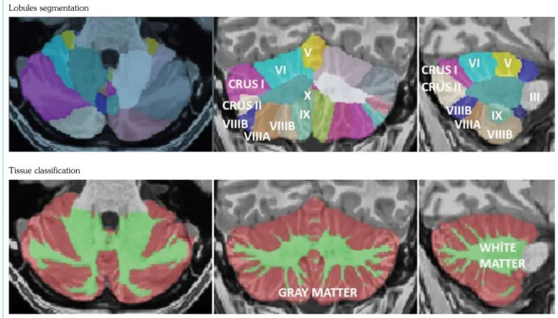
MRICloud provides a fully automated cloud service for brain par-cellation of MPRAGE images based on multiple atlas likelihood fusion algorithm, an ontology level control technology The at-las used for the processing of our data was the adult_286labels_ 11atlases_V5L and JHU multi-atlas inventories with 286 defined structures (Fig. 1, 2) (18).
ImageJ
ImageJ is a public domain Java image processing program created by NIH Image for the Macintosh. It runs, as a downloadable
appli-cation or either as an online applet, on whichever computer with a Java 1.4 or later virtual machine. Downloadable distributions are available for Linux, Windows, Mac OS X and Mac OS. It can print 8-bit, 16-bit and 32-bit images and display, edit, analyze, process, save. It can read plenty of image formats, including FITS, GIF, BMP, JPEG, DICOM, TIFF, and “raw. Its assistance “stacks”, a series of images that share a single window (19).
In the images opened in the ImageJ program, the cerebellum vol-ume was calculated by adding CTRL + M areas of cerebellum. Statistical Analysis
In the data obtained, statistical analysis was conducted on the com-puter using IBM SPSS 22.0 program. In the data obtained, five parameters were evaluated kurtosis, skewness, mean-standard de-viation ratio, histogram plot, Shapiro-Wilko test and normality test were performed. Non-parametric chi-square test was chosen for the comparison between the groups. In statistical analysis, α=0.05 was taken and p<α was significant and p>α was considered statis-tically insignificant.
RESULTS
Using the VolBrain (CERES) web-based program, the brain and cerebellum volumes are calculated automatically, as well as the ra-tio of white matter and gray matter (Fig. 3).
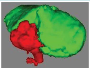
Figure 1. Appearance of cerebellum
Cerebellum volumes were measured using MRICloud, VolBrain and ImageJ programs. There was no statistically significant differ-ence between these three different methods (p>0.05). The results of these three methods were close to each other. The methods can be used interchangeably.
Graphical comparisons of MRICloud-ImageJ, MRICloud-VolBrain and VolBrain-ImageJ Bland-Altman were performed (Fig. 4). Cerebellar segmentation was accomplished with CERES (25). T1 data were loaded at http://VolBrain.upv.es web address and re-sults were obtained.
Internal software extracted volumetric data from downloaded CERES results tables. The data obtained on the CERES website included left and right measurements for the whole cerebellum volume.
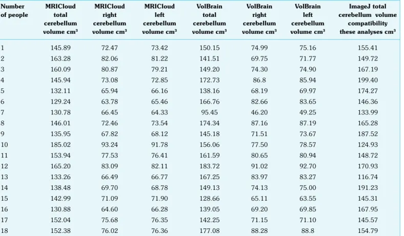
In the MRICloud program, total cerebellum volume, right cere-bellum volume and left cerecere-bellum volume were calculated in 18 men (Tables 1, 2).
When the mean cerebellum volume in this study is compared with the three methods, the results are similar. Cerebellum volume was measured as 152.101 cm3 in VolBrain (CERES) software, 154.504
Differ ence (MRICloud-ImageJ) Differ ence (MRICloud-V olBrain) Differ ence (V olBrain-ImageJ)
Mean (MRICloud-ImageJ) Mean (MRICloud-VolBrain) Mean (VolBrain-ImageJ)
75.00 40.00 50.00 50.00 20.00 25.00 25.00 0.00 0.00 0.00 -25.00 -20.00 -25.00 -50.00 -40.00 -50.00 180.00 180.00 200.00 170.00 160.00 160.00 180.00 150.00 140.00 140.00 140.00 160.00 130.00 120.00 120.00 100.00 120.00 a b c
Figure 4. (a) MRICloud-ImageJ Bland-Altman graphic. (b) MRICloud-VolBrain Bland-Altman graphic. (c) VolBrain-ImageJ Bland-Altman graphic
Lobules segmentation
Tissue classification
Figure 3. In our study, the image of a 24-year-old male brain parcel was examined. Lobules can be seen in the V-X middle (coronal) slice and lobules III-IX in the right (sagittal) slice. Gray matter and white matter structure are examined in the following 3-up image
cm3 in MRICloud software, and 158.077 cm3 in the ImageJ pro-gram. As a result of the studies obtained, there was no difference between the three different measurement parameters.
DISCUSSION
One of the significant features of cloud-based analysis methods is that the user does not have to install the software on his personal computer, so it does not put any strain on the version or operating system compatibility. These analysis methods also make it easy to access shared high-level computing resources and do not have to be limited by the memory capacity of the user’s own computer. In addition, cloud-based analysis methods allow more efficient imple-mentation of the software update and error correction (20).
This study aims to calculate and compare these methods using three different methods in measuring cerebellum volume. Vol-Brain, MRICloud and ImageJ programs were used for measure-ment. Findings obtained from this study are highly reliable and time-saving. With its role in balance, other motor skills and coordi-nation, the cerebellum is believed to make significant contributions to various cognitive and emotional functions (2). Regarding the clinical significance of non-motor functions of the cerebellum, it becomes important in patients who demonstrate the relationship between behavioral changes and cerebellar injury. Cerebellum has many cognitive roles, such as dysprosodia, agrammatism, visual impairment, diffuse vision, verbal fluency, reduced working memo-ry and abstract reasoning. Our findings can help inform another in-vestigator about the considerable advantages and caveats the same noted in adult populations, as in advance reported. These core properties of spatial, executive and linguistic describe the cerebellar cognitive affective syndrome and accepted notions of dementia. Cerebellar cognitive affective syndrome is distinguishable from oth-er subcortical syndromes by virtue of the symptom complex con-sisting of disturbances in spatial, executive affective functions and linguistic, routinely evaluated on MRI in diseases, such as X-linked adrenoleukodystrophy, 4H syndrome (hypomyelination, hypodon-tia and hypogonadotropic hypogonadism) and metachromatic leu-kodystrophy Pelizaeus-Merzbacher’s disease (21). MRI images can be assessed for determined criteria like localization of lesions, pro-gression or repro-gression of the disease, along with changes in brain volume. In recent years, brain volumes have been investigated for diseases, such as hydrocephalus, atrophy, Huntington and Parkin-son’s disease, multiple sclerosis, human immunodeficiency virus Table 1. Cerebellum volume measurement through three separate programs
Number MRICloud MRICloud MRICloud VolBrain VolBrain VolBrain ImageJ total
of people total right left total right left cerebellum volume
cerebellum cerebellum cerebellum cerebellum cerebellum cerebellum compatibility
volume cm3 volume cm3 volume cm3 volume cm3 volume cm3 volume cm3 these analyses cm3
1 145.89 72.47 73.42 150.15 74.99 75.16 155.41 2 163.28 82.06 81.22 141.51 69.75 71.77 149.72 3 160.09 80.87 79.21 149.20 74.30 74.90 167.19 4 145.94 73.08 72.85 172.73 86.8 85.94 199.40 5 132.11 65.94 66.16 138.16 68.19 69.97 174.27 6 129.24 63.78 65.46 166.76 82.66 83.65 146.36 7 130.78 66.45 64.33 95.45 46.20 49.25 133.99 8 146.01 72.46 73.54 174.34 87.16 87.19 165.28 9 135.95 67.82 68.12 145.18 71.51 73.67 187.52 10 185.02 93.24 91.78 156.06 77.50 78.57 124.93 11 153.94 77.53 76.41 161.59 80.65 80.94 148.72 12 165.20 83.09 82.11 183.72 91.02 92.70 170.93 13 133.26 66.49 66.77 167.25 83.97 83.27 116.74 14 138.48 69.70 68.78 149.13 74.13 75.00 191.23 15 142.99 71.09 71.90 128.66 65.11 63.55 145.31 16 130.88 64.60 66.28 139.05 69.20 69.85 167.95 17 152.04 75.68 76.35 142.25 71.15 71.10 145.57 18 152.38 76.02 76.36 177.08 88.28 88.8 154.79
Table 2. Average values of measurements
Measurements Mean±SD Min.–Max.
MRICloud total cerebellum volume cm3 146.81±14.49 129.24–185.02
MRICloud right cerebellum volume cm3 72.79±8.09 60.47–93.24
MRICloud left cerebellum volume cm3 72.50±7.88 57.42–91.78
VolBrain total cerebellum volume cm3 152.12±20.40 95.45–183.72
VolBrain right cerebellum volume cm3 75.69±10.41 46.20–91.02
VolBrain left cerebellum volume cm3 76.40±10.00 49.25–92.70
ImageJ total cerebellum volume cm3 158.07±21.54 116.74–199.40
(HIV) -related leukoencephalopathy and progressive supranucle-ar palsy (22). Caviness et al. measured cerebellum volumes in 15 males and 15 females between 7 and 11 years of age. They report-ed that the cerebellum volume of the boys was less than that of the cerebellum in females but that the cerebellum volume of the girls was close to the cerebellum of the adults. The reason for this was that males’ cerebellum volumes developed later than females (23). Baykan et al. reported that cerebellum volume was 103.3 cm3 for infants, 148.8 cm3 for children and 153 cm3 for adolescents (24). Acer et al. (2008) used the point-counting and planimetry methods for cerebellum volume estimation. They found that the planime-try method was 116.69±10.1 and 114.41±9.3 cm3 in males and females, respectively. The mean results of the point-counting method were 116.34±10.6 and 113.48±8.8 cm3 in males and females, respectively. Total cerebellar volumes obtained by two dif-ferent methods were not statistically difdif-ferent (25).
Tiemeier et al., in a study of 50 people aged 5–24 years, reported that cerebellar volume was 10 to 13% higher in males (26). Sullivan et al. examined cerebellum segmentation in alcohol users and correlations between 6 and 10 lobules were determined using high glu-glutamyl transferase (GGT) levels, CERES and suites that reflect liver function (27).
Romero et al. another important feature of CERES in another study performed its adaptability. As it uses the similarity of manu-ally segmented templates as an information segment, it can learn new anatomy by adding new cases to the library (28).
In individuals with the pathology of the anterior superior delivery of the cerebellum, postural balance is disturbed. Sullivan et al., in another study they conducted, they pointed out this balance, which provides a suitable model to study the relationship between known vermian pathology and chronic alcoholism, cerebellar pathology, and postural stability.
CONCLUSION
In this study, cerebellum volumes were calculated using three dif-ferent methods using MR images of 18 people between the ages of 22–30. The results of three different methods used were seen close to each other. Thus, rapid automated methods for measuring cerebellum volume in MR images can be used. The use of brain volume determination with automatic calculation can be a rapid radiological guide to diagnose or monitor disease status in the pos-terior fossa and brain stem.
Ethics Committee Approval: The Erciyes University Clinical Research Ethics Committee granted approval for this study (date: 21.02.2014, num-ber: 2014/122).
Informed Consent: Written informed consent was obtained from patients who participated in this study.
Peer-review: Externally peer-reviewed.
Author Contributions: S. Bastepe-Gray, N. Acer designed the study. L. Degirmencioglu, H. Donmez and N. Acer collected the data. S. Yılmaz, N. Acer, A. Tokpınar and Ş. Ateş analyzed the data. All authors wrote this pa-per. All authors contributed to the data interpretation, review and revision of this manuscript.
Conflict of Interest: The authors have no conflict of interest to declare.
Financial Disclosure: This work was supported by the Erciyes University Scientific Research Projects Coordination Unit under grant number TIR-2017-5045.
REFERENCES
1. Vulturar D, Fărcăşanu A, Turcu F, Boitor D, Crivii C. The volume of the cerebellum in the second semester of gestation. Clujul Med 2018; 91(2): 176–80. [CrossRef]
2. Hashimoto N, Michaels TI, Hancock R, Kusumi I, Hoeft F. Maternal cerebellar gray matter volume is associated with daughters’ psychotic experience. Psychiatry Clin Neurosci 2020; 74(7): 392–7. [CrossRef]
3. Manto M. Cerebellar motor syndrome from children to the elderly. Handb Clin Neurol 2018; 154: 151–66. [CrossRef]
4. Akudjedu TN, Nabulsi L, Makelyte M, Scanlon C, Hehir S, Casey H, et al. A comparative study of segmentation techniques for the quantifi-cation of brain subcortical volume. Brain Imaging Behav 2018; 12(6): 1678–95. [CrossRef]
5. Kocaman H, Acer N, Köseoğlu E, Gültekin M, Dönmez H. Evaluation of intracerebral ventricles volume of patients with Parkinson’s disease using the atlas-based method: A methodological study. J Chem Neuro-anat 2019; 98: 124–30. [CrossRef]
6. Ünalmış D, Acer N, Yılmaz S, Tokpınar A, Doğan S, Demir, et al. The Calculation of the Femoral Condyle Cartilage Volume and Sur-face Area in Patients with Osteoarthritis. Erciyes Med J 2020; 42(2): 178–84.
7. Manjón JV, Coupé P. volBrain: An Online MRI Brain Volumetry Sys-tem. Front Neuroinform 2016; 10: 30. [CrossRef]
8. Makowski C, Béland S, Kostopoulos P, Bhagwat N, Devenyi GA, Malla AK, et al. Evaluating accuracy of striatal, pallidal, and thalamic segmen-tation methods: Comparing automated approaches to manual delinea-tion. Neuroimage 2018; 170: 182–98. [CrossRef]
9. Aydin S, Ozoner B. Comparative Volumetric Analysis of the Brain and Cerebrospinal Fluid in Chiari Type I Malformation Patients: A Morpho-logical Study. Brain Sci 2019; 9(10): 260. [CrossRef]
10. Sakamoto R, Marano C, Miller MI, Lyketsos CG, Li Y, Mori S, et al. Cloud-Based Brain Magnetic Resonance Image Segmentation and Par-cellation System for Individualized Prediction of Cognitive Worsening. J Healthc Eng 2019; 2019: 9507193. [CrossRef]
11. Rentería ME, Schmaal L, Hibar DP, Couvy-Duchesne B, Strike LT, Mills NT, et al. Subcortical brain structure and suicidal behaviour in major depressive disorder: a meta-analysis from the ENIGMA-MDD working group. Transl Psychiatry 2017; 7(5): e1116. [CrossRef]
12. Mamah D, Alpert KI, Barch DM, Csernansky JG, Wang L. Subcortical neuromorphometry in schizophrenia spectrum and bipolar disorders. Neuroimage Clin 2016; 11: 276–86. [CrossRef]
13. van Erp TG, Hibar DP, Rasmussen JM, Glahn DC, Pearlson GD, An-dreassen OA, et al. Subcortical brain volume abnormalities in 2028 individuals with schizophrenia and 2540 healthy controls via the ENIG-MA consortium. Mol Psychiatry 2016; 21(4): 547–53. Erratum in: Mol Psychiatry 2016; 21(4): 585.
14. Hibar DP, Westlye LT, van Erp TG, Rasmussen J, Leonardo CD, Fas-kowitz J, et al. Subcortical volumetric abnormalities in bipolar disorder. Mol Psychiatry 2016; 21(12): 1710–6. [CrossRef]
15. O’Callaghan C, Hornberger M, Balsters JH, Halliday GM, Lewis SJ, Shine JM. Cerebellar atrophy in Parkinson’s disease and its implication for network connectivity. Brain 2016; 139(Pt 3): 845–55. [CrossRef]
16. Sussman D, Leung RC, Chakravarty MM, Lerch JP, Taylor MJ. The developing human brain: age-related changes in cortical, subcortical, and cerebellar anatomy. Brain Behav 2016; 6(4): e00457. [CrossRef]
17. Huhtaniska S, Jääskeläinen E, Heikka T, Moilanen JS, Lehtiniemi H, Tohka J, et al. Long-term antipsychotic and benzodiazepine use and brain volume changes in schizophrenia: The Northern Finland Birth Cohort 1966 study. Psychiatry Res Neuroimaging 2017; 266: 73–82. 18. Otsuka Y, Chang L, Kawasaki Y, Wu D, Ceritoglu C, Oishi K, et al.
A Multi-Atlas label fusion tool for neonatal brain MRI parcellation and quantification. J Neuroimaging 2019; 29(4): 431–9. [CrossRef]
19. Tokpınar A, Ülger H, Yılmaz S, Acer N, Ertekin T, Görkem SB, et al. Examination of inclinations in spine at childhood and adolescence stage. Folia Morphol 2019; 78(1): 47–53.
20. Li Y, Liu P, Li Y, Fan H, Su P, Peng SL, et al. ASL-MRICloud: An online tool for the processing of ASL MRI data. NMR Biomed 2019; 32(2): e4051. [CrossRef]
21. Vrij-van den Bos S, Hol JA, La Piana R, Harting I, Vanderver A, Barkhof F, et al. 4H Leukodystrophy: A Brain Magnetic Resonance Imaging Scoring System. Neuropediatrics 2017; 48(3): 152–60. 22. Butzkueven H, Kolbe SC, Jolley DJ, Brown JY, Cook MJ, van der
Mei IA, et al. Validation of linear cerebral atrophy markers in multiple sclerosis. J Clin Neurosci 2008; 15(2): 130–7. [CrossRef]
23. Caviness VS Jr, Kennedy DN, Richelme C, Rademacher J, Filipek PA.
The human brain age 7–11 years: a volumetric analysis based on mag-netic resonance images. Cereb Cortex 1996; 6(5): 726–36. [CrossRef]
24. Baykara S, Atmaca M, Yıldırım H. Comparison of Cerebellum Vol-umes of the Patients With Healthy Controls’ in Conversion Disorder. KSU Med J 2017; 12(3): 12–8.
25. Acer N, Sahin B, Usanmaz M, Tatoğlu H, Irmak Z. Comparison of point counting and planimetry methods for the assessment of cerebel-lar volume in human using magnetic resonance imaging: a stereological study. Surg Radiol Anat 2008; 30(4): 335–9. [CrossRef]
26. Tiemeier H, Lenroot RK, Greenstein DK, Tran L, Pierson R, Giedd JN. Cerebellum development during childhood and adolescence: a longitudinal morphometric MRI study. Neuroimage 2010; 49(1): 63–70. [CrossRef]
27. Sullivan EV, Zahr NM, Saranathan M, Pohl KM, Pfefferbaum A. Con-vergence of three parcellation approaches demonstrating cerebellar lobule volume deficits in Alcohol Use Disorder. Neuroimage Clin 2019; 24: 101974. [CrossRef]
28. Romero JE, Coupe P, Giraud R, Vinh-Thong Ta, Fonov V, Park MTM, et al. CERES: A new cerebellum lobule segmentation method. Neuro-Image 2017; 147: 916–24. [CrossRef]