Clues to the diagnosis of biliary atresia in neonatal cholestasis
Mehmet Ağın1, Gökhan Tümgör1, Murat Alkan2, Önder Özden2, Mehmet Satar3, Recep Tuncer2 1Department of Pediatric Gastroenterology, Çukurova University Medical Faculty, Adana, Turkey
2Department of Pediatric Surgery, Çukurova University Medical Faculty, Adana, Turkey 3Department of Neonatalogy, Çukurova University Medical Faculty, Adana, Turkey
INTRODUCTION
Cholestasis is a clinical condition characterized by a de-creased flow of canalicular bile and direct hyperbilirubi-nemia. Decreased bile flow leads to the accumulation of substances, such as bile acids, that would normally be expelled in the bile. Cholestasis developing in early life can arise from a hepatic response to exogenous agents or because of a specific congenital pathology (1). The evaluation of patients with cholestasis is difficult be-cause of the variety of cholestatic syndromes, the fact that the pathogeneses are not fully understood, and that the clinical findings are not specific to the disease. Because of the severity of conditions leading to neona-tal cholestasis, it is essential to diagnose cholestasis and its underlying cause early. Differential diagnosis helps
clinicians identify diseases that do not respond to spe-cific treatments and provide appropriate general sup-portive therapy. The most common causes of choles-tatic jaundice in the first month of life are biliary atresia (BA) and neonatal hepatitis. Diagnostic evaluation must be performed within 45–60 days in the early period of life, and BA must be excluded (2).
Biliary atresia is one of the most common causes of cholestasis in newborns and infants, being responsible for approximately one in three of all cases of neonatal cholestasis and for at least 90% of all cases of obstruc-tive cholestasis (3). It is a destrucobstruc-tive, inflammatory cholangiopathy affecting both the intra- and extrahe-patic bile ducts (4). Time is of great importance in BA: if the Kasai procedure is not performed early in life, Address for Correspondence: Gökhan Tümgör E-mail: [email protected]; [email protected]
Received: October 05, 2015 Accepted: December 04, 2015
© Copyright 2016 by The Turkish Society of Gastroenterology • Available online at www.turkjgastroenterol.org • DOI: 10.5152/tjg.2015.150379
BILIARY
Or
iginal Ar
ticle
ABSTRACT
Background/Aims: The purpose of this study was to identify important clues in differentiating biliary atresia (BA) from causes of neonatal cholestasis other than BA (non-BA) and establishing the reliability of current tests. Materials and Methods: Thirty-four patients with BA and 27 patients with non-BA cholestasis being monitored at the Çukurova University Medical Faculty, the Pediatric Gastroenterology Department and the Pediatric Sur-gery Department between 2009 and 2015 were retrospectively assessed.
Results: Cases of early onset jaundice, acholic stool, gamma-glutamyl transferase (GGT) elevation, and absent or small gallbladder on ultrasonography (USG) were greater in the BA group, while the levels of consanguinity and splenomegaly were higher in the non-BA group. The highest positive predictive value and specificity was determined for a GGT level greater than 197 in addition to absent or small gallbladder on USG and acholic stool in the BA group. Moreover, the presence of acholic stool (97%) exhibited the highest sensitivity and accuracy in the diagnosis of BA.
Conclusion: Pale stool, GGT elevation, and absent or small gallbladder on USG are the most reliable tests for diagnosing BA. We recommend that intraoperative cholangiography should be performed without waiting for further test results when a neonate or infant presents with acholic stool, high GGT values, and absent or small gallbladder on abdominal USG.
death can occur within 2 years from biliary cirrhosis and liver failure (5). However, it is difficult to differentiate the causes of BA and other causes of neonatal cholestasis (non-BA) based on histopathology, biochemistry, clinical examination, or imaging alone, with no single test known to provide a definitive diag-nosis of BA (6). Clinicians must, therefore, make their differential diagnosis by combining several parameters.
The purpose of this retrospective study was to determine the sensitivity and specificity of the tests used in the differential di-agnosis of BA, assessed both separately and in combination; to better explain the requirement for intraoperative cholangiog-raphy; and to facilitate earlier diagnosis.
MATERIALS AND METHODS
This study was conducted at the Çukurova University Faculty of Medicine, the Department of Pediatric Gastroenterology, Hep-atology, and Nutrition, and the Department of Pediatric Sur-gery. The records of 61 patients diagnosed with neonatal cho-lestasis were retrospectively assessed for the period between 2009 and 2015. For the neonates and infants, age, sex, time of onset of jaundice, presence and time of onset of acholic stools, birth weight, birth week, and physical examination findings were recorded. In addition, we recorded the number of ma-ternal pregnancies, parental consanguinity, and a history of a similar disease in the family.
Details of the following tests were assessed: liver function tests for cholestasis, complete blood count, reticulocyte count, blood gas analysis, serum biochemistry [transaminase levels, total and conjugated bilirubin levels, alkaline phosphatase, gamma-glutamyl transferase (GGT), total protein, albumin, blood sugar, triglyceride, and cholesterol levels], ammonia, lactate, pyruvate, alpha-1-antitripsin, ferritin, alpha-fetoprotein, prothrombin time, activated partial prothrombin time (aPTT), the international normalized ratio, thyroid function tests, sero-logical examinations (hepatitis A, B, and C virus, Herpes simplex types 1 and 2, Epstein–Barr virus, cytomegalovirus, and toxo-plasma), sweat test, urine and blood amino acids, reducing substances in urine, tandem mass and urine organic acids, and blood and urine cultures. Abdominal ultrasonography (USG) and echocardiography were used as diagnostic imaging tools. The gallbladder was considered abnormal if it was not visual-ized and if it was less than 1.9-cm long (7). All patients in the study underwent ocular examination to identify metabolic dis-eases or syndromes. Babies with syndromes, born prematurely and hospitalized in the neonatal unit, with anatomical bile tract problems other than BA, sepsis, or receiving total parenteral nutrition were excluded from the study. The diagnosis of BA was made by intraoperative cholangiography (n=34). Cases without BA at intraoperative cholangiography and those with other causes of cholestasis were included in the non-BA group (n=27; idiopathic neonatal hepatitis 21, Niemann–Pick 2, cystic fibrosis 1, congenital toxoplasma 1, glycogen storage disease 1, and progressive familial intrahepatic cholestasis 1). Cases were
evaluated in terms of history and physical examination, labora-tory, and radiology findings.
Statistical analysis
Statistical tests were performed using the Statistical Package for the Social Sciences (SPSS) version 20.0 software (IBM; Armonk, NY, USA). The results are expressed as mean±standard devia-tion (SD). The chi-square test and Student’s t-test were used for comparisons. A p-value of <0.05 was regarded as significant. The accuracy of a test was calculated using the following for-mula: [(accurately diagnosed cases/performed cases)×100]. Sensitivity, specificity, and likelihood ratios were calculated with their 95% confidence intervals (CIs). The optimal cutoff level for serum GGT activity to diagnose BA was established using receiver operating characteristic curves.
The protocol was approved by the Ethics Committee in Çuku-rova University, Turkey.
RESULTS
The mean age of patients was 59 days; 29 patients were male and 32 patients were female. The most common symptom at presentation was jaundice, which was seen in all cases, followed by acholic stool (n=44; 72%), restlessness (n=20; 32%), nutrition disorder (n=15; 24%), delayed weight gain (n=11; 18%), and ab-dominal swelling (n=1; 1.6%). Parental consanguinity was pres-ent in 23 cases (23/61; 37%). However, none of the families re-ported a history of a similar disease or death of a sibling. BA was diagnosed in 34 (77%) of the 44 patients that under-went intraoperative cholangiography, and Kasai portoenteros-tomy was performed in all these cases. Cholangiography was normal in 10 cases, and these were included in the non-BA group. Female gender was slightly dominant in the BA group (BA: 15 males versus 19 females; non-BA: 14 males versus 13 females); however, the difference was not statistically different between the groups (p>0.05).
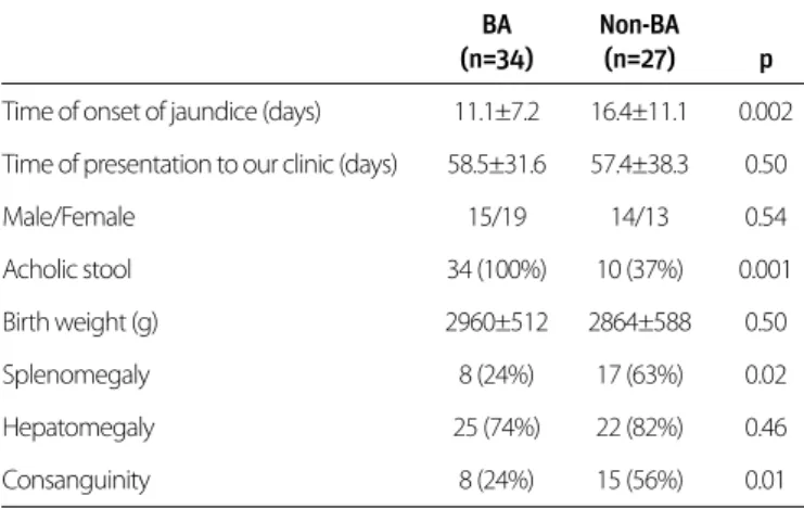
Consanguinity was significantly higher in the non-BA group (n=15; 55%) than in the BA group (n=8; 23%; p<0.01). Jaundice was recognized after a mean 11.1±7.2 days in the BA group and after 16.4±11 days in the non-BA group (p<0.002). No sta-tistically significant difference was observed in the liver size between the BA and non-BA groups on physical examination, although the spleen size was significantly greater in the non-BA group (p<0.02). Acholic stool was observed in all patients in the BA group but only in 10 cases (37%) in the non-BA group. The difference was statistically significant (p<0.001). On echo-cardiography, atrial isomerism and a persistent left superior vena cava were observed in one case with BA. In addition, atrial and ventricular septal defects were present in one case with BA. In the non-BA group, an atrial septal defect was observed in one case, left ventricular hypertrophy in another, and multiple cardiac anomalies in two cases. No statistically significant dif-ferences were present between the two groups (Table 1).
Or
iginal Ar
No significant difference was determined between groups with respect to serum aspartate aminotransaminase (AST), ala-nine aminotransaminase (ALT), total bilirubin, direct bilirubin, coagulation, triglyceride, or cholesterol values. However, serum GGT values were significantly higher in the BA group than in the non-BA group (p<0.001) (Table 2).
Abdominal USG was performed in all cases with cholestasis. The gallbladder either was not visualized or small in 23 cases (67%) in the BA group. Polysplenia was determined in one case. The gallbladder was small in eight (8/27; 29%) cases in the non-BA group. When comparing abdominal USG findings between the groups, there was a statistically significant difference in the presence of a gallbladder (p<0.002).
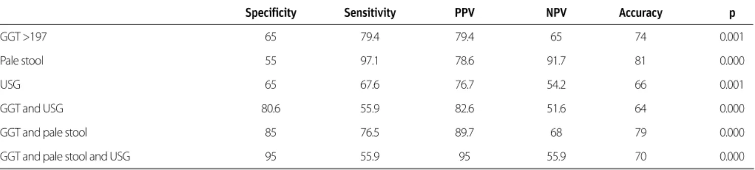
In brief, early onset jaundice, acholic stool, GGT elevation, and absent or small gallbladder on USG were greater in the BA group, while the levels of consanguinity and splenomegaly were higher in the non-BA group. Therefore, we calculated the specificity, sensitivity, positive predictive value (PPV), negative predictive value (NPV), and the accuracy of acholic stool, GGT elevation, and absent or small gallbladder on USG for the dif-ferential diagnosis of BA. The results are summarized in Table 3. DISCUSSION
The North American Society for Pediatric Gastroenterology, Hep-atology and Nutrition guideline for the evaluation of cholestatic jaundice in neonates and infants recommends that any neonate observed to be jaundiced at the 2-week well child visit should be evaluated for cholestasis (8). Early diagnosis of the various metabolic diseases that lead to cholestasis, such as sepsis, hy-pothyroidism, panhypopituitarism, and galactosemia, can be life saving for babies (9). The earlier that BA can be diagnosed, the greater the chance of a successful Kasai procedure (10).
Despite advances in molecular biology, biochemistry, serology, and imaging techniques, no method by itself is sufficient to di-agnose the cause of neonatal cholestasis. Indeed, diagnosis can
only be established using all available methods (9). Our clinic is a reference center for cases with cholestasis. Large numbers of cases of cholestasis are referred to our clinic from the East-ern Anatolia and Southeast Anatolia regions. We therefore saw large numbers of cases of biliary atresia over the 6 years. In Turkey, hepatobiliary scintigraphy is not available; therefore, because the histopathologies of BA and several other diseases that cause neonatal cholestasis are similar, we perform early intraoperative cholangiography whenever BA is suspected. BA was diagnosed in 34 of 44 cases by this method. The decision to perform intraoperative cholangiography is based on case history, physical examination, the presence of acholic stool, GGT elevation, and USG results. However, the decision to de-cide whether to perform intraoperative cholangiography can remain problematic despite these tests; therefore, we aimed to investigate the sensitivity and specificity of these tests when used separately and in combination.
Similar to our study, other researchers have determined that jaundice begins earlier in patients with BA (9,11,12). In our study, the presence of acholic stool and early onset jaundice were key features suggestive of BA, while parental consanguin-ity and splenomegaly were suggestive of non-BA cholestasis. In the literature, acholic stool was present in 83%–94.7% of pa-tients with BA compared with 45.7%–56.5% of papa-tients with non-BA cholestasis (12-14). The specificity and sensitivity of acholic stool in cases of BA have also been reported at 74%– 86% and 83%–100%, respectively (11, 15, 16). Here, we report that acholic stool was present in all cases with BA and in 19 (37%) of the non-BA cases, with a sensitivity of 97.1%,
specific-Or
iginal Ar
ticle
BA Non-BA (n=34) (n=27) p Time of onset of jaundice (days) 11.1±7.2 16.4±11.1 0.002 Time of presentation to our clinic (days) 58.5±31.6 57.4±38.3 0.50
Male/Female 15/19 14/13 0.54 Acholic stool 34 (100%) 10 (37%) 0.001 Birth weight (g) 2960±512 2864±588 0.50 Splenomegaly 8 (24%) 17 (63%) 0.02 Hepatomegaly 25 (74%) 22 (82%) 0.46 Consanguinity 8 (24%) 15 (56%) 0.01
BA: biliary atresia; non-BA: non-biliary atresia
Table 1. Comparison of demographic characteristics and physical examination findings in the biliary atresia and non-biliary atresia cases
BA Non-BA (n=34) (n=27) p White cell, /mm3 11710±5100 11991±269 0.936 Hemoglobin, g/dL 10.2±1.5 10.1±1.3 0.227 Platelet, /mm3 364647±140,721 365814±196,744 0.100 AST, IU/L 284±260 279±208 0.690 ALT, IU/L 140±98 146±152 0.309 T.Bil, mg/dL 10.1±4 10.3±4.5 0.879 D.Bil, mg/dL 6.7±2.8 6.3±3.2 0.282 ALP, IU/L 889±656 651±502 0.182 GGT, IU/L 606±561 251±252 0.001 Total protein, g/dL 5.3±0.7 5.3±0.8 0.374 Albumin, g/dL 3.3±0.4 3.3±0.5 0.628 Triglyceride, mg/dL 185±59 169±76 0.381 Total cholesterol, mg/dL 225±77 198±68 0.352
AST: aspartate aminotransaminase; ALT: alanine aminotransaminase; T. bil; total bilirubin; D. Bil: direct bilirubin; ALP: alkaline phosphatase; GGT: gamma glutamyl transferase
Table 2. Comparison of biliary atresia and non-biliary atresia patients’ laboratory values
ity of 55%, PPV of 78.9%, NPV of 91.7%, and an accuracy rate of 81% in diagnosing BA.
The mean time to presentation at our clinic was 2 months, most being spent outside hospital, which contrasts with re-ported presentations between 62 and 112 days in the literature (11-13, 17). This is late for the cases of BA and other treatable causes of cholestasis. Therefore, to improve the early diagnosis and treatment of BA, it is necessary to ensure a proper assess-ment of stool color and jaundice at outpatients clinic checks in the first week after birth and during presentation to basic health services for vaccinations in the first and second months. Reports in the literature suggest that BA is diagnosed earlier with stool color cards and by instructing family and healthcare workers on their proper use (18).
No single biochemical test can be successfully used to differen-tiate BA and non-BA cases. Indeed, clinical and laboratory find-ings resemble those of BA in 10% of non-BA cases (19). Despite this, several studies have shown that GGT is the most reliable differentiating parameter (17, 20-22). For example, Liu et al. (20) reported an accuracy level of 85% for GGT levels above 300 U/L when BA presented before 10 weeks of age, while Tang et al. (21) reported a sensitivity of 83.1%, a specificity of 98.1%, and an accuracy level of 65.6% for GGT levels above 300 U/L. In this study, there was no statistically significant difference in the GGT levels between the BA and non-BA groups. GGT values above 197 U/L exhibited 79.4% sensitivity, 65% specificity, 79.4% PPV, 65% NPV, and an accuracy level of 74% for differentiating BA and non-BA cases.
Imaging techniques occupy an important place in the differen-tial diagnosis of BA. Abdominal USG is an easily applied, non-in-vasive, and economical technique in the differential diagnosis of neonatal cholestasis. Insufficient pre-procedural fasting, ex-cessive intestinal gas, and operator experience can each affect the results. Under the appropriate conditions, abdominal USG can provide relevant information about the morphological ap-pearance of the hepatic parenchyma, gallbladder, and intra- and extrahepatic bile ducts (23). The sensitivity of abdominal USG in the diagnosis of BA ranges from 50% to 86.7% in the literature, with a specificity of 71%–100%, PPV of 73.9%–90%,
NPV of 56%–92%, and an accuracy level of 65.2%–95% (12, 16, 24-26). In our clinic, abdominal USG is performed by members of the radiology department teaching staff. Nonetheless, we only determined a sensitivity of 67.6%, a specificity of 65%, a PPV of 76.7%, an NPV of 54.2%, and an accuracy of 66%, sug-gesting the technique provided inadequate information for the differentiation of BA and non-BA in our cohort.
Although history, physical examination, and relevant tests can assist in making a diagnosis, as in this report, it may be difficult to differentiate the two diseases. Diagnosis also relies on the observations and experience of the clinician when combined with tests; however, we encountered no studies in the litera-ture showing the sensitivity and specificity of such combina-tions. However, we think that a better understanding of these combinations would be important when diagnosing BA. As shown in Table 3, the highest PPV and specificity was obtained for the following combination: GGT level >197, absent or small gallbladder on abdominal USG, and the presence of acholic stool. Moreover, the presence of acholic stool exhibited the highest sensitivity and accuracy in the diagnosis of BA.
One limitation of this study is that hepatobiliary scintigraphy, one highly important technique employed in the diagnosis of biliary atresia, has for many years been unavailable in Turkey. In addition, liver biopsy was not employed in every patient because we do not consider it as useful in the differential di-agnosis of biliary atresia in neonatal cholestasis, and intraop-erative cholangiography was performed in the early period in suspected cases of biliary atresia.
In conclusion, although patients with and without BA exhibit various statistically significant diagnostic differences, we found that differentiation was problematic. Specifically, it remains difficult for the clinician to identify cases that should undergo intraoperative cholangiography. Exposing a newborn with cholestasis to unnecessary anesthesia and surgery is traumatic for both the baby and family. Therefore, we recommend that intraoperative cholangiography should be performed without waiting for further test results when a neonate or infant pres-ents with acholic stool, high GGT values, and absent or small gallbladder on abdominal USG.
Or
iginal Ar
ticle
Specificity Sensitivity PPV NPV Accuracy p
GGT >197 65 79.4 79.4 65 74 0.001
Pale stool 55 97.1 78.6 91.7 81 0.000
USG 65 67.6 76.7 54.2 66 0.001
GGT and USG 80.6 55.9 82.6 51.6 64 0.000
GGT and pale stool 85 76.5 89.7 68 79 0.000
GGT and pale stool and USG 95 55.9 95 55.9 70 0.000
PPV: positive predictive value; NPV: negative predictive value; GGT: gamma-glutamyl transferase; USG: ultrasonography
Ethics Committee Approval: Ethics committee approval was received for this study from the ethics committee of Çukurova University. Informed Consent: Informed consent was not received due to the retrospective nature of the study.
Peer-review: Externally peer-reviewed.
Author Contributions:Concept - M.A., G.T.; Design - M.A.; Supervision
- M.S., R.T.; Materials - M.A.; Data Collection and/or Processing - M.A., Ö.Ö.; Analysis and/or Interpretation - G.T.; Literature Review - M.A., G.T.; Writer - M.A., G.T.; Critical Review - M.A.
Conflict of Interest: No conflict of interest was declared by the au-thors.
Financial Disclosure: The authors declared that this study has re-ceived no financial support.
REFERENCES
1. Ohmuna N, Takahashi H, Tanabe M, Yoshida H, Iwai J. The role of ERCP in biliary atresia. Gastrointest Endosc 1997; 45: 365-70.
[CrossRef]
2. Hussein M, Howard ER, Mieli-Vergani G, et al. Jaundice at 14 days of age: exclude biliary atresia. Arch Dis Child 1991; 66: 1177-9.
[CrossRef]
3. Emerick KM, Whitington PF. Neonatal liver disease. Pediatr Ann 2006; 35: 280-6. [CrossRef]
4. Karakayali H, Sevmis S, Ozçelik U, et al. Liver transplantation for biliary atresia. Transplant Proc 2008; 40: 231-3. [CrossRef]
5. Hays DM, Snyder WH Jr. Life-span in untreated biliary atresia. Sur-gery 1963; 54: 373-5.
6. El-Guindi MA, Sira MM, Konsowa HA, El-Abd OL, Salem TA. Value of hepatic subcapsular flow by color Doppler ultrasonography in the diagnosis of biliary atresia. J Gastroenterol Hepatol 2013; 28: 867-72. [CrossRef]
7. Humphrey TM, Mark D. Stringer. Biliary Atresia: US Diagnosis. Radi-ology 2007; 244: 845-51. [CrossRef]
8. Moyer V, Freese DK, Whitington PF, et al. Guideline for the evalu-ation of cholestatic jaundice in infants: recommendevalu-ations of the North American Society for Pediatric Gastroenterology, hepatol-ogy and Nutrition. J Pediatr Gastroenterol Nutr 2004; 39: 115-28.
[CrossRef]
9. McLin VA, Balistereri WF. Approach to neoonatal cholestasis. In: Walker WA, Goulet O, Kleinman RE, Sherman PM, Shneider BL, Sanderson IR. Pediatric Gastrointestinal Disease: Pathopsychol-ogy, Diagnosis, Management. 4th ed. Ontario: BC Decker; 2004. p. 1079-93.
10. Ohi R. Surgery for biliary atresia. Liver 2001; 21: 175-82. [CrossRef]
11. Poddar U, Thapa BR, Das A, et al. Neonatal cholestasis: differen-tiation of biliary atresia from neonatal hepatitis in a developing country. Acta Paediatrica 2009; 98: 1260-64. [CrossRef]
12. Yang JG, Ma DQ, Peng Y, et al. Comparison of different diagnostic methods for differentiating biliary atresia from idiopathic neona-tal hepatitis. Clinical Imaging 2009; 33: 439-46. [CrossRef]
13. Dehghani SM, Haghighat M, Imanieh MH, Geramizadeh B. Com-parison of different diagnostic methods in infants with Cholesta-sis. World J Gastroenterol 2006; 12: 5893-6.
14. Mowat AP, Psacharopoulos HT, Williams R. Extrahepatic biliary atresia versus neonatal hepatitis. Review of 137 prospectively in-vestigated infants. Arch Dis Child 1976; 51: 763-70. [CrossRef]
15. Brown SC, Househam KC. Visual stool examination: a screening test for infants with prolonged neonatal cholestasis. South Afr Med J 1990; 77: 358-9.
16. Sarı S, Egritas Ö, Barıs Z, et al. Infantile cholestatic liver diseases: ret-rospective analysis of 190 cases. Turk Arch Ped 2012; 47: 167-73. 17. Macias MER, Keever MAV, Mucino GC et al. Improvement in
accu-racy of gamma-glutamyl transferase for differential diagnosis of biliary atresia by correlation with age. Turk J Pediatr 2008; 50: 253-9. 18. Tseng JJ, Lai MS, Lin MC, Fu YC. Stool color card screening for
bili-ary atresia. Pediatrics 2011; 128: 1209-15. [CrossRef]
19. Suchy FJ. Approach to the infant with cholestasis. In: Suchy FJ, So-kol RJ, Balistreri WF, (eds). Liver disease in children. 3rd ed. New York: Cambridge University Press; 2007. p. 179-89. [CrossRef]
20. Liu CS, Chin TW, Wei CF. Value of gamma-glutamyl transpeptidase for early diagnosis of biliary atresia. Zhonghua Yi Xue Za Zhi (Tai-pei) 1998; 61: 716-20.
21. Tang, KS, Huang LT, Huang YH, et al. Gamma-glutamyl transferase in the diagnosis of biliary atresia. Acta Paediatr Taiwan 2007; 48: 196-200.
22. Kuloğlu Z, Ödek C, Kırsaclıoglu CT, Kansu A, et al. Yenidoğan kolestazı olan 50 vakanın değerlendirilmesi. Çocuk Sağlığı ve Hastalıkları Dergisi 2008; 51: 140-6.
23. Paltiel HJ. Imaging of neonatal cholestasis. Semin Ultrasound CT MR 1994; 15: 290-305. [CrossRef]
24. Lin WY, Lin CC, Changlai SP, Shen YY, Wang SJ. Comparison tech-netium of Tc-99m disofenin cholescintigraphy with ultrasonog-raphy in the differentiation of biliary atresia from other forms of neonatal jaundice. Pediatr Surg Int 1997; 12: 30-3. [CrossRef]
25. Park WH, Choi SO, Lee HJ, Kim SP, Zeon SK, Lee SL. A new diag-nostic approach to biliary atresia with emphasis on the ultraso-nographic triangular cord sign: comparison of ultrasonography, hepatobiliary scintigraphy, and liver needle biopsy in the evalu-ation of infantile cholestasis. J Pediatr Surg 1997; 32: 1555-9.
[CrossRef]
26. Azuma T, Nakamura T, Nakahira M, Harumoto K, Nakaoka T, Moriu-chi T. Pre-operative ultrasonographic diagnosis of biliary atresia-with reference to the presence or absence of the extrahepatic bile duct. Pediatr Surg Int 2003; 19: 475-7. [CrossRef]
Or
iginal Ar