Arthroscopic stabilization of anterior shoulder
instability using a single anterior portal
Correspondence: H. Çağdaş Basat, MD. Özel Etimed Hastanesi, Ortopedi ve Travmatoloji Kliniği, Elvan Mah., 1934. Sok., No:4, Etimesgut, Ankara, Turkey. Tel: +90 505 – 772 14 89 e-mail: [email protected]
Submitted: January 15, 2014 Accepted: May 21, 2014 ©2015 Turkish Association of Orthopaedics and Traumatology
Available online at www.aott.org.tr doi: 10.3944/AOTT.2015.14.0035 QR (Quick Response) Code
doi: 10.3944/AOTT.2015.14.0035
Mehmet ARMANGİL1, H. Çağdaş BASAT2, Burak AKAN3, Mert KARADUMAN4, Mehmet DEMİRTAŞ1
1Department of Hand Surgery, Faculty of Medicine, Ankara University, Ankara, Turkey; 2Department of Orthopedics and Traumatology, Etimed Hospital, Ankara, Turkey;
3Department of Orthopedics and Traumatology, Faculty of Medicine, Ufuk University, Ankara, Turkey; 4Department of Orthopedics and Traumatology, Keçiören Training and Research Hospital, Ankara, Turkey
Objective: The aim of this study was to compare the effects of the use of a single anterior portal in the arthroscopic surgery treatment of traumatic anterior shoulder instability with those in the literature. Methods: The study included 72 patients (60 males, 12 females; mean age: 23.9 years) who underwent surgery using a single arthroscopic anterior portal for the treatment of traumatic anterior shoulder in-stability between 2002 and 2011. Clinical outcomes were assessed using the Rowe and Oxford scales, forward flexion range and external rotation limitation. Redislocation was considered failure.
Results: Mean follow-up was 49.3 months. Bankart lesion was determined in 38 patients and Bankart and SLAP lesions in 34. An average of 3.7 (range: 2 to 5) anchors were used. Redislocation was ob-served in 4 (5.6%) patients in the postoperative period. Postoperative Rowe and Oxford scores were 93.4 and 42.6, respectively.
Conclusion: Instability surgery performed using a single arthroscopic anterior portal provided find-ings comparable with the literature regarding clinical outcomes, postoperative shoulder movements and low recurrence rates, emphasizing the importance of appropriate patient selection rather than the number of the portals. The use of a single portal is less invasive and reduces the surgical period. Key words: Arthroscopy; Bankart; shoulder instability; single anterior portal.
Bankart lesions are defined as the tear occurring in the glenoid labrum following traumatic shoulder dislocation.
[1] However, studies have shown that stand-alone labral
tear does not cause instability, which is caused by capsu-lar prolongation, with or without this tear.[2] In the past,
open Bankart repair was considered the gold standard for the treatment of instability occurring after traumatic dislocation. However, arthroscopic repair methods have become popular in the last decade. Despite the negative outcomes in the early practice of arthroscopic treatment,
improvements in anchors and manual tools in parallel with technological developments have reduced negative outcomes.[3,4]
Studies have reported the importance of patient se-lection in arthroscopic instability surgery. The most im-portant factor to be considered for treatment selection is the presence of bone defects in the glenoid cavity and humeral head and currently open surgical methods are preferred for the treatment of patients with such defects.
treatment using two anterior portals has been reported to provide highly successful outcomes.[6]
However, in cases with young and small patients, it may not be possible to place and effectively use two separate cannulas. In such cases, the use of a single portal may be more advantageous.
The aim of the present study was to evaluate the re-sults of labroligamentous complex repair performed us-ing a sus-ingle anterior portal.
Patients and methods
The study included 72 patients (60 males, 12 females; mean age: 23.9 years) who underwent surgery using a single arthroscopic anterior operating portal for the treatment of traumatic anterior shoulder instability. All patients were admitted with a diagnosis of posttraumat-ic anterior shoulder instability between 2002 and 2011. Inclusion criteria were posttraumatic shoulder disloca-tion, less than 25% glenoid bone loss and no anterior glenohumeral entrapment caused by the humeral Hill-Sachs lesion. Glenoid bone loss was determined during arthroscopy using a bare spot reference point located at the center of articular margin of the inferior glenoid.[7]
Patients with non-traumatic, voluntary dislocations and multidirectional instability were excluded.
Table 1 shows the demographic characteristics of the patients. Informed consent was obtained from all pa-tients. Detailed anamnesis on how the first dislocation occurred, how many dislocations occurred and how re-duction was performed following these dislocations was obtained from the patients prior to surgery. A detailed physical examination was performed for each patient
fol-lowing anamnesis and included the amount of anterior translation of the shoulder in addition to the anterior relocation and apprehension tests. Detailed neurologi-cal examinations were performed. In addition, shoulder muscle strength was evaluated and shoulder laxity was assessed based on bilateral shoulder external rotation greater than 90 degrees with the patient in the supine position with his/her shoulder in adduction.
Shoulder X-ray, axillary X-ray and MR imaging were obtained for each patient prior to surgery. These images were evaluated regarding the Bankart lesion of the glenoid cavity and bone defects in the glenoid cavity and humerus.
All surgeries were performed by the same senior surgeon. Patients were prepared in the modified beach chair position under general anesthesia. Shoulder sta-bility and joint motion space were reevaluated and re-corded under general anesthesia. A standard 30° scope was used. The posterior portal was established 1.5 cm medial and inferior to the posterolateral corner of the acromion. The best angle and position to allow access to the glenoid labrum and anteroinferior capsule was determined by means of an epidural needle before es-tablishing the anterosuperior portal. The anterosuperior portal was established through rotator interval ensuring a close position to the anterior corner of acromion by means of epidural needle guidance with the outside-in technique. The presence of anterior labral lesion, glenoid bone deficit, Hill-Sachs lesion and quality of capsular tissue were recorded. The size of the Hill-Sachs lesion and labrum tear were assessed by means of a 3-mm probe to measure the width, length and depth using the
Table 1. Demographic characteristics of the patients expressed in mean±SD. Values in parentheses show the range unless otherwise stated.
Bankart Bankart + SLAP p Total
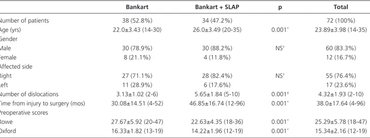
Number of patients 38 (52.8%) 34 (47.2%) 72 (100%) Age (yrs) 22.0±3.43 (14-30) 26.0±3.49 (20-35) 0.001* 23.89±3.98 (14-35) Gender Male 30 (78.9%) 30 (88.2%) NS† 60 (83.3%) Female 8 (21.1%) 4 (11.8%) 12 (16.7%) Affected side Right 27 (71.1%) 28 (82.4%) NS† 55 (76.4%) Left 11 (28.9%) 6 (17.6%) 17 (23.6%) Number of dislocations 3.13±1.02 (2-6) 5.65±1.84 (5-10) 0.001‡ 4.32±1.93 (2-10)
Time from injury to surgery (mos) 30.08±14.51 (4-52) 46.85±16.74 (12-96) 0.001* 38.0±17.64 (4-96)
Preoperative scores
Rowe 27.67±5.92 (20-47) 22.63±4.35 (18-36) 0.001* 25.29±5.78 (18-47)
Oxford 16.33±1.82 (13-19) 14.22±1.96 (12-19) 0.001* 15.34±2.16 (12-19) *: Student’s t-test. †: Continuity correction (Yates) test. ‡: Mann-Whitney U test.
clock-face method. Capsular tissue quality and capsular tension were assessed using a grasper. The labroligamen-tous complex was completely separated from the glenoid and mobilized towards the superior and lateral using a rasp and radiofrequency (RF). The glenoid neck was de-corticated using a burr and the bleeding bone required for tissue recovery was revealed. The glenoid rim was perforated at a 45° medial angle including the 2 mm of the glenoid anterior lip at the 5:30 o’clock position and a mm absorbable or 3-mm metal anchor (Mitek 2.9-mm LUPINE® anchor or 3-2.9-mm FASTIN® threaded anchor; DePuy Mitek Inc., Raynham, MA, USA) was placed. Using a suture transferring system (Mitek IDE-AL™ Suture Shuttle; DePuy Mitek Inc., Raynham, MA, USA), a no. 2 polydioxanone suture (PDS) was passed through the labrum and inferior glenohumeral ligament at 5 to 7 mm inferior to the anchor. Subsequently, the anchor threads were transferred and passed through the labrum and ligament using the PDS. During fixa-tion, the shoulder was kept between the neutral posi-tion and 45° of external rotaposi-tion to avoid fixaposi-tion with excessive tension. Fixation was then secured using the Revo knot technique. Similarly, threads of other anchors were transferred and subsequently fixated at the 4:30 and 3:30 o’clock positions using the no. 2 PDS passed through labroligamentous tissue at an appropriate
posi-tion. Following the knot, the repair was assessed based on the presence of an arthroscopic ‘drive-through sign’. Figure 1 shows the images obtained during arthroscopy and Figure 2 illustrates the schematic diagram of the procedure. In the presence of a SLAP lesion, two more anchors were placed in the anterior and posterior of the biceps anchor and their threads were transferred and fix-ated using the PDS passed through at an appropriate position.
All patients were kept in internal rotation with a Velpeau bandage for four weeks. Passive pendulum ex-ercise was initiated on the first postoperative day and continued at home 5 times a day for 10 minutes. Phys-iotherapy was started at postoperative Week 4. External rotation was limited to 45° until the end of the 6th week. Strengthening exercises were initiated between the 8th and 12th weeks. Sportive activities were allowed after six months. Follow-up was performed at the postoperative Weeks 3 and 6 and at the 3rd, 6th and 12th month. Patients
were then invited for annual visits.
Shoulder range of motion and instability tests were performed during physical examination. Pre- and post-operative assessments were performed using the Rowe and Oxford scales. A Rowe score of 100 to 90 was con-sidered excellent, 89 to 75 good, 74 to 51 fair and 50
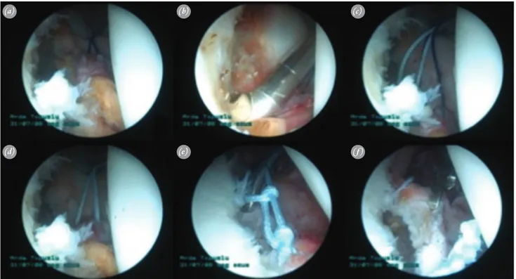
Fig. 1. The arthroscopic procedure. (a) Examination of the mobilization in the labroligamentous complex by pulling with strap stitching. (b)
Mobili-zation of the labroligamentous complex by means of radiofrequency. (c, d) Transfer of the anchor thread placed with the PDS thread. (e, f)
Upward and lateral fixation of the labroligamentous complex by stitches. [Color figures can be viewed in the online issue, which is available at www.aott.org.tr] (a) (d) (b) (e) (c) (f)
or less poor. In the Oxford scale, scores of 48 to 40 was considered excellent, 39 to 30 good, 29 to 20 fair and 19 to 0 poor. Redislocation was accepted as failure criteria.
Statistical analysis was performed using the Student’s t-test, Mann-Whitney U test, continuity correction (Yates) test, Fisher’s exact test, and Pearson correlation coefficient method. The confidence level was 95% and significance was set at p<0.05. Analyses were performed using SPSS v.15 for Windows (SPSS Inc., Chicago, IL, USA) software.
Results
Mean follow-up period was 49.31±24.72 (range: 12 to 108) months. Mean time from the first dislocation to surgical treatment was 38.0±17.64 (range: 4 to 96) months. Mean number of dislocations at presentation was 4.32±1.93 (range: 2 to 10) (Table 1). Mean number of anchors used was 3.69±0.71 (range: 2 to 5). Metal anchors were used in 38 patients and absorbable anchors
in 34. Examinations performed under general anesthesia revealed Grade 2 anterior translation in 26 (36.1%) pa-tients and Grade 3 anterior translation in 46 (63.9%). In-traoperative examinations demonstrated Bankart lesion in 38 (52.8%) patients and Bankart and SLAP lesions in 34 (47.2%). Mean surgical duration was 68.15±7.01 (range: 55 to 94) minutes. Intraoperative findings are summarized in Table 2.
Redislocation was seen in 4 (5.6%) patients in the postoperative period. These patients received open treat-ment with the Latarjet method and their functional out-comes were excluded from the evaluation. Three (4.2%) patients had ongoing postoperative apprehension test and did not experience redislocation or receive any ad-ditional treatment during follow-up.
Preoperative mean Rowe and Oxford scores of patients (n=68) were 25.29±5.78 (range: 18 to 47) and 15.34±2.16 (range: 12 to 19), respectively. Mean Rowe and Oxford scores at their final follow-up were
Table 2. Intraoperative findings of the patients expressed in mean±SD. Values in parentheses show the range unless otherwise stated.
Bankart Bankart + SLAP p Total
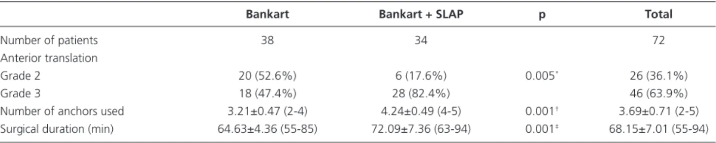
Number of patients 38 34 72
Anterior translation
Grade 2 20 (52.6%) 6 (17.6%) 0.005* 26 (36.1%)
Grade 3 18 (47.4%) 28 (82.4%) 46 (63.9%) Number of anchors used 3.21±0.47 (2-4) 4.24±0.49 (4-5) 0.001† 3.69±0.71 (2-5)
Surgical duration (min) 64.63±4.36 (55-85) 72.09±7.36 (63-94) 0.001‡ 68.15±7.01 (55-94) *: Continuity correction (Yates) test. †: Mann-Whitney U test. ‡: Student’s t-test.
Statistically significant p values are written in bold.
Fig. 2. Schematic diagram of the method. (a)
Placement of the scope via posterior portal and the operating cannula via anterior portal of the shoulder. (b)
Ex-amination of the torn labrum. (c)
Pas-sage of the PDS through the labrum.
(d) Examination of mobilization in the
labrum by pulling with strap stitch-ing. (e) Mobilization of the
labroliga-mentous complex by means of radio-frequency. (f) Transfer of the anchor
thread placed with the PDS thread. (g)
Fixation of the labroligamentous com-plex with stitches. [Color figures can be viewed in the online issue, which is available at www.aott.org.tr] (a) (b) (e) (c) (f) (d) (g)
93.37±3.42 (range: 84 to 100) and 42.56±2.72 (range: 36 to 48), respectively. Physical examination during the fi-nal follow-up revealed a mean exterfi-nal rotation limitation of 4.22±1.67 (range: 2 to 9) degrees and a mean forward flexion range of 173.67±4.41 (range: 163 to 180) degrees. Mean time to redislocation was 20.50±7.77 (range: 9 to 26) months in patients with redislocation. The findings obtained during the final follow-up are summarized in Table 3. Superficial wound infection was detected and re-solved with appropriate oral antibiotic therapy in one pa-tient. No other complication was observed in our patients.
Comparison of pre- and postoperative Rowe and Oxford scores showed a statistically significant differ-ence (p<0.05) (Table 4). There was no significant dif-ference between the postoperative outcomes (Rowe and Oxford scores, external rotation limitation and forward flexion range) and the number of dislocations dur-ing the preoperative period, between the postoperative outcomes and time to surgical treatment, between the postoperative outcomes and grade of translations ob-tained during the physical examination under general anesthesia, and between the postoperative outcomes and
Table 3. Final visit results expressed in mean±SD. Values in parentheses show the range unless otherwise stated.
Bankart Bankart + SLAP p Total
Duration of follow-up (mos) 48.11±25.77 (12-96) 50.65±23.81 (17-108) NS* 49.31±24.72 (12-108)
Postoperative scores
Rowe 94.86±2.21 (90-100) 91.69±3.78 (84-100) 0.001† 93.37±3.42 (84-100)
Oxford 44.36±1.38 (42-48) 40.53±2.42 (36-48) 0.001† 42.56±2.72 (36-48) Redislocation 2 (5.3%) 2 (5.9%) NS‡ 4 (5.6%)
Apprehension test 1 (2.6%) 2 (5.9%) NS 3 (4.2%) Time to redislocation (mos) 23.50±0.71(23-24) 17.5±12.02 (9-26) NS 20.50±7.77 (9-26) Limited external rotation (deg) 3.45±1.03 (2-6) 5.09±1.83 (3-9) 0.001† 4.22±1.67 (2-9)
Forward flexion (range of motion) 175.05±3.73 (166-180) 172.12±4.64 (163-180) 0.001† 173.67±4.41 (163-180) *: Mann-Whitney U test. †: Student’s t-test. ‡: Fisher’s exact test.
NS: Non-significant. Statistically significant p values are written in bold.
Table 4. Pre- and postoperative Rowe and Oxford scores of the patients.
Bankart Bankart + SLAP p*
Mean±SD Mean±SD
Rowe score Preoperative 27.67±5.92 22.63±4.35 0.001 Postoperative 94.86±2.21 91.69±3.78 0.001
p† 0.001 0.001
Oxford score Preoperative 16.33±1.82 14.22±1.96 0.001 Postoperative 44.36±1.38 40.53±2.42 0.001
p† 0.001 0.001
*: Student’s t-test. †: Paired-samples t-test. Statistically significant p values are written in bold.
Table 5. Comparison of biostatistics findings of the patients who underwent arthroscopic repair.
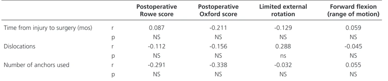
Postoperative Postoperative Limited external Forward flexion
Rowe score Oxford score rotation (range of motion)
Time from injury to surgery (mos) r 0.087 -0.211 -0.129 0.059
p NS NS NS NS
Dislocations r -0.112 -0.156 0.288 -0.045
p NS NS ns NS
Number of anchors used r -0.291 -0.338 -0.032 0.055
p NS NS NS NS
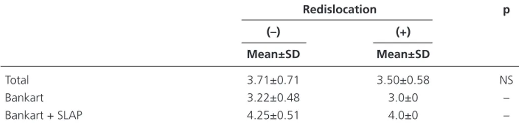
number of anchors used (p>0.05) (Tables 5 and 6). The number of dislocations during the preoperative period, time to surgery and grade of anterior translation during general anesthesia did not adversely affect the postopera-tive outcomes. There was no statistically significant dif-ference regarding postoperative dislocation rates accord-ing to the number of anchors used in patients (p>0.05) (Table 7). A statistically significant difference was found in postoperative outcomes between patients with Ban-kart lesion and patients with BanBan-kart and SLAP lesions (p<0.001). While, mean outcomes were better in pa-tients who received treatment for Bankart lesion alone, results were excellent in both groups.
Discussion
In the current study, the redislocation rate was 5.6% fol-lowing arthroscopic repair of Bankart and Bankart and SLAP lesions. Redislocation rate was high during the first two years following surgery. Our redislocation rate was lower than those in the literature.[3,6,8] Gartsman
et al. followed 53 patients for two years and reported a redislocation rate of 8%,[6] while Mahiroğulları et al.
reported a redislocation rate of 5.9% in 34 patients fol-lowed for two years.[3]
According to the literature, one of the most impor-tant factors affecting outcome is the number of anchors used for treatment. The mean number of anchors used in our study was 3.7, with up to 5 anchors applied in pa-tients with SLAP lesion. Stability requires a minimum
of 3 anchors. Studies show an increased rate of redislo-cation with the use of 2 or fewer anchors.[8-11]
An additional risk factor is the presence of humeral bone or glenoid defects. Success rates in arthroscopic Bankart repair is particularly low in patients with an inverted pear glenoid with glenoid bone defects greater than 30% and in patients with Hill-Sachs lesion with a bone defect large enough to cause complete entrapment to the anterior glenoid at the humeral head. Redisloca-tion rates are considerably high in these patients.[9,10,12,13]
Arthroscopic Bankart repair was not used in our study in patients with a large Hill-Sachs lesion or in those with a glenoid defect greater than 25%. These patients were treated using the Latarjet method.
Another important risk factor of redislocation is joint laxity and considerably high redislocation rates have been reported in the literature. External rotation greater than 90 degrees with the shoulder in adduction during preoperative bilateral shoulder examination is a particularly important indicator. In such cases, open repair appears to produce more satisfactory outcomes than arthroscopic methods.[5,10,11,14] Arthroscopic repair
methods were not used for patients with hyperlaxity in this study.
Outcomes obtained in the present study were suc-cessful compared to those in the literature.[15-17] Our
study demonstrated that postoperative outcomes are not adversely affected by the number of preoperative disloca-tions, time to surgical treatment and grade of anterior
Table 6. Biostatistics findings of the patients after arthroscopic repair.
Anterior translation grade p
Grade 2 Grade 3
Mean±SD Mean±SD
Postoperative Rowe score 95.80±2.59 91.95±3.04 NS Postoperative Oxford score 44.64±2.04 41.35±2.32 NS Limited external rotation (deg) 3.42±1.14 4.67±1.77 NS Forward flexion (range of motion) 175.73±3.58 172.5±4.44 NS
Student’s t-test. NS: Non-significant.
Table 7. Number of anchors used in patients who underwent arthroscopic repair.
Redislocation p (–) (+) Mean±SD Mean±SD Total 3.71±0.71 3.50±0.58 NS Bankart 3.22±0.48 3.0±0 – Bankart + SLAP 4.25±0.51 4.0±0 –
translation observed during preoperative examination during general anesthesia. Gartsman et al.[6] reported
that increased number of preoperative dislocations, time to surgery and increased rate of anterior translation do not adversely affect postoperative outcomes. Mobilizing the anterior capsuloligamentous complex precisely from the glenoid neck, then shifting upward and towards the lateral, and fixation with an appropriate number of an-chors provides successful arthroscopic outcomes.
The outcomes obtained in the current study in patients with Bankart lesion alone were better than those of pa-tients with Bankart and SLAP lesions. As emphasized in the literature, appropriate capsule mobilization and shift-ing followed by SLAP repair provides comparable findshift-ings to those achieved in patients with Bankart lesion alone, even in cases in which tissue quality is disrupted.[6,18,19]
Although it has become the standard practice in ar-throscopic instability surgery, the frequently used double anterior portals have certain limitations. In young pa-tients and in those in which an appropriate preoperative plan for the administration of two portals cannot be es-tablished, the placement and efficient use of two separate cannulas may not be possible. However, there is no such limitation in single portal surgery. In single portal sur-gery, while it may not be possible to place the anchor at the 5:30 o’clock position through a portal established in a too superior location, a percutaneous anchor may be placed via the transsubscapular route.[20]
In conclusion, outcomes obtained using a single ante-rior portal for the arthroscopic surgery treatment of trau-matic anterior shoulder appear to be comparable to those achieved in double portal repair. Proper patient selection contributes to the success of the procedure. This reduc-tion in the surgical period of this less invasive procedure may be considered an advantage over double portals.
Acknowledgement: The authors would like to thank
Cihan KIRÇIL for drawing the figures in this article and Ebru OSMANOĞLU AKYOL for making the statisti-cal assessments.
Conflicts of Interest: No conflicts declared.
References
1. Bankart AS. The pathology and treatment of recurrent disloca-tion of the shoulder joint. Br J Surg 1938;26:23-9. CrossRef
2. Bigliani LU, Pollock RG, Soslowsky LJ, Flatow EL, Pawluk RJ, Mow VC. Tensile properties of the inferior glenohumeral liga-ment. J Orthop Res 1992;10:187-97. CrossRef
3. Mahiroğulları M, Ozkan H, Akyüz M, Uğraş AA, Güney A, Kuşkucu M. Comparison between the results of open and ar-throscopic repair of isolated traumatic anterior instability of the shoulder. Acta Orthop Traumatol Turc 2010;44:180-5. CrossRef
4. Cole BJ, Warner JJ. Arthroscopic versus open Bankart repair for traumatic anterior shoulder instability. Clin Sports Med 2000;19:19-48. CrossRef
5. Boileau P, Villalba M, Héry JY, Balg F, Ahrens P, Neyton L. Risk factors for recurrence of shoulder instability after arthroscopic Bankart repair. J Bone Joint Surg Am 2006;88:1755-63. CrossRef
6. Gartsman GM, Roddey TS, Hammerman SM. Arthroscopic treatment of anterior-inferior glenohumeral instability. Two to five-year follow-up. J Bone Joint Surg Am 2000;82-A:991-1003. 7. Burkhart SS, Debeer JF, Tehrany AM, Parten PM. Quantifying
glenoid bone loss arthroscopically in shoulder instability. Ar-throscopy 2002;18:488-91. CrossRef
8. Ee GW, Mohamed S, Tan AH. Long term results of arthroscopic Bankart repair for traumatic anterior shoulder instability. J Or-thop Surg Res 2011;6:28. CrossRef
9. Provencher MT, Ghodadra N, Romeo AA. Arthroscopic man-agement of anterior instability: pearls, pitfalls, and lessons learned. Orthop Clin North Am 2010;41:325-37. CrossRef
10. Atalar AC, Bilsel K, Eren I, Celik D, Cil H, Demirhan M. Modi-fied Latarjet procedure for patients with glenoid bone defect ac-companied with anterior shoulder instability. Acta Orthop Trau-matol Turc 2013;47:393-9. CrossRef
11. Voos JE, Livermore RW, Feeley BT, Altchek DW, Williams RJ, Warren RF, et al. Prospective evaluation of arthroscopic bankart repairs for anterior instability. Am J Sports Med 2010;38:302-7. 12. Ahmed I, Ashton F, Robinson CM. Arthroscopic Bankart repair and capsular shift for recurrent anterior shoulder instability: functional outcomes and identification of risk factors for recur-rence. J Bone Joint Surg Am 2012;94:1308-15. CrossRef
13. Porcellini G, Campi F, Pegreffi F, Castagna A, Paladini P. Pre-disposing factors for recurrent shoulder dislocation after ar-throscopic treatment. J Bone Joint Surg Am 2009;91:2537-42. 14. Chechik O, Maman E, Dolkart O, Khashan M, Shabtai L,
Mozes G. Arthroscopic rotator interval closure in shoulder in-stability repair: a retrospective study. J Shoulder Elbow Surg 2010;19:1056-62. CrossRef
15. Milano G, Grasso A, Santagada DA, Saccomanno MF, Deriu L, Fabbriciani C. Comparison between metal and biodegradable su-ture anchors in the arthroscopic treatment of traumatic anterior shoulder instability: a prospective randomized study. Knee Surg Sports Traumatol Arthrosc 2010;18:1785-91. CrossRef
16. Mauro CS, Voos JE, Hammoud S, Altchek DW. Failed anterior shoulder stabilization. J Shoulder Elbow Surg 2011;20:1340-50. 17. Shah AS, Karadsheh MS, Sekiya JK. Failure of operative treat-ment for glenohumeral instability: etiology and managetreat-ment. Arthroscopy 2011;27:681-94. CrossRef
18. Hantes ME, Venouziou AI, Liantsis AK, Dailiana ZH, Malizos KN. Arthroscopic repair for chronic anterior shoulder instabil-ity: a comparative study between patients with Bankart lesions and patients with combined Bankart and superior labral anterior posterior lesions. Am J Sports Med 2009;37:1093-8. CrossRef
19. Gutierrez V, Monckeberg JE, Pinedo M, Radice F. Arthroscopi-cally determined degree of injury after shoulder dislocation re-lates to recurrence rate. Clin Orthop Relat Res 2012;470:961-4. 20. Chambers KL, Kremen TJ, Snell CJ, Gamradt SC. Arthroscopic anterior shoulder stabilization in the beach chair position using trans-subscapularis drilling of the 5:30 anchor. Tech Should Elb Surg 2011;12:56-61. CrossRef