DEVELOPMENT AND CHARACTERIZATION OF PEPTIDE
NANOFIBERS FOR CARTILAGE REGENERATION
A DISSERTATION SUBMITTED TO
THE GRADUATE SCHOOL OF ENGINEERING AND SCIENCE OF BILKENT UNIVERSITY
IN PARTIAL FULFILLMENT OF THE REQUIREMENTS FOR THE DEGREE OF
DOCTOR OF PHILOSOPHY IN
MATERIALS SCIENCE AND NANOTECHNOLOGY
By
SEHER YAYLACI
i
DEVELOPMENT AND CHARACTERIZATION OF PEPTIDE
NANOFIBERS FOR CARTILAGE REGENERATION
By Seher YaylacıSeptember, 2015
We certify that we have read this thesis and that in our opinion it is fully adequate, in scope and in quality, as a thesis of the degree of Doctor of Philosophy.
………. Asst. Prof. Dr. Ayşe Begüm Tekinay (Advisor) ………. Assoc. Prof. Dr. Mustafa Özgür Güler (Co-Advisor) ………. Assoc. Prof. Dr. Aykutlu Dana ………. Assoc. Prof. Dr. Hüseyin Özkan ……….
Assoc. Prof. Dr. Çağdaş Son ………. Assoc. Prof. Dr. Hatice Duran Approved for the Graduate School of Engineering and Science:
……….
Prof. Dr. Levent Onural
ABSTRACT
DEVELOPMENT AND CHARACTERIZATION OF PEPTIDE
NANOFIBERS FOR CARTILAGE REGENERATION
Seher Yaylacı
PhD in Materials Science and Nanotechnology Supervisor: Asst. Prof. Dr. Ayşe Begüm Tekinay Co-Supervisor: Assoc. Prof. Dr. Mustafa Özgür Güler
September, 2015
Articular cartilage is a tissue that is continuously exposed to cyclical compressive stresses, but exhibits no capacity for self-healing following trauma. Cartilage has a dense extracellular matrix that is sparsely populated with cells, and the whole tissue lacks blood and lymphatic vessels, which complicates the cell infiltration response that ordinarily occurs during inflammation. In addition, the only cell type capable of synthesizing new cartilage matrix lies deeper in the tissue, near the bone boundary, and due to the dense extracellular matrix, chondrocytes cannot migrate to the defect site following injury. Consequently, cartilage tissue cannot effectively respond to treatment options. Treatment options exist for the short-term reduction of pain in smaller defects, but larger injuries necessitate tissue donation, and there is a severe shortage of articular cartilage that can be donated for autografting.
Microfracture and autologous chondrocyte implantation are the current treatment options that use cellular therapy for the repair of cartilage. However, the cartilage
iii
tissue that forms in the course of these treatments is not the functional hyaline cartilage, but rather fibrous cartilage, which is mechanically weaker and degenerates over time. Tissue engineering studies using biodegradable scaffolds and autologous cells are gaining importance as effective long-term treatment options for the post-injury production of hyaline cartilage. Such scaffold systems are designed to be biodegradable and bioactive, which allows them to induce new tissue formation in shorter periods of time.
In this dissertation, peptide nanofibers mimicking glycosaminoglycan molecules, which are important constituents of cartilage extracellular matrix, are designed and the effectiveness of these materials in terms of chondrocyte differentation are tested under in vitro conditions. As a follow-up study to in vitro experiments, the capacity of bioactive peptide nanofibers to support cartilage regeneration is evaluated in the rabbit osteochondral defect model. Structural and mechanical properties of newly deposited cartilage are highly dependent on the quality and quantity of its extracellular matrix, which also has a major impact on the integration of replacement cartilage into the surrounding healthy tissue. Signals provided by bioactive peptide nanofibers to cells at the defect site can strongly alter the quality of the newly synthesized extracellular matrix. Consequently, we designed glycosaminoglycan-mimetic peptide nanofibers that closely imitate the structure of the native cartilage extracellular matrix and demonstrated that these nanofiber networks are able to induce the synthesis of collagen II and aggrecan molecules, which are the main constituents of cartilage tissue, during chondrogenic differentiation.
Keywords: Cartilage Regeneration, Mesenchymal Stem Cells, Biomaterials, Peptide Amphiphile Nanofibers, Glycosaminoglycans
ÖZET
KIKIRDAK REJENERASYONU İÇİN PEPTİT
NANOFİBERLERİN GELİŞTİRİLMESİ VE
KARAKTERİZASYONU
Seher Yaylacı
Malzeme Bilimi ve Nanoteknoloji, Doktora Tez Danışmanı: Yrd.Doç.Dr. Ayşe Begüm Tekinay
Eş Danışman: Doç.Dr. Mustafa Özgür Güler Eylül, 2015
Artiküler kıkırdak dokusu sürekli olarak tekrarlayan kompresif strese maruz kalan fakat küçük boyutlu yaralanmalarda dahi kendini yenileyemeyen bir dokudur. Kıkırdak dokusu yoğun hücrelerarası matrise ve sınırlı sayıda kondrosit hücresine sahip bir dokudur. Doku genelinde kan ya da lenf damarları bulunmamakta ve herhangi bir enflamasyon durumunda dokuya projenitör hücre girişi olmamaktadır. Bunun yanında, yeni dokunun oluşmasını sağlayacak tek hücre tipi olan kondrosit hücreleri kıkırdak dokusunun daha derinlerinde kıkırdak kemik sınırına yakın olarak bulunmakta ve yoğun hücrelerarası matristen dolayı hasar bölgesine göç edememektedir. Bundan dolayı günümüzde uygulanan ve küçük boyutlu hasarlarda acıyı ortadan kaldıran ancak kısa süreli çözüm olabilen tedavi yöntemlerine rejenerasyonla cevap verememektedir. Ağır kıkırdak hasarları ise doku transferini
v
gerektirmekte fakat otograft olarak bağışlanacak kıkırdak dokusu azlığı nedeniyle herkese uygulanamamaktadır.
Mikrokırık ve otolog kondrosit implantasyonu günümüzde uygulanan rejenerasyon amaçlı hücre terapisinin kullanıldığı yöntemlerdir. Fakat bu yöntemler sonucu oluşan kıkırdak dokusu fonksiyonel doku olan hiyalin kıkırdak değil zamanla dejenere olan fibröz kıkırdak dokudur. Bu sebeplerle uzun vadeli etkili çözüm üretebilecek biyobozunur iskele ve otolog hücrelerin kullanıldığı çalışmalar önem kazanmaktadır.
Bu tezde, kıkırdak hücrelerarası matrisinin önemli bileşenlerinden olan glikozaminoglikan moleküllerin taklit eden malzemeler geliştirilmiş ve bu malzemelerin in vitro ortamda kıkırdak farklılaşmasını desteklediği gösterilmiştir.
İn vitro deneyleri takip eden in vivo deneylerde, biyoaktif peptit nanofiber sistemler kıkırdak doku hasarı tamiri yetisi bakımından tavşan osteokondral hasar modelinde test edilmiştir. Yeni oluşan dokunun yapısal ve mekanik özellikleri ve çevresindeki doku ile entegrasyonu içerdiği hücrelerarası matrisin niteliğine ve miktarına bağlıdır. Hasar bölgesinde biyoaktif peptit nanofiberlerle sağlanan sinyaller, hücrelerin üreteceği ektraselüler matrisin niteliğini belirlemiştir. İn vitro çalışmada gözlendiği üzere glikozaminoglikan benzeri peptitler hücrelerin farklılaşmasını ve bunu takiben kıkırdak dokusunun temel bileşenleri olan kollajen II ve agrekan moleküllerinin sentezini tetiklemiştir. İn vivo çalışmamızda da hasarlı kıkırdak bölgesinde peptit nanofiberler yardımıyla kollajen II ve agrekan molekülleri bakımından zengin hiyalin kıkırdak dokusu üretimi gözlenmiştir.
Anahtar Kelimeler: Kıkırdak Rejenerasyonu, Mezenkimal Kök Hücre, Biyomalzemeler, Peptit Amfifil Nanofiberleri, Glikozaminoglikanlar
ACKNOWLEDGEMENTS
The completion of this doctoral dissertation was possible with the mental and emotional support of several people. I would like to express my sincere gratitude to all of them.
Firstly, I would like to sincerely thank my advisors Prof. Tekinay and Prof. Guler for their guidance, motivation, care, understanding and immense knowledge I received through my Ph.D research. They provided me with an excellent atmosphere for doing research. Besides my advisors, I would like to thank the rest of my thesis committee for their insightful comments.
I would like to acknowledge the financial supports of The Scientific and Technological Research Council of Turkey (TÜBİTAK); BİDEB 2214/A for PhD scholarship and projects 111M710 and 113T045 for experimental works.
A very special thanks goes out to my husband Ali Kemal Yaylacı, without whose motivation, encouragement and hearty support I would not have completed this thesis. I am also grateful to Lütfiye Yaylacı, Musa Yaylacı and Furkan Yaylacı for their hearty support.
I would like to express my most sincere thanks to Hakan Ceylan, Hilal Gülsüner and Zeynep Ülger for their friendship, moral support and motivation, which made my lab hours more enjoyable.
Special thanks to Merve Sen, Özlem Bulut, Melis Sardan and Aysegul Tombuloglu for their excellent collaboration in projects we took part together. I am especially grateful Alper Özkan for his willingness for editing papers, reviews and even this thesis. I would also like to thank all labmates of the Nanobiotechnology Lab and Biomimetics Lab for providing a warm and peaceful research environment.
vii
Finally, I would like to express my heartfelt thanks to my parents, Habibe Üstün, Hasan Üstün, to my brother Gökhan Üstün, to my brother’s wife Özlem Üstün, and to my nephews Hakan Kerem Üstün and Enes Üstün for supporting me spiritually throughout of my life and encouraging me with their best wishes.
CONTENTS
CONTENTS ... viii
LIST OF FIGURES ... xii
LIST OF TABLES ... xv
ABBREVIATIONS ... xvi
CHAPTER 1 ... 1
1 Introduction: Cartilage Tissue Engineering ... 1
1.1 Cartilage Tissue ... 1
1.1.1 Basic Properties of Cartilage Tissue ... 1
1.1.2 Cartilage Structure and Composition ... 2
1.1.3 Chondrocyte-‐ECM Interactions ... 6
1.2 Issues Related to Cartilage Repair and Their Clinical Significance ... 9
1.2.1 Cartilage Repair Strategies ... 9
1.2.2 Tissue Engineering Efforts ... 15
1.2.2.1 Natural Polymers ... 17
1.2.2.2 Synthetic Polymers ... 19
1.2.2.3 Composite Materials ... 20
1.2.2.4 Physical Stimuli ... 22
CHAPTER 2 ... 25
2 Growth and Differentiation of Pre-‐Chondrogenic Cells on Bioactive Self-‐Assembled Peptide Nanofibers ... 25
2.1 Synopsis ... 25
2.2 Introduction ... 26
ix
2.3.1 Self-‐Assembled Peptide Amphiphile Nanofiber Formation ... 29
2.3.2 Viability, Adhesion and Spreading of Cells on Peptide Nanofiber Networks… ... 35
2.3.3 Morphological Effects of Nanofiber Networks on ATDC5 Cells ... 39
2.3.4 Cartilaginous Matrix Deposition ... 45
2.3.5 Gene Expression Profiles ... 53
2.4 Conclusion ... 58
2.5 Experimental Section ... 58
2.5.1 Materials ... 58
2.5.2 Synthesis, Purification and Characterization of Peptide Amphiphile Molecules ... 59
2.5.3 Analysis of Structural and Mechanical Characteristics of Peptide Nanofibers ... 60
2.5.4 Cell Culture ... 61
2.5.4.1 Monolayer Culturing ... 61
2.5.4.2 Cell Seeding and Cultivation on Peptide Nanofiber Networks ... 61
2.5.5 In vitro Adhesion, Spreading and Cell Viability ... 61
2.5.6 Differentiation Analysis ... 63
2.5.6.1 Morphology Screening ... 63
2.5.6.2 Analysis of Sulfated Glycosaminoglycan Production ... 63
2.5.6.3 Immunofluorescence Staining and Imaging ... 64
2.5.6.4 Gene Expression Analysis ... 65
2.5.7 Statistical Analysis ... 65
CHAPTER 3 ... 67
3 Glycosaminoglycan-‐Mimetic Peptide Networks Promote the Chondrogenic Differentiation of Mesenchymal Stem Cells ... 67
3.1 Synopsis ... 67
3.2 Introduction ... 68
3.3 Results and Discussion ... 70
3.3.1 Characterization of Peptide Amphiphile Nanofibers ... 70
3.3.2 Cellular Behavior on Nanofiber Networks ... 74
3.3.3 Nanofiber Networks Promote MSC Aggregation and Deposition of Cartilage ECM Components ... 76
3.3.4 Gene Expression Profiles of MSCs Confirm Chondrogenic Lineage Commitment ... 79
3.4 Conclusion ... 84
3.5 Experimental Section ... 85
3.5.1 Materials ... 85
3.5.2 Synthesis, Purification and Characterization of Peptide Amphiphile Molecules ... 85
3.5.3 rMSC Culture Conditions and the Preparation of Nanofibrous Networks for In Vitro Culture ... 87
3.5.4 Viability and Proliferation Assays ... 88
3.5.5 Glycosaminoglycan Imaging and Quantification ... 89
3.5.6 Gene Expression Analysis ... 90
3.5.7 Immunostaining and Imaging ... 91
3.5.8 Statistical Analysis ... 92
CHAPTER 4 ... 93
4 Supramolecular Glycosaminoglycan-‐like Self-‐Assembled Glycopeptide Nanofibers Induce Chondrogenesis and Cartilage Regeneration ... 93
4.1 Synopsis ... 93
xi
4.3 Results and Discussion ... 96
4.3.1 Interactions of mMSCs with Glycopeptide Nanofibers is Mediated Through CD44 Receptors ... 96
4.3.2 Early Chondrogenesis of mMSCs is Promoted on Glycopeptide Nanofibers and Hyaluronic Acid Networks ... 99
4.3.3 CD44 Blockade Downregulates the Expression of Sox 9 on Both Glycopeptide and Hyaluronic Acid Nanofibers ... 106
4.3.4 Regeneration of Osteochondral Defects was Improved by Glycopeptide Nanofiber Gel Treatment ... 109
4.4 Conclusion ... 115
4.5 Experimental Section ... 117
4.5.1 mMSC Culturing and Preparation of Nanofibrous Networks for In Vitro Culture ... 117
4.5.2 Viability, Proliferation and SEM imaging ... 118
4.5.3 Glycosaminoglycan Quantification ... 119
4.5.4 Gene Expression Analysis ... 119
4.5.5 Immunostaining and Imaging ... 120
4.5.6 In Vivo Osteochondral Defect Model and Treated with Microfracture Treatment ... 121
4.5.7 Histological and Immunohistochemical Stainings of Tissue Sections ... 122
4.5.8 Statistical Analysis ... 123
CHAPTER 5 ... 124
5 CONCLUSION and FUTURE PERSPECTIVES ... 124
BIBLIOGRAPHY ... 127
LIST OF FIGURES
Figure 1.1 The Organization of Normal Articular Cartilage. ... 5
Figure 1.2 Cartilage Tissue ECM ... 7
Figure 1.3 Cartilage Regeneration Techniques. ... 12
Figure 2.1 Self-Assembled Peptide Amphiphile Nanofibers. ... 33
Figure 2.2 Liquid Chromatography of PA molecules. ... 34
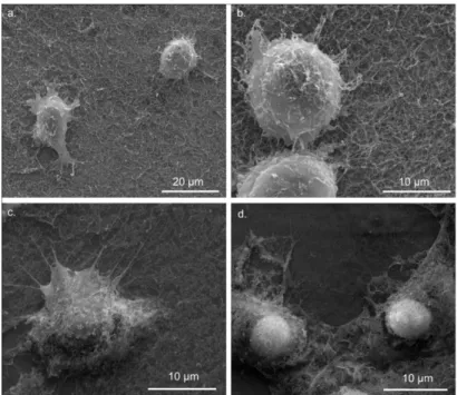
Figure 2.3 ATDC5 Cells Encounter the Nanofibrous Structure of Peptide Amphiphile Nanofibers During Cell Culture Experiments. ... 36
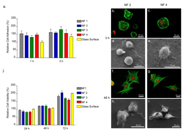
Figure 2.4 Adhesion, Spreading and Viability of ATDC5 Cells Cultured on Peptide Nanofibers. ... 37
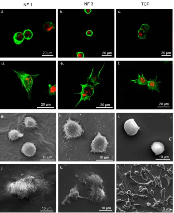
Figure 2.5 Comparable Spreading of ATDC5 Cells on Peptides vs. TCP. ... 38
Figure 2.6 Aggregate Formation of ATDC5 Cells in the Absence of Insulin on Scaffolds. ... 41
Figure 2.7 Aggregate Formation of ATDC5 Cells in the Presence of Insulin on Scaffolds. ... 42
Figure 2.8 ATDC5 cells on TCP. ... 43
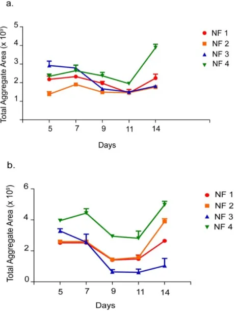
Figure 2.9 Total Aggregate Area of ATDC5 Cells Cultured on PA-Coated Surfaces. ... 44
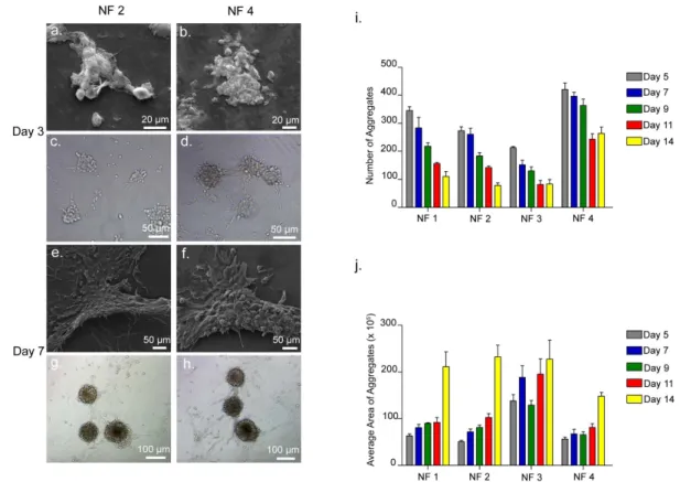
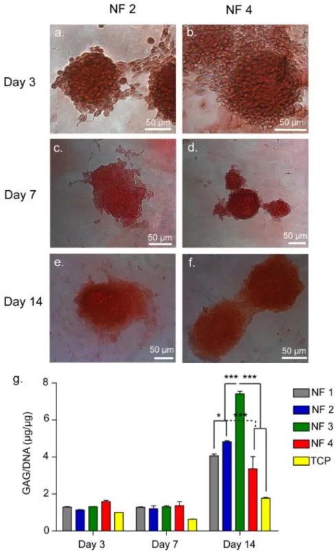
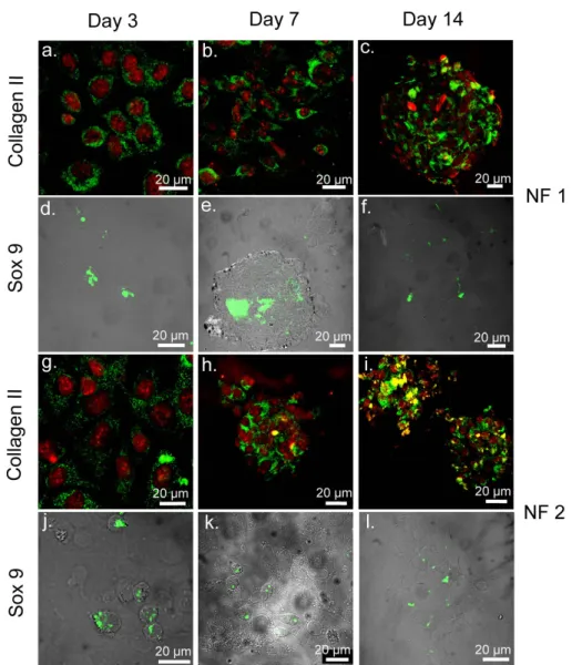
Figure 2.10 Safranin-O and DMMB Staining of ATDC5 Cells on NF 2 and NF 4, Showing GAG Incorporation. ... 47
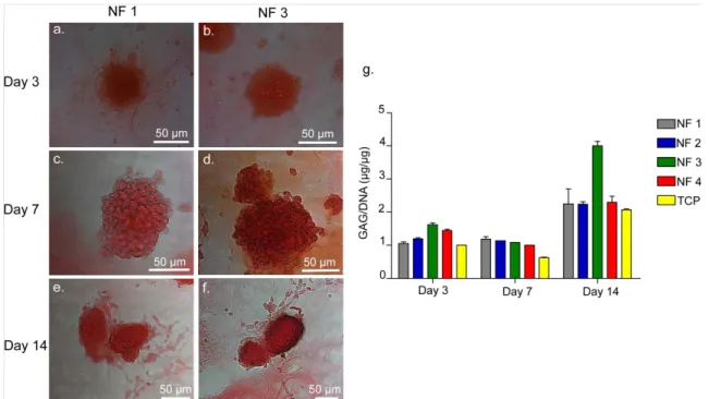
Figure 2.11 Safranin-O and DMMB Staining by ATDC5 Cells on NF 1 and NF 3 Showing GAG Incorporation. ... 48
xiii
Figure 2.12 ATDC5 Cells Cultured on All Scaffolds Express Cartilage Specific
Proteins on Day 3, 7, and 14. ... 49
Figure 2.13 ATDC5 Cells Cultured on All Scaffolds Express Cartilage Specific Proteins on Day 3, 7, and 14. ... 50
Figure 2.14 ATDC5 Cells Cultured on TCP Stained with Collagen II and Sox 9 Antibodies on Day 3, 7, and 14. ... 51
Figure 2.15 Localization of Sox 9 at or around Nucleus on Day 3. ... 52
Figure 2.16 Gene Expression Analysis in the Absence of Insulin. ... 56
Figure 2.17 Gene Expression Analysis in the Presence of Insulin. ... 57
Figure 3.1 Self-assembly of Peptide Amphiphile Molecules into Nanofibrous Networks. ... 72
Figure 3.2 Liquid Chromatography-Mass Spectrometry (LC-MS) Analysis of PA Molecules. ... 73
Figure 3.3 Viability and Proliferation of MSCs on Nanofiber Networks at 24 h. ... 75
Figure 3.4 Morphological Changes of MSCs on Day 7 Cultured in Either Maintenance or Chondrogenic Medium. ... 77
Figure 3.5 Glycosaminoglycan Deposition of MSCs on Nanofiber Networks or TCP on Day 7. ... 78
Figure 3.6 Cartilage-Specific Gene and Protein Expression in Chondrogenic Medium at Day 3 and 7. ... 81
Figure 3.7 Cartilage-Specific Gene Expression in Maintenance Medium at Day 3 and 7. ... 82
Figure 3.8 Cartilage-Specific Gene Expression on Bioactive Nanofibers with Different Epitope Concentrations. ... 83
Figure 4.1 In vitro Viability, Proliferation and Morphology of mMSCs Cultured on Glc-PA/E-PA, K-PA/E-PA, and HA/K-PA. ... 98 Figure 4.2 mMSC Morphology and Cluster Formation on Glc-PA/E-PA, K-PA/E-PA and HA/K-PA ... 101 Figure 4.3 Glycopeptide Nanofibrous Networks Enhance the Chondrogenic Differentiation of mMSCs. ... 102 Figure 4.4 Glycosaminoglycan (GAG) Accumulation by mMSCs Cultured on Glc/E-PA, K-PA/E-Glc/E-PA, HA/K-PA and Culture Plate on Day 3, 7 and 14. ... 103 Figure 4.5 Gene Expression Levels of Collagen II, Aggrecan and Sox 9 of mMSCs Cultured in Maintenance Medium on Days 3, 7 and 14. ... 104 Figure 4.6 Immunostaining of Collagen II, Aggrecan and Sox 9 Expressed by mMSCs Cultured on K-PA/E-PA, HA/K-PA and Culture Plate on Day 14 in Chondrogenic Differentiation Medium. ... 105 Figure 4.7 CD44 Blocking Downregulates Sox 9 Expression of mMSCs on Glycopeptide Nanofibrous Networks on Day 1. ... 107 Figure 4.8 Effect of CD44 Inhibition on the Expression of Sox 9 by mMSCs Cultured on Glc/E-PA, K-PA/E-PA, HA/K-PA and Culture Plate on Day 3. ... 108 Figure 4.9 Hyaline Cartilage Formation Predominates in Groups Treated with Glycopeptide Nanofiber Gels Following Microfracture. ... 112 Figure 4.10 Immunofluorescence Collagen II Staining on Sections of mMSC Cultures in 3D Glc/E-PA, HA and Treatment-Free Sections. ... 114
xv
LIST OF TABLES
Table 2.1 List of Nanofiber Combinations Used in Differentiation of Chondroprogenitor ATDC5 Cells ... 31 Table 3.1 Molar Ratios Used in the Preparation of Peptide Amphiphile Networks. . 72 Table 3.2 Molar Ratios Used in the Preparation of Peptide Amphiphile Scaffolds. . 80 Table 3.3 Primers Used for qRT-PCR Expression Analyses ... 91 Table 4.1 Histological Scores for Sham, Hyalgan and Glycopeptide Hydrogel Groups ... 113 Table 4.2 Primers Used for qRT-PCR Expression Analysis ... 120
xvi
ABBREVIATIONS
ACI : Autologous chondrocyte implantation
AEMA : 2-aminoethyl methacrylate
ATDC5 : C h o n d r o g e n i c C e l l
Mouse 129 teratocarcinoma AT805 derived cell line
Boc : Tert-butoxycarbonyl
BSA : Bovine serum albumin
CD : Circular dichroism
CH : Chondrogenic medium
DCM : Dichloromethane
DIEA : N-ethyl-diisopropylamine
DMF : N, N-Dimethylformamide
DMEM : Dulbecco’s modified Eagle’s medium
DMMB : d İ M E T
Dimethyl methylene blue
ECM : Extracellular matrix
EdU : 5-ethynyl-2’-deoxyuridine
EDTA : Ethylenediaminetetraacetic acid
E-PA : Lauryl-VVAGE
FBS : Fetal bovine serum
FDA : Food and Drug Administration
Fmoc : 9-Fluorenylmethoxycarbonyl GAG : Glycosaminoglycan GAG-PA : L a u r y l -Lauryl-VVAGEGD-K(p-sulfobenzoyl)-S-Am
Glc-PA : Lauryl-VVAGKS(β-Glc)-Am
β
HA : Hyaluronic acid
HBTU :
O-Benzotriazole-N,N,N’,N’-tetramethyl-uronium-hexafluoro-phosphate
xvii
K-PA : Lauryl-VVAGK-Am
LC-MS : Liquid chromatography-mass spectrometry
MACI : Matrix-induced autologous chondrocyte implantation
mMSC : Mouse mesenchymal stem cell
MT : Maintenance medium
MTT : 3-(4,5-dimethylthiazol-2-yl)-2,5-diphenyltetrazolium bromide
OA : Osteoarthritis
PA : Peptide amphiphile
PLA : Polylactic acid
PGA : Polyglycolic acid
PBS : Phosphate-buffered saline
PCM : Pericellular matrix
PG : Proteoglycan
rMSC : Rat mesenchymal stem cell
SEM (Microscopy) : Scanning electron microscope
SEM (Statistics) : Standard error of the mean
TIS : Triisopropylsilane
TCP : TCP
TEM : Transmission electron microscope
TFA : Trifluoroacetic acid
1
CHAPTER 1
1 Introduction: Cartilage Tissue Engineering
1.1 Cartilage Tissue
1.1.1 Basic Properties of Cartilage Tissue
Cartilage is a type of connective tissue found in both vertebrates and invertebrates. Connective tissues form the majority of the body mass, in terms of both cellular and extracellular components. They are responsible for a variety of functions, such as mechanical support, cell migration and differentiation and wound healing, which strongly depend on mostly cellular elements for nerve, epithelial or muscle tissues. However, the properties and functions of cartilage tissue are primarily determined by the arrangement, type and amount of its extracellular matrix (ECM) components. Vertebrate cartilage is consequently classified on the basis of its predominant ECM elements. Accordingly, hyaline cartilage is primarily composed of glycosaminoglycans and found in joints between bones, sternum and ribs; elastic cartilage is composed of elastic fibers and present in ear pinna, eustachian tubes and the epiglottis; and fibrous cartilage is composed mainly of type I collagen and found in intervertebral discs and the pubic symphysis. Lastly, articular hyaline cartilage is mostly known for its function in joints, where it permits the movement of one bone against another while absorbing impact forces by imparting the joint with low friction coefficients and high load resistance1.
Cartilage can function as a transitional tissue during human bone development or as a permanent tissue to provide mechanical rigidity while retaining a degree of
flexibility. It is also capable of defining the external morphology of some organs, such as the ear and the nose.
During embryogenesis, cartilage arises from the mesodermal germ layer. Cartilage development involves a series of dynamic and strictly regulated processes involving mesenchymal cell recruitment, progenitor cell condensation and chondrogenic differentiation, which ultimately leads to the formation of various types of cartilage 2. Cellular condensation is the pivotal stage during chondrogenesis and results in production of specialized chondroblast cells that secrete the characteristic cartilage ECM. Further deposition of ECM elements increases the distance between individual chondroblasts, leading to their encapsulation within the tough, dense ECM and stimulating their differentiation into mature chondrocytes 3.
1.1.2 Cartilage Structure and Composition
The composition of cartilage tissue changes as the tissue develops, with mature articular cartilage ECM being composed mainly of water, collagen fibers and PGs (though noncollagenous proteins and glycoproteins also exist in smaller amounts). Water is the most abundant component of articular cartilage, accounting for approximately 70-80% of its wet weight. Most of the water is associated with the interfibrillar space of collagens, while the remainder is contained in small pores within the intracellular space. Adult cartilage is characteristically aneural, alymphatic and avascular, and nourishment and waste transport is primarily performed through long-range diffusion in synovial fluid.
Chondrocytes are the only metabolically active units of cartilage, and facilitate the turnover and synthesis of the tissue despite occupying only around 5% of the tissue. Chondrocytes have different morphologies and expression patterns depending on the
3
zone in which they reside. Cells near the tissue edge display a flattened morphology, while those in the middle and deep zones are more spherical 4.
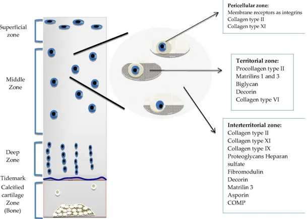
Cartilage tissue shows two distinct levels of organization, being stratified in a depth-dependent manner and exhibiting a second layer of matrix compartmentalization within each strata (Figure 1.1). Consequently, features such as chondrocyte morphology, biochemical composition and collagen fiber orientation differ between the surface and deeper regions of the tissue. The superficial zone represents the first 10-20% of articular cartilage by depth, and is a region in which collagen fibers are aligned in parallel to the cartilage surface. This zone is responsible for most of the tensile properties of cartilage and protects deeper regions from forces imposed by articulation. The middle zone makes up the middle 40-60% of cartilage; collagen fibers are oriented obliquely in this region. Chondrocytes are found in lower amounts in the middle zone, and display a spherical morphology. The deep zone is responsible for resisting the bulk of the compressive forces exerted on cartilage, and performs this duty through the assistance of its radially oriented collagen fibers and high PG content. Chondrocytes are arranged in columnar structures in this zone. The second layer of organization is observed in the ECM surrounding chondrocytes, which is divided into zones depending on its distance from cells 5. The pericellular matrix (PCM) is the closest to chondrocytes, possesses a distinct collagen fiber type and bears a rich aggrecan content. Signaling molecules such as growth factors are also concentrated in this region 6–8. The territorial matrix, which surrounds the PCM, is
thicker than the PCM and has been suggested to play a role in protecting chondrocytes from mechanical stress. The interterritorial region is the most distal from the cells and the largest of 3 matrices. It is responsible for most of the biomechanical properties of cartilage.
As previously mentioned, the dry weight of cartilage tissue is mainly composed of collagen and PGs, and these two molecules form the networks that imbue cartilage tissue with its mechanical properties.
Cartilage tissue is known to contain collagen types II, VI, IX, X, and XI, although type II collagen comprises the 90-95% of total collagen in the tissue 9. Different from other collagen types, type II collagen has a greater amount of bound carbohydrate groups, which allows it to establish stronger interactions with water molecules. Collagen fiber orientation also differs in each zone: Tangential fibers are found in superficial zones, while the deep zone contains radially oriented fibers 10.
The predominant PG found in articular cartilage is aggrecan, which exists in aggregates of hyaluronan and chondroitin sulfate through connections formed by link proteins 11. Aggrecan is responsible for providing the osmotic pressure cartilage requires resisting compressive loads. Other than aggrecan, small PGs such as decorin, perlecan and biglycan are also present in cartilage. Similar to aggrecan, these PGs are also comprised of a core protein that is associated with various glycosaminoglycan species attached as side chains 12.
5
Figure 1.1 The Organization of Normal Articular Cartilage.
The organization of cartilage tissue is divided depthwise into superficial, middle and deep zones, while the cartilage ECM is classified depending on its distance from the chondrocytes. Each zone has specific ECM components and a characteristic chondrocyte morphology. (© 2013 García-Carvajal ZY, Garciadiego-Cázares D, Parra-Cid C, Aguilar-Gaytán R, Velasquillo C, Ibarra C, Castro Carmona JS. Published in [179] under CC BY 3.0 license.).
1.1.3 Chondrocyte-ECM Interactions
Chondrocytes have cell long processes that allow their interaction with ECM molecules; however, they have no direct contact with other cells within the tissue. Thus, the regulation of tissue homeostasis depends heavily on cell-matrix interactions.
ECM serves not only as structural support for cells, but also acts as a bioregulatory niche that modulates tissue formation, organization and maintenance. ECM is therefore an extraordinarily dynamic and versatile environment; as cells embedded in the matrix actively remodel its components through enzymatic or non-enzymatic pathways. The physical and chemical diversity of ECM components determine tissue characteristics and can vary from one tissue to another, or even between two different physiological states of the same tissue. The reciprocal and dynamic signaling between cell and ECM compartments has attracted special attention in cartilage, where cell-matrix interactions predominate and cell-cell interaction are minimal due to the sparsity of cells in this tissue. Most cell-matrix interactions are mediated by transmembrane receptors, and this interaction helps the cells to sense changes in the ECM 13–15.
Articular chondrocytes have been shown to express transmembrane extracellular receptors such as CD44 and integrins 16–18. Both of these receptors interact with
adjacent extracellular macromolecules and detect changes in matrix composition and state. It is known that disruption of either CD44- or integrin-mediated interactions may have drastic effects on the cartilage metabolism.
7 Figure 1.2 Cartilage Tissue ECM
Cartilage ECM is composed of three major components; collagens, proteoglycans and water. The predominant collagen type is type II, and the most abundant proteoglycan is aggrecan. (Reproduced from Ref. 180 with permission from Nature Publishing Group).
CD44 is the primary receptor for hyaluronic acid (HA) molecules in many cells 19. In cartilage tissue ECM, HA molecules are found interacting with aggrecan at a ratio of about 50 HA to one core protein, with link proteins serving to connect the two 20. Thus, the binding of HA to CD44 in cartilage mediates the retention of large HA/PG/link protein aggregates at the surface of chondrocytes 21. Several experiments with anti-CD44 antibodies and CD44 antisense oligonucleotides support the idea that CD44 is an important mediator of chondrocyte cell-matrix interactions that involve proteoglycan (PG)/HA/link protein aggregates 22.
The unique organization of CD44 at the cell surface may function to establish and also to regulate the structure of the PCM in conjunction with a hyaluronan scaffold. The interaction of the cytoplasmic domain of CD44 with components of the cytosolic proteins, and the interaction of the extracellular domain of CD44 with the matrix, are indicative of both inside-out and outside-in communication patterns. It was shown that the inhibition of CD44-matrix interactions through the use of antisense oligonucleotides or HA oligosaccharides results in a chondrocytic chondrolysis cascade 23.
Adult articular chondrocytes also express members of integrin subfamilies. These integrins mediate adhesion to several different ECM proteins found in cartilage. Chondrocytes were found to use integrins to attach to fibronectin, collagen types II and VI, vitronectin, osteopontin, and bone sialoprotein II 24. The functional role of integrins in cartilage has not been determined in full. Chondrocyte integrins might be expected to help regulate processes involved in cell survival, growth, and differentiation, as well as matrix remodeling. Chondrogenesis was inhibited in mouse limb bud cells by blocking integrin with integrin antibodies 25. In one study, RGD
9
peptides were used to inhibit integrin-ECM interactions and it was shown that differentiation of epiphyseal chondrocyte was ceased 26.
1.2 Issues Related to Cartilage Repair and Their Clinical
Significance
Defects in articular cartilage can result from traumatic injury or pathological degeneration. The capacity of cartilage to repair injury is limited, and when not treated, such defects can progress to degenerative arthritis. The increasing prevalence of osteoarthritis (OA) is correlated with an aging population and the growing problem of obesity. 9% of the United States population aged 30 and older suffer from OA of the hip or knee, and more than 250,000 knee and hip replacements are performed each year for end stage disease joint failure 27. In addition, cartilage damage resulting from sports injuries can also result in premature cartilage degeneration in the younger or more active individuals.
1.2.1 Cartilage Repair Strategies
Various repair strategies are currently used in the treatment of cartilage defects. Arthroscopic techniques, such as lavages and debridements, aim to clean out the defect site and alleviate pain associated with the inflammation of the cartilage tissue
28. These procedures are considered to be palliative, as they only provide short-term
relief by removing unstable cartilage flaps and preventing inflammation; they do not restore the function of the damaged site 29. An arthroscopic lavage involves the washing of the joint space for the removal of blood or loose debris, while debridement is the removal of unstable cartilage flaps. The main targets of these procedures are patients with very small cartilage defects 30.
Other arthroscopic techniques aim to utilize the intrinsic repair capacity of cartilage, and include drilling and microfractures. Both of these techniques consist of the penetration and subsequent bleeding of subchondral bone to induce fibrocartilage formation. They rely on the formation of a blood clot at the defect site by stimulating bone marrow, which triggers the deposition of mechanically weak fibrocartilage tissue at the defect site. The microfracture technique is used for defects smaller than 2.5 cm2, and involves the removal of the unstable cartilage for the creation of a well-defined defect environment that is surrounded by healthy cartilage 31. In this procedure, a microscopic awl is used to penetrate subchondral bone without damaging its integrity. The defect is then filled with clot material from the subchondral bone, which contains mesenchymal stem cells (MSCs) that will oversee the subsequent repair of cartilage tissue. The outcome of the treatment depends mainly on how well the patient can avoid overexerting the cartilage tissue with excessive weight loads. Clinical data suggest that 75% of patients display short-term clinical improvements; however, functional deterioration of the defect site begins within 24 months in 48-80% of cases 32. Low numbers of MSCs, or their dilution by synovial fluid, can result in low regeneration capacity and early deterioration following the procedure. Adequate regeneration therefore depends on the optimization of the number of cells extracted from subchondral bone, which is performed by controlling the depth and numbers of the holes produced on the cartilage surface.
Grafting is the preferred way of treating larger cartilage defects and consists of the removal of healthy cartilage tissue from non-weight bearing areas of the knee, followed by the transplantation of the removed tissue into the defect site 33. Multiple plugs may be required depending on the depth and scale of the defect site. Grafts are
11
self-secured to the defect site without using any adhesives; their success depends heavily on whether draft plugs from non-load bearing regions are able to withstand the stresses applied to the weight-bearing parts of the tissue. Another concern about the treatment of cartilage injuries through osteochondral grafting is the negative effect the procedure has on the viability of cells at the margins of the grafts. Cells in these regions are more prone to be affected by stress, and the resulting cell death may lead to the degeneration of the tissue over time 34. In addition, the dead space between the graft and native tissue affects the integrity of repaired cartilage.
A new generation of tissue repair strategies involving the external administration of autologous or donor cells into the defect site have been developed to replace damaged cartilage with biomechanically stronger and functional hyaline cartilage. Autologous chondrocyte implantation is one such strategy and utilizes the patient’s own chondrocytes. It is conducted in three stages: Chondrocytes are first arthroscopically extracted from non-load bearing parts of cartilage, subsequently expanded in an in vitro environment until sufficient numbers of cells are obtained, and finally reimplanted back into the defect site and secured with a periosteal graft taken from the tibia 35. The use of the patient’s own chondrocytes prevents immune responses and the isolation of chondrocytes can be performed through a minor biopsy operation, which reduces potential complications at the donor site. However, the operations involved are complex and the procedure necessitates a longer recovery time to allow the maturation of new cartilage tissue 36. Graft hyperthrophy is one of the most pronounced post-surgery complications of this technique.
Figure 1.3 Cartilage Regeneration Techniques.
a) A full-thickness chondral lesion. b) The lesion is cleaned to have stable margins for integration of the host tissue. c) Microfracture is used to bleed subchondral bone and subsequent flow of blood and progenitor cells start to repair processes. d) ACI utilizes the patient’s own chondrocytes to fill defect site and it is covered with periosteal flap e) In MACI restorable collagen matrices are used instead of periosteal flaps different from ACI. (ACI, autologous chondrocyte implantation; MACI, matrix-assisted autologous chondrocyte implantation). (Reproduced from Ref. 50 with permission from Nature Publishing Group)
13
Second-generation autologous chondrocyte implants (ACI) use restorable collagen matrices instead of periosteal flaps. A combination matrix of collagens I and III has been shown to reduce graft hypertrophy and the number and depth of incisions necessary for the operation 37. However, the use of artificial scaffolds increases the risk of immune reaction. Technical constraints during the culture and transplantation of cells, such as non-homogeneous distribution of chondrocytes, leakage of the transplanted cell suspension due to gravity and the risk of chondrocyte dedifferentiation, limit the usefulness of this technique for the production of functional hyaline cartilage and create large variations in the success of the operation
38.
These difficulties have led to the development of a third generation of autologous chondrocyte implants, composed of biomechanically stable and functional tissue matrices that integrate better into host tissues 39. Use of scaffold-based techniques has some major advantages over scaffold-free techniques: Scaffolds can better assume the 3D structure of the defect, provide a suitable substrate for cellular adhesion, ensure the homogenous distribution of cells and prevent leakage during transplantation. Matrix-induced ACI (MACI) is an example of a scaffold-based technique in clinical use 40,41. Unlike second-generation ACI methods, cells in this technique are seeded onto 3D porcine-derived type I/III collagen matrices and cultured for an additional three days prior to implantation. These matrices are designed in such a way that one side is porous and allows the infiltration of MSCs, while the other side exhibits high mechanical strength and has a low-friction surface that allows easier integration into the chondral cavity. The membrane patch is secured by fibrin glue during implantation, eliminating the need for tedious watertight sutures. Materials used in MACI include collagen type I/III, hyaluronan
(Hyalograft®-C, HYAFF®11; Fidia Advanced Biopolymers, Abano Terme, Italy) and fibrin (Tissucol, Baxter, Austria) scaffolds 42. In vitro studies have demonstrated that bilayer collagen membranes also present an adequate environment for cell attachment and chondrogenic differentiation 43. However, there are no clinical findings that clearly prove the superiority of MACI over existing techniques 44: Indeed, no differences were found in the arthroscopic appearances, histological observations and rates of tissue hypertrophy between collagen I/III-based ACI-C and bilayer collagen-based MACI grafts used in the treatment of symptomatic cartilage defect cases 45. MACI is nonetheless an attractive treatment method, as it provides a means of arthroscopic implantation that reduces invasiveness and operation times. Nonetheless, chondrocytes tend to lose their differentiated states during clonal expansion, which adversely affects their ability to synthesize functional cartilage ECM and results in large variances in the outcomes of ACI and MACI treatments. 3 days of culture in collagen matrices may result in the formation of only an immature tissue environment, leading to the dedifferentiation of cells and a corresponding decrease in their matrix synthesis capability 46. Longer culture periods may permit chondrocytes to synthesize their own matrix, resulting in more mature and stable tissue replacements.
15 1.2.2 Tissue Engineering Efforts
Existing treatment techniques are insufficient for the long-term repair of cartilage and need to be complemented with engineered systems containing cartilage-mimetic material architectures, inductive signals or stem cells to produce reproducible, safe and functional tissue constructs. There are four main approaches for cartilage regeneration: cultured cell implantation, engineered tissue construct implantation, scaffoldless tissue regeneration and guided tissue regeneration. Cartilage growth, development and repair are dependent on both biomechanical and biological signals, and in vitro culture environments that attempt to induce cartilage repair or recapitulate the structural and biochemical elements of cartilage environment can therefore increase the success of implantation. Scaffold materials can directly alter the sensory landscape of the cells they contain, in addition to providing the mechanical cues necessary for chondrogenic differentiation. As such, a great variety of factors should be considered in tandem for the design of optimal cartilage scaffolds. In a general sense, the material should allow the transport of nutrients and waste products, facilitate the migration and development of cells, be intrinsically biocompatible and provide the biochemical signals necessary for the survival and maintenance of chondrocytes.
Scaffold surface characteristics have a direct role on controlling the adhesion, migration, and differentiation of cells 47. Material properties such as scaffold morphology, hydrophilicity and surface charge should therefore be considered for the development of functional scaffolds that can interact with the surrounding environment. An additional factor in the design of cartilage scaffolds is the time of implantation, i.e. whether the construct is set to be implanted immediately or following an in vitro culture period. Constructs implanted immediately should be
stable in the physical environment of cartilage and protect the seeded cells until neotissue formation is completed. However, this kind of structural integrity and durability is not particularly necessary for cell-laden constructs matured in in vitro environments, since newly formed tissue itself may be able to withstand the native, high shear cartilage environment. The material should also be able to fill the defect site and integrate with the surrounding native tissue for the implantation to succeed. In addition, degradation rates of scaffolds in native tissue should match the formation rate of neo-tissue, as faster degradation results in shape retention issues while slow degradation intervenes with the ingrowth of neo-tissue.
Material source (natural or synthetic), physical properties, biofunctionality and tissue integration capability can affect the choice and design parameters of a scaffold. A wide range of natural and synthetic polymers have been investigated in cartilage tissue engineering efforts 48–50. Natural materials are preferred for their low costs and similarity to the cartilage ECM, which allows their natural degradation, facilitates their interaction with cells through functional groups and ensures that the scaffold will be biocompatible. Agarose, alginate, HA, fibrin glue, chitosan, type I and II collagen and reconstituted tissue matrices are among the natural materials used in cartilage tissue engineering 51. However, natural polymers lack the mechanical properties necessary to withstand the high-stress environment of cartilage and tend to undergo rapid degradation following implantation. Synthetic polymers, in contrast, provide tailorable physical and chemical properties through different synthesis methods and can be processed in different sizes and shapes. In addition, they present low immunogenic responses and toxicity, and tend to have lower batch-to-batch variances compared to natural materials.
17 1.2.2.1 Natural Polymers
Agarose and alginate are both derived from algae and provide a biocompatible three-dimensional environment that has been shown to preserve the rounded morphology of chondrocytes. Alginate and agarose are continuous, hydrogel-based materials that can transmit applied forces to encapsulated chondrocytes and allow the uniform distribution of seeded cells; this property has led to their use in studies investigating effects of dynamic loading on cell behaviour 52–55. However, alginate is not degraded rapidly in the body and interferes with growth of neotissue. Covalent crosslinking can be performed to tune the mechanical properties, degradation kinetics and swelling ratio of alginate 56. Photocrosslinkable alginate systems allow the non-invasive implantation of scaffolds that can fill defects of any size and geometry. In a recent study, alginate was modified with 2-aminoethyl methacrylate (AEMA) and crosslinked under 365 nm UV light with the help of a photoinitiator, creating hydrogels with adjustable degradation kinetics based on the rate of methacrylation 57. Collagen types I and II are the most abundant proteins in the native cartilage ECM and have been used in the development of bioactive scaffolds. Collagen–derived materials bear inherent biological cues that facilitate cell attachment and can be remodeled by cells during their development. Like other natural materials, collagen has to be processed to decrease its antigenicity before use. This process removes immunogenic components within collagen and crosslinks the remaining telopeptides by aldehyde or carbodiimide chemistry. The resulting material can easily be processed into various physical configurations, such as tubes, sheets, fleeces and sponges; however, gels and fibers are the forms most commonly studied in cartilage tissue engineering 58. The predominant collagen of cartilage is type II collagen, and chondrocytes seeded in type II collagen scaffolds were previously demonstrated to
maintain their phenotype. However, poor availability and undesirable mechanical properties of type II collagen limit its use in tissue engineering studies.
Hyaluronan is a major glycosaminoglycan component of cartilage and synovial fluid, and helps in the lubrication of joints. It is a nonsulfated glycosaminoglycan and is involved in cell differentiation, ECM organization and cell motility 58. Hyaluronan can be derived from various animal tissues or produced by microbial fermentation. Hyaluronan can be injected to fill defects in any shape, and is therefore suited for less invasive applications however, it is quickly degraded in vivo by cell-secreted hyaluroidases. As with agarose and alginate, hyaluronan and HA can be crosslinked to produce porous solid platforms with higher resistances to degradation 59. Aqueous solutions of HA can be crosslinked through covalent crosslinking or photocrosslinking. HYAFF®’7 and HYAFF®11 are the ethyl and the benzyl esters of hyaluronan, respectively, and remain intact in the body for around two months prior to their degradation by the hydrolysis of their ester bonds 60.
Fibrin is a protein involved in the blood clotting process and sees common use as an adhesive in surgery. It is composed of fibrinogen monomers, which possess two sets of three polypeptide chains that are solidified through the binding of thrombin. The stability and mechanical integrity of the resulting fibrin gel are dependent on pH and the concentrations of fibrinogen and calcium ions. Fibrin gels can be derived from the patient’s own blood and subsequently utilized as an autologous scaffold. Because of its complete biodegradability and availability in injectable forms, it is popular in various tissue-engineering applications. However, it needs to be blended with different materials to enhance its mechanical properties.
Chitosan is the deacetylated form of chitin, one of the most abundant polysaccharides in nature. This polysaccharide has an intrinsic antibacterial ability, triggers mild
19
immunological reactions and has a structure similar to glycosaminoglycan molecules. Its highly cationic nature and resemblance to GAGs allows it to bind growth factors and adhesion proteins. Chitosan is degraded by deacetylation, and the host tissue progressively metabolizes the sugar units detached from its structure. The degradation rate of chitosan scaffolds can be modified during processing by altering the number of acetyl units. Similar to fibrin, it can be injected into target tissues due to its unique temperature-dependent gelation property: It is liquid at room temperature and gel at physiological temperature. Generally, chitosan is combined with other materials, such as PLGA and hyaluronan, to improve chondrocyte attachment, proliferation and matrix synthesis 61.
1.2.2.2 Synthetic Polymers
Synthetic polymers show predictable chemical and physical properties and can be customized easily to meet specific requirements, including degradation rates, mechanical properties and biological activities. In addition, synthetic polymers are generally cheaper than natural polymers, can be produced in large quantities and have longer shelf lives 62,63.
Short chain saturated aliphatic polyesters, including poly (glycolic acid) (PGA), poly (lactic acid) (PLA) and poly(lactic-co-glycolic acid) (PLGA) copolymers, are commonly used for scaffold materials because of their biodegradability and the US Food and Drug Administration (FDA) approval for their clinical use. Their degradation products are monomeric glycolic or lactic acids and can be resorbed in vivo. However, these products alter the local pH, resulting in insufficient tissue ingrowth and triggering inflammatory responses. Total degradation takes place within 24 months and accompanies the loss of mechanical durability. These
polymers can be produced in different shapes and forms, but for cartilage tissue engineering purposes, they are generally utilized as non-woven meshes or felt-like forms 62.
PGA is a hydrophilic polymer and enables the attachment of chondrocytes onto its surface. However, its acidic degradation products adversely affect cell proliferation and polymers implanted into the defect site undergo rapid degradation, losing 50% of their mass within two weeks 64. For these reasons, applications of PGA are limited. PLA is similar to PGA, although its extra methyl group renders it more hydrophobic. PLA is also degraded slowlier than PGA, allowing the deposition of a replacement ECM in the defect site before the total loss of its mechanical integrity. PLGA (Poly-lactic-co-glycolic acid) is a copolymer composed of PGA and PLA monomers and can also be used as a scaffold in cartilage tissue engineering. Its overall material properties are dependent on the ratio of each polymer 65. Polycaprolacton (PCL) is another polymer approved by FDA. It has a longer degradation time compared to PGA/PLA/PLGA; however, it also has poor wettability characteristics and has cellular interaction potential 64.
1.2.2.3 Composite Materials
Single-phase homogeneous scaffolds cannot fully replicate the structure and function of cartilage, or withstand the mechanical stresses at the defect site until the formation of new tissue. The disadvantages associated with natural and synthetic materials can be overcome by combining two or more polymers into a single material. The use of multiple polymers can improve the interaction of the scaffold material with native tissue and therefore enhance cartilage regeneration. Inert polymers in particular are coated or blended with biofunctional natural polymers in order to enhance cellular
21
attachment, proliferation and matrix synthesis. For example, it has been reported that filling the empty fraction of PLGA with chondrocytes in fibrin glue creates a material that exhibits homogenous cell distribution and better infiltration capacity during neotissue formation 66. In another study, the immobilization of HA on PLGA scaffolds was shown to enhance chondrocyte attachment and differentiation, and also to prevent the dedifferentiation of chondrocytes 67.
The bioactivity of scaffolds can also be improved by the incorporation of bioactive molecules such as growth factors, adhesion proteins and short peptide sequences into their structure. The most important growth factors used in cartilage regeneration and tissue engineering approaches are the transforming growth factor β (TGF-β) family members, particularly TGF-β1 and TGF-β3. Various in vitro and in vivo studies have demonstrated that the use of TGF-beta assists in the maintenance of the chondrocyte phenotype and stimulates the synthesis of a collagen II-rich ECM 68,69. Other growth factors used for the enhancement of cartilage regeneration are insulin-growth factor I (IGF-I), basic fibroblast growth factor (FGF-2), bone morphogenetic growth factors (BMPs), and Hedgehog (hh), wingless (Wnt) proteins.
In situ delivery of growth factors involves the covalent or non-covalent binding of the factors to the scaffold. Non-covalent binding is achieved by the physical entrapment of the growth factor within the scaffolding material. In one study, for example, basic FGF was immobilized onto the surface of PLGA after carbon dioxide plasma treatment, which facilitated the formation of ionic binding sites for the positively-charged FGF and greatly enhanced its binding to the polymer matrix 70. Covalent binding allows the prolonged release, slow degradation and cellular internalization of the growth factor. Functional groups that are incorporated to
polymers by physical/chemical binding or copolymerization can be used for the conjugation of growth factors onto the scaffold's material 71,72.
Many research efforts focus on replicating tissue function in terms of both the structural architecture and mechanical and biological properties. Self-assembling peptides are used extensively to provide short amino acid-based biological signals to cells. Unlike long chains, the use of short chains does not sterically hinder active domains and increases the stability of the structure 73. In one study, peptide amphiphile molecules were designed to display a TGFβ-binding epitope to sequester endogenously released TGFβ growth factor, and when implanted with MSCs, the TGFβ-binding PA significantly enhanced the recovery of microfracture-treated cartilage defects without the addition of exogenous growth factors 74. As an alternative approach, the incorporation of cell-interacting sequences to the scaffold material is also used to elicit specific cellular responses. Methacrylated HA hydrogels have been functionalized with N-cadherin mimetic peptides to induce chondrogenic differentiation of MSCs, and results showed that the conjugation of N-cadherin mimetic peptides promoted the chondrogenesis and cartilage-specific ECM production of MSCs 75.
1.2.2.4 Physical Stimuli
Conventional cell culture methods lack the biochemical and structural characteristics of the ECM. As such, it is hard to obtain highly organized, layered and differentiated constructs. Nutrient diffusion and waste transport issues, and especially the accumulation of anabolic products, result in fluctuations in the metabolic state of the medium and affects the homogeneity of scaffolds in static cultures 76. However, mechanical stimulation can be used during the conditioning of cell-seeded constructs
23
in ex vivo environments to improve the mechanical and functional properties of the resulting constructs 77. Cartilage is a tissue that is constantly exposed to mechanical stimuli, and its maintenance and development are dependent on strong mechanical forces. It has been shown that extracellular matrices synthesized by chondrocytes under dynamic conditions display better compositions and zonal organization 77. Bioreactors have been developed to apply mechanical loading regimens and create reproducible and homogeneous tissue constructs. Functional tissue engineering efforts have consequently focused on the modulation of dynamic compression, shear stress and hydrostatic pressure through bioreactors, with the aim of producing cartilage-mimetic constructs under a well-defined set of mechanical conditions 78. Compressive loading is the main mechanical stimulus in the native cartilage environment, and especially in articulating joints. In joints, the cartilage tissue on one side physically compresses the opposing cartilage surface in a continuous manner. The most commonly used bioreactors are compression bioreactors, which generally use plates to imitate this form of contact. Dynamic compression, in which the loading is cyclical, has better outcomes than static compression in terms of ECM synthesis and chondrocyte proliferation rate. The combination of dynamic loading and growth factor application on agarose constructs have demonstrated that the two stimuli have a synergistic effect on the ECM synthesis 79.
The other type of loading exerted on chondocytes in their native environment is hydrostatic pressure. As mentioned previously, cartilage is a highly hydrated tissue and mature cartilage is composed primarily of water (70-80% by weight). During joint function, stress imparted on cartilage is distributed on cartilage surface homogeneously by water entrapped in the tissue; this effect is enhanced by the small effective pore size of cartilage tissue. As stress continues to be applied on the joint
surface, water is expelled from tissue and synovial fluid transmits the mechanical stress to water, thus decreasing friction and dissipating energy. During its normal function, cartilage is exposed to 3-10 MPa of stress at a frequency of around 1 Hz. Tissue engineering efforts using 0.1 and 15 MPa and 0.05 and 1 Hz pressures and frequencies have produced positive results for chondrocyte phenotype maintenance
80. However, the application of constant hydrostatic pressure over long periods of
time results in low cellular viability and matrix synthesis 81. For that reason, in addition to pressure and frequency, the duration of hydrostatic pressure needs to be optimized for each construct or explant system.
The research described in this thesis aims to improve cartilage regeneration using bioactive peptide nanofibers. In order to achieve this, the composition and structural properties of native cartilage tissue was mimicked through bioactive peptide nanofibers. Glycosaminoglycan molecules are important components of both mature and developing cartilage and many regulator molecules acting on chondrogenesis rely on glycosaminoglycans. Based on the functional role of glycosaminoglycans in native cartilage tissue, in this work glycosaminoglycan mimicking peptide nanofibers were used to construct a chondrogenesis-triggering environment for progenitor cells.
25
CHAPTER 2
2 Growth and Differentiation of Pre-Chondrogenic Cells
on Bioactive Self-Assembled Peptide Nanofibers
Reproduced with permission from [Ustun, S.; Tombuloglu, A.; Kilinc, M.; Guler, M. O.; Tekinay, A. B. Growth and differentiation of prechondrogenic cells on bioactive self-assembled peptide nanofibers. Biomacromolecules 2013, 14 (1)] Copyright [2014] American Chemical Society
2.1 Synopsis
Cartilage defects are difficult to heal and current treatments are incapable of restoring the integrity of the tissue following injury. Consequently, development of novel treatment methods for cartilage tissue injuries, such as those caused by common joint diseases, is vital. Sulfated glycosaminoglycan molecules are fundamental components of both developing and mature cartilage extracellular matrices, and the interaction between regulator proteins and glycosaminoglycan molecules is of great importance in coordinating the differentiation, expansion and patterning of chondrocytes during cartilage development. In this study, we investigated the functional role of ECM on chondrogenic differentiation by emulating the cartilage ECM both chemically by presenting the functional groups of native glycosaminoglycans, and structurally through a self-assembled peptide nanofiber network. For this purpose, sulfonate, carboxylate and hydroxyl groups were integrated into the structure of self-assembled peptide nanofibers. We observed that prechondrogenic cells in insulin-free medium were able to aggregate into
cartilage-like nodule formations and deposit sulfated GAGs when cultured on GAG-mimetic peptide nanofibers. Collagen II and aggrecan expressions were likewise upregulated in these cells, further supporting the idea that the fibers stimulated chondrogenic differentiation. We therefore demonstrated that these GAG-mimetic fibers are able to modulate the maturation of prechondrogenic cells and can therefore be utilized for effective cartilage regeneration therapies.
2.2 Introduction
Cartilage tissue is regularly exposed to strong mechanical forces, but unlike other load-bearing tissues of the body, it lacks circulatory, nervous and lymphatic system input and is almost incapable of regenerating following injury. As such, diseases and injuries of cartilage tissue are exceptionally debilitating, and options for their treatment are limited 82. Healthy cartilage is a highly structured tissue that is composed of chondrocytes and is surrounded with a specialized ECM that is largely composed of collagen and PGs. Chondrocytes are the only metabolically active units of cartilage, and are responsible for the turnover, maintenance and remodeling of the tissue. The solid fraction of cartilage tissue, which is composed of a dense collagen fibril network intertwined with a high concentration of negatively charged PGs, provides cartilage with its unique mechanical features in addition to offering biochemical signals to dictate complex cellular responses. The network of collagen fibers provides tensile strength to the tissue and counteracts the swelling pressure of PGs present in the cartilage ECM.83
PGs are composed of a core protein and a variable number of covalently attached glycosaminoglycan units. Glycosaminoglycans in cartilage are found in a variety of forms, including chondroitin sulfate, heparan sulfate, keratan sulfate, dermatan
27
sulfate and heparin.84 Glycosaminoglycans bear a large number of sulfate and carboxyl groups, and the strongly negative charges imparted by these moieties are responsible for mediating specific protein-glycosaminoglycan interactions.85,86 Due to the importance of protein-glycosaminoglycan interactions on cellular behavior, glycsoaminoglycans were reported as important regulator elements that guide the cell response towards migration, attachment, and differentiation during development by modulating the activity, concentration and presentation of several growth factors15,87,88. Many regulator molecules acting on chondrogenesis rely on heparan sulfate glycosaminoglycans 89–92. Perlecan, a heparan sulfate PG 93, functions as a growth factor reservoir, thereby increasing local concentration of growth factors 94. It provides signals to trigger chondrogenic differentiation 95–98. In addition to their biological functions, PGs are highly negatively charged biomacromolecules. The carboxylate and sulfate groups on their structure provide fixed negative charge to the cartilage ECM and each PG-associated negative charge requires a mobile counter-ion to maintain tissue electroneutrality 83,99. The mobile counter-ions (e.g. Na+) also attract water to the tissue, resulting in a high swelling pressure that is critical for the mechanical integrity of cartilage. As such, negatively-charged groups are vital for regulating both the signaling networks and structural properties associated with cartilage, and are important models for matrix-mimicking scaffolds.
A number of studies have attempted to recapitulate the native cell microenvironment that is formed during chondrogenesis under in vitro conditions by manipulating a variety of signals 100–102. In the present study, we utilized bioactive peptide nanofibers in order to construct a chondrogenesis-triggering environment. Self-assembled peptide amphiphile nanofibers are versatile scaffolds that enable the direct incorporation of various functional peptide moieties. Peptide amphiphile molecules