BIOACTIVE PEPTIDE NANOFIBERS FOR TISSUE REGENERATION
A DISSERTATION SUBMITTED TO
THE GRADUATE SCHOOL OF ENGINEERING AND SCIENCE OF BILKENT UNIVERSITY
IN PARTIAL FULFILLMENT OF THE REQUIREMENTS FOR THE DEGREE OF
DOCTOR OF PHILOSOPHY IN
MATERIALS SCIENCE AND NANOTECHNOLOGY
By
Gözde Uzunallı
ii
BIOACTIVE PEPTIDE NANOFIBERS FOR TISSUE REGENERATION By Gözde Uzunallı
January 2016
We certify that we have read this thesis and that in our opinion it is fully adequate, in scope and in quality, as a thesis for the degree of Doctor of Philosophy.
Ayşe Begüm Tekinay (Advisor)
Mustafa Özgür Güler Bahri Aydın Çağlar Elbüken Fatih Büyükserin
Approved for the Graduate School of Engineering and Science:
Levent Onural
iii
ABSTRACT
BIOACTIVE PEPTIDE NANOFIBERS FOR TISSUE REGENERATION
Gözde Uzunallı
Ph.D. in Materials Science and Nanotechnology Advisor: Ayşe Begüm Tekinay
January, 2016
Defects in the tissues or organs caused by trauma or diseases can have detrimental effects on all aspects of patients’ life quality. During the last three decades, considerable developments have been made in tissue engineering and regenerative medicine in order to find alternative treatment methods to recover tissue function after injury. These methods are based on the development of materials that are uniquely suited to the specific requirements of the tissue type and the repair process itself. Consequently, the implanted biomaterial must be compatible with biological systems and capable of delivering the signals necessary to facilitate tissue repair. In the present thesis, peptide amphiphile molecules were used to meet these requirements and develop next-generation biomaterials that are able to enhance the repair process while minimally affecting the integrity of surrounding tissues. Peptide amphiphiles are molecules that naturally self-assemble into nanofibrous hydrogel structures that closely emulate the composition of the extracellular matrix. As peptide amphiphiles contain amino acid sequences, bioactive signals can also be integrated into their structure to create a biocompatible environment and enhance the survival and proliferation of the resident cell population. In the scope of the present thesis, peptide amphiphile systems
iv
were utilized in three distinct applications. The first chapter covers the fundamentals of regenerative medicine and tissue engineering, the interactions between biomaterials and cells and extracellular materials, and the materials that are commonly used for these applications. The second chapter details the use of fibronectin- and laminin-derived peptide amphiphiles for the regeneration of corneal injuries. The third chapter investigates the ability of heparin-mimetic peptide hydrogels to facilitate the survival of pancreatic islets in vitro and demonstrates that islets transplanted in tandem with peptide gels trigger a local angiogenic response, decrease blood glucose levels and retain these functionalities even after 28 days of observation. The fourth chapter concerns the application of heparin-mimetic peptide amphiphile molecules for the recovery of acute wound injuries through the establishment of a well-ordered collagen matrix and the enhancement of the re-epithelialization process. Distinct peptide amphiphiles bearing bioactive signals conductive to tissue development were developed and utilized in all three studies, and the use of these materials has been demonstrated to serve as an adequate means of enhancing tissue repair.
Keywords: Extracellular matrix, peptide amphiphiles, self-assembly, cornea
v
ÖZET
DOKU REJENERASYONUNDA BİYOAKTİF PEPTİT NANOFİBERLER
Gözde Uzunallı
Malzeme Bilimi ve Nanoteknoloji, Doktora Tez Danışmanı: Ayşe Begüm Tekinay
Ocak, 2016
Doku ve organlardaki hastalık ya da travma kaynaklı hasarlar, hastaların yaşam kalitesi üzerinde ciddi etkilere yol açmaktadır. Son 30 yılda, doku mühendisliği ve rejeneratif tıp alanında hasar sonrası doku fonksiyonunun geri kazanılmasına alternatif tedavi yöntemleri bulmak adına önemli gelişmeler kaydedilmiştir. Bu yöntemler, özellikle doku tipine özgü gereksinimlere ve iyileşme sürecine elverişli malzemelerin geliştirilmesine dayanmaktadır. Bu nedenle implante edilecek biyomalzemelerin, biyolojik sistemlerle uyumlu ve doku tamirini gerçekleştirecek önemli sinyalleri iletmeye yetkin olması gerekmektedir. Bu tez çalışmasında, belirtilen gereksinimleri karşılamak için peptit amfifil molekülleri kullanılmış ve tamir sürecini hızlandırırken çevre dokuların bütünlüğünü minimal düzeyde etkileyen yeni nesil biyomalzemeler geliştirilmiştir. Peptit amfifil molekülleri, hücreler arası ortamın komposizyonunu taklit edebilen ve doğal olarak kendiliğinden bir araya gelebilen nanofiber hidrojel yapılardır. Peptit amfifiller aminoasit dizileri içerdikleri için yapılarına, biyouyumlu ortam yaratmak ve yerleşik hücre popülasyonunun sağkalımı ve çoğalmasını arttırmak için biyoaktif sinyaller entegre edilebilir. Bu tez çalışması kapsamında üç uygulama alanı için farklı biyoaktif peptit amfifil hidrojel sistemleri incelenmiştir. İlk bölümde rejeneratif
vi
tıp ve doku mühendisliğinin temelleri, bunların hücreler arası ortam ile etkileşimleri ve temelde kullanılan malzemeler hakkında bilgiler verilmiştir. İkinci kısımda, fibronektin ve lamininde bulunan önemli biyoaktif dizileri içeren peptit amfifil moleküllerinin kornea stroma hasarı üzerindeki rejeneratif etkileri tartışılmıştır. Üçüncü kısımda, heparin benzeri peptit hidrojellerin in vitro ortamda pankreatik adacıkların sağkalımı üzerindeki etkileri incelenmiştir. Bununla beraber, peptit jelleri ile birlikte transplante edilen adacıkların bölgesel anjiyogenez yanıtını tetiklediği, kan glikoz seviyesini düşürdüğü ve bu fonksiyonelliği 28 boyunca koruduğu gösterilmiştir. Dördüncü bölüm ise anjiyogenik etkisi olduğu bilinen heparin benzeri peptit amfifil molekülleri uygulamasının, akut yara hasarında düzenli kollajen yapısı oluşturması ve epitelizasyonu hızlandırması üzerinedir. Doku gelişiminde etken biyoaktif sinyalleri taşıyan farklı peptit amfifiller bu iç çalışma içinde geliştirilmiş ve değerlendirilmiştir. Bu malzemelerin kullanımlarının elverişli bir şekilde doku tamiri hızlandırılmasına hizmet ettiği gösterilmiştir.
Anahtar kelimeler: Hücreler arası ortam, peptit amfifiller, kendiliğinden birleşme, kornea rejenerasyonu, adacık transplantasyonu, yara iyileşmesi, anjiyogenez
vii
ACKNOWLEDGEMENT
Though only my name appears on the cover of this dissertation, a great many people who deserved more than I can say have made this dissertation possible and because of whom my graduate experience has been one that I will cherish during my way to this long journey.
First and foremost my sincerest gratitude is to my advisor Prof. Ayşe Begüm Tekinay for her knowledge and patience, whilst allowing me the room to work in my own way. She supported me not only by academically but also emotionally through the rough road to move on. Her support helped me overcome many crisis situations. My co-advisor, Prof. Mustafa Özgür Güler, has been always there to listen and give advice. I am deeply grateful to him for his knowledge, guidance and perspective.
I would like to thank Prof. Bahri Aydın and Prof. Fatma Yülek Turgut for their guidance and support during my PhD studies.
I would like to thank Rashad Mammadov, Turan Selman Erkal, Yasin Tümtaş, Melis Şardan, Çağla Eren and Öncay Yaşa for especially valuable discussions, mostly sleepless nights and their friendship. I also would like to acknowledge Büşra Mammadov, Burcu Gümüşçü, Melike Sever, Didem Mumcuoğlu, Melis Göktaş, Ruslan Garifullah, M. Aref Khalily, Göksu Çınar, Ceren Garip, Mustafa Beter, Gökhan Günay, Egemen Deniz Eren, Zeynep Orhan, Fatih Yergöz, Merve Şen, Seren Hamsici, Meryem Hatip, Aygül Zengin, Canelif Yılmaz, İbrahim Çelik, Idil Uyan, Şehmus Tohumeken, Ayşe Özdemir,
viii
Nuray Gündüz, Gülcihan Gülseren, Nurcan Haştar, Özüm Günel, Oğuz Tuncay and Begüm Kocatürk for the their help and for all the fun we have had in these years. I am also so grateful to Alper Devrim Özkan for his help and suggestions for all my manuscripts. It has been my pleasure to know and work with all of you.
I would like to thank The Scientific and Technological Research Council of Turkey (TÜBİTAK BIDEB-2211C, 213M682, 110S010) for funding my PhD research.
Süleyman Gülsüner, Ali Hashemi, Deniz Özel… There has been no courage of me to start over without your support. Thank you for mentoring and trusting me.
Zeynep Aytaç, Aslı Çelebioğlu, David Slattery... Thank you for help me to stay sane through these years. Having such open hearted and incredible people like you is one of my greatest chances in my life. I am so grateful having you as a good friend and being my cheerfulness in this journey.
Gülnihal Coşkuntürk, Gülşah Kayserili, Gültaç Coşkuntürk, Nilay Göçet…When I look to my last 23 years, I see incredible memories which make me stronger even we are far away from each other. Physical presence does not matter, because I will always know that we are here for each other forever.
Betül Akpınar, Yıldız Gözde Sağlam, Sinem Gürbüz… Sometimes just a word explains the mystery of the whole world, so thank you for being my home.
ix
Ahmet Kalır, Ozan Şişman, Alper Mortaş… My big or little brothers… Your keen interest was a great help throughout the course of this work. Growing with you is an invaluable experience in my life even though our crazy idiosyncrasies, immature personality and weird habits.
Ezgi Çalışkan, Cansu Özdemir and foremost Yamanitom… I feel myself as a luckiest person in the world since you spent all these years putting up with my everlasting ventures. Thank you for encouragements and bonding that will forever be my treasure.
Yılmaz Yıldız, Leman Akcan… We have felt at home since we cried, laughed and made fun of everything we went through. Thank you for being there for me in all forms. And Barış, it will be difficult to live in a world like this. I know that you will be upset, or filled with despair, or wish to rebel against it, but don’t – Just remember, you are a part of a beautiful story with the greatest parents ever!
Rüya Turna, Pınar Erdem, Can Turan, Songül Kaynak Bayar, Özlem Telci Özkaya, Cem Erkan, Pervin İvegen, Çağrı Acar, Mübeccel Koyutürk, Seval İnceoğlu, Tuğba Okatan Aykar, Ümit Dedeoğlu. I owe a debt of gratitude to those incredibly amazing people who are my lighthouse for ‘any step’ of my life. I want to give my all appreciation for providing freedom to explore everything on my own with your full support and guidance to recover when my steps faltered.
Kazım Cihan Can, Nadir Çakır (Çona’m), Emiralp Emre… It is always difficult for one to bare their inner self to the world and live among others with
x
no fear or pain or regret. I deeply appreciate you doing this for me. Thank you for being my superheroes, mostly my İkinci “Yeni”, my tears and my laughter.
Lastly, Fırat Uzunallı, Ümit Demir, Levent Alkan, Emir Özçalışkan, Deniz Eyice (or Cep Rüya’m)… I would like to express my most sincere gratitude to my family. I am eternally grateful for supporting and allowing me to pursue my ambitions throughout my life. Thank you for letting me be the person I am today. I would like to let you to know that you all are my inspiration and motivation for entire my life. Without your endless support, enduring love, guidance and encouragement, I could not have made it this far. I love you all from the bottom of my hypothalamus or degraded megakaryocytes.
xi
CONTENTS
ABSTRACT ... iii ÖZET ... v ACKNOWLEDGEMENT ... vii CONTENTS ... xiLIST OF FIGURES ... xvi
Abbreviations ... xxii
Chapter 1 ... 1
INTRODUCTION ... 1
1.1 The Fountain of Regeneration: An approach in biology and engineering 2 1.2 Basic Principles of Tissue Engineering and Regenerative Medicine ... 7
1.2.1 Cell Types ... 9
1.2.2 Soluble macromolecules in ECM ... 12
1.2.3 Scaffolds ... 14
1.2.3.1 The extracellular matrix and the importance of its interactions with cells ... 14
1.2.3.2 Requirements of scaffolds ... 20
1.2.3.3 Biomaterials utilized for tissue engineering and regenerative medicine ... 22
1.2.3.3.1 Naturally derived biomaterials ... 22
1.2.3.3.2 Synthetic biomaterials ... 25
xii
BIOACTIVE SELF-ASSEMBLED PEPTIDE NANOFIBERS FOR CORNEAL
STROMA REGENERATION... 30
2.1 INTRODUCTION ... 31
2.2 MATERIALS & METHODS ... 35
2.2.1 Materials ... 35
2.2.2 Synthesis and Purification of PA Molecules ... 35
2.2.3 Physical and Chemical Characterizaiton of PA Molecules…….37
2.2.3.1. Atomic Force Microscopy ... 38
2.2.3.2 Transmission electron microscopy ... 38
2.2.3.3 Circular Dichroism ... 38
2.2.3.4 Scanning Electron Microscopy ... 39
2.2.4 Cell Culture & Maintenance ... 39
2.2.4.1 Analysis of In Vitro Effects of PA Molecules ... 40
2.2.4.2 Spreading ... 41
2.2.5 In Vivo Experiments ... 42
2.2.5.1 Surgical Procedure ... 43
2.2.5.2 Immunohistochemistry ... 44
2.2.6 Statistical analyses ... 45
2.3. RESULTS AND DISCUSSION ... 46
2.3.1 Synthesis of PA molecules ... 46
2.3.2 Characterization of Self-assembled PA Nanofibers ... 52
2.3.3. In vitro Characterization of Cellular Responses on PA Nanofibers ... 56
xiii
2.4 CONCLUSION ... 74
Chapter 3 ... 75
IMPROVING PANCREATIC ISLET TRANSPLANTATION EFFICIENCY USING HEPARIN MIMETIC PEPTIDE AMPHIPHILE NANOFIBER GEL .. 75
3.1 INTRODUCTION ... 76
3.2 MATERIALSANDMETHOD…..………79
3.2.1 Materials ... 79
3.2.2 Synthesis of Peptide Amphiphile Molecules ... 79
3.2.3 Purification of Peptide Amphiphile Molecules ... 80
3.2.4 Physical, Mechanical and Chemical Characterization of Self assembled Nanofiber Network ... 81
3.2.4.1 Transmission Electron Microscopy [30] ... 81
3.2.4.2 Atomic Force Microscopy (AFM) ... 81
3.2.4.3 Scanning Electron Microscopy (SEM) ... 82
3.2.4.4 Circular Dichroism (CD) ... 82
3.2.4.5 Oscillatory Rheology ... 83
3.2.5 Animals ... 83
3.2.6 Islet Isolation and Culture ... 83
3.2.6.1 Pancreatic Islet Viability ... 84
3.2.6.2 Glucose Stimulated Insulin Release ... 85
3.2.7 Transplantation ... 86
3.2.8 Scaffold Function Assessment ... 87
3.2.9 Histological Analysis ... 88
xiv
3.3 RESULTS ... 89
3.3.1 Heparin Mimetic PA Molecules Self-assembles Into Nanofibers ... 89
3.3.2 HM-PA Improves Function of Islets In Vitro ... 95
3.3.3 Transplantation within Bioactive PA Scaffold Improves Glucose Responsiveness of the Transplanted Islets ... 100
3.3.4 PA Scaffold Supports Islet Integrity and Enhance Vascular Density of the Transplantation Site ... 103
3.4 DISCUSSION ... 110
3.5 CONCLUSIONS ... 114
Chapter 4 ... 116
ANGIOGENIC HEPARIN-MIMETIC PEPTIDE NANOFIBER GEL IMPROVES THE REGENERATIVE HEALING OF ACUTE WOUNDS ... 116
4.1 INTRODUCTION ... 117
4.2 MATERIALS & METHOD ... 121
4.2.1 Materials ... 121
4.2.2 Synthesis and Purification of Peptide Amphiphile Molecules . 121 4.2.3 Physical and Mechanical Characterization of Self-assembled Nanofiber Networks ... 122
4.2.3.1 Scanning Electron Microscopy (SEM) ... 122
4.2.3.2 Oscillatory Rheology ... 122
4.2.4 Wound Healing Animal Model ... 123
4.2.5. Histological Analyses ... 124
xv
4.3 RESULTS ... 125
4.4 DISCUSSION ... 144
4.5 CONCLUSION ... 147
Chapter 5 ... 149
CONCLUSION AND FUTURE PERSPECTIVES ... 149
Bibliography ... 155
xvi
LIST OF FIGURES
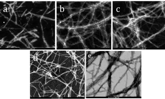
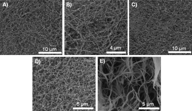
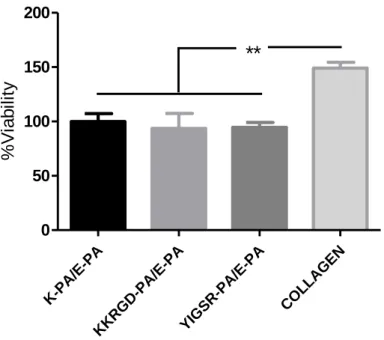
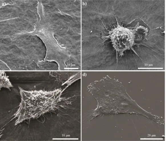
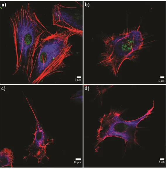
Figure 1.1 Commercially available tissue engineering products. ... 5 Figure 1.2 Basics of tissue engineering. ... 8 Figure 1.3 Interactions between cells and their surrounding extracellular matrix organize the biological dynamic ... 16 Figure 1.4 Glycosaminoglycans and their structural units. ... 19 Figure 1.5 Schematic representations of peptide amphiphile molecules and nanofibers. (A) Chemical structure, (B) different regions of structure, (C) nanofibrous structure of peptide amphiphiles ... 28 Figure 2.1 Chemical structures of PA nanofibers. (A) YIGSR-PA, (B) KKRGD-PA, (C) K-KKRGD-PA, (D) E-PA ... 48 Figure 2.2 (A) HPLC chromatogram of purified YIGSR-PA molecule at 220 nm. (B) Mass spectrometry analysis of YIGSR-PA molecule. ... 49 Figure 2.3 (A) HPLC chromatogram of purified KKRGD-PA molecule at 220 nm. (B) Mass spectrometry analysis of KKRGD-PA molecule. ... 50 Figure 2.4 (A) HPLC chromatogram of purified E-PA molecule at 220 nm. (B) Mass spectrometry analysis of E-PA molecule ... 51 Figure 2.5 (A) HPLC chromatogram of purified K-PA molecule at 220 nm. (B) Mass spectrometry analysis of K-PA molecule. ... 52 Figure 2.6 Circular dichroism spectra of the peptide nanostructures. ... 54 Figure 2.7 Transmission electron micrographs of (A) YIGSR-PA/E-PA, (B) PA/E-PA, (C) K-PA/E-PA, (D) YIGSR-PA/CS, and (E) KKRGD-PA/CS. ... 55
xvii
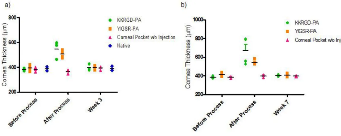
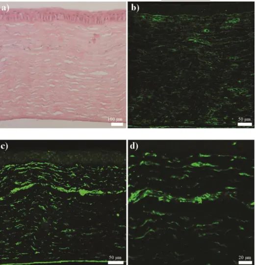
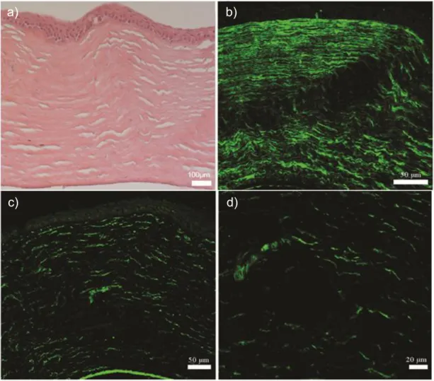
Figure 2.8 Atomic force microscopy of (A) YIGSR-PA/E-PA, (B) KKRGD-PA/E-PA, (C) K-PA/E-KKRGD-PA/E-PA, (D) YIGSR-PA/CS and (E) KKRGD-PA/CS. ... 55 Figure 2.9 Scanning electron micrographs of (A) YIGSR-PA/E-PA, (B) KKRGD-PA/E-PA, (C) K-PA/E-PA, (D) YIGSR-PA/CS and (E) KKRGD-PA/CS. ... 56 Figure 2.10 Viability results of corneal fibroblast at 24 h. PA scaffolds do not exhibit any toxic effect on HTK cell line. ... 58 Figure 2.11 Proliferation results of corneal fibroblasts at 72 h. The YIGSR-PA nanofibers exhibited a similar proliferation profile compared with collagen. . 58 Figure 2.12 SEM images of corneal fibroblasts cultured on (A) YIGSR-PA coated surface, (B) KKRGD-PA coated surface, (C) K-PA coated surface and, (D) collagen coated surface show the adhesion profiles of cells. ... 59 Figure 2.13 Spreading and cellular morphology of corneal fibroblasts acquired by vimentin (blue), TRITC-conjugated phalloidin (red) and TO-PRO®-3 (green) iodide staining on (A) YIGSR-PA coated surface, (B) KKRGD-PA coated surface, (C) K-PA coated surface, (D) collagen coated surface. ... 60 Figure 2.14 Representative images of injection process of KKRGD-PA. ... 61 Figure 2.15 Thickness of the rabbit corneas at (A) week 3 and (B) week 7. 62 Figure 2.16 IHC analyses of YIGSR-PA injected samples at week 3. ... 64 Figure 2.17 IHC analyses of KKRGD-PA injected samples at week 3. ... 65 Figure 2.18 IHC analyses of damaged rabbit cornea samples at week 3. ... 66 Figure 2.19 IHC analyses of YIGSR-PA injected samples at week 7. ... 68 Figure 2.20 IHC analyses of KKRGD-PA injected samples at week 7. ... 69 Figure 2.21 IHC analyses of damaged rabbit cornea samples at week 7 .... 70
xviii
Figure 2.22 Histological and immunohistochemical analyses of native rabbit corneas ... 70 Figure 3.1 Chemical structures of peptide amphiphiles used in this study ... 90 Figure 3.2 (A) HPLC chromatogram of purified Heparin-mimetic PA molecule at 220 nm. (B) Mass spectrometry analysis of Heparin-mimetic PA molecule. ... 91 Figure 3.3 (A) HPLC chromatogram of purified K-PA molecule at 220 nm. (B) Mass spectrometry analysis of K-PA molecule ... 92 Figure 3.4 Circular dichroism spectra of the peptide amphiphile molecules. 93 Figure 3.5 Characterization of peptide amphiphile nanofiber and scaffolds at pH 7.4 ... 94 Figure 3.6 Morphology of pancreatic islets cultured with HM-PA, HM-PA with growth factors (GF), HM-PA/K-PA, HM-PA/K-PA growth factors at day 7. .. 97 Figure 3.7 Viability analysis of the islets seeded on different PA hydrogels with or without growth factor (GF) by using the Alamar blue assay at (A) day 1, (B) day 3 and (C) day 7. ... 98 Figure 3.8 Results of glucose stimulation test at (A) day 1, (B) day 3, and (C) day 7 ... 99 Figure 3.9 Technique used to implant peptide amphiphile gel into the omentum of rats.. ... 101 Figure 3.10 Blood glucose levels were measured 5 days before and throughout 28 days after transplantation. ... 102 Figure 3.11 Change of weight of animals during 28 days. ... 102
xix
Figure 3.12 Intraperitoneal glucose tolerance test (IPGTT) at the end of 28
days. ... 103
Figure 3.13 H&E staining of (A) Sham, (B) PA hydrogel, (C) Islet, and (D) Islet + PA hydrogel groups. ... 105
Figure 3.14 Masson’s trichrome staining of (A) Sham, (B) PA hydrogel, (C) Islet, and (D) Islet + PA hydrogel groups. ... 106
Figure 3.15 Immunohistological staining of (A) Sham, (B) PA hydrogel, (C) Islet, and (D) Islet + PA hydrogel groups using anti-insulin antibody. ... 107
Figure 3.16 Immunohistochemical staining of macrophages in omenta. .... 108
Figure 3.17 Immunohistochemical staining of blood vessels by using anti-von Willebrand Factor in the omenta and quantification of blood vessels. ... 109
Figure 4.1 Chemical structure of HM-PA molecule. ... 126
Figure 4.2 Chemical structure of K-PA molecule. ... 126
Figure 4.3 Chemical structure of E-PA molecule. ... 127
Figure 4.4 Purification and characterization of HM-PA. (A) HPLC chromatogram of purified heparin-mimetic PA molecule at 220 nm. (B) Mass spectrometry analysis of heparin-mimetic PA molecule ... 127
Figure 4.5 Purification and characterization of E-PA. (A) HPLC chromatogram of purified E-PA molecule at 220 nm. (B) Mass spectrometry analysis of E-PA molecule. ... 128
Figure 4.6 Purification and characterization of K-PA. (A) HPLC chromatogram of purified K-PA molecule at 220 nm. (B) Mass spectrometry analysis of K-PA molecule. ... 129
xx
Figure 4.7 Scanning electron microscopy images of (A) HM-PA/K-PA and (B) E-PA/K-PA reveal the ECM-like morphology of the scaffolds. ... 130 Figure 4.8 Rheology results showed that HM-PA/K-PA and E-PA/K-PA combinations formed gels and that their mechanical properties were similar. ... 130 Figure 4.9 Representative images of wounds following hydrogel application ... 132 Figure 4.10 Wound areas of (A) sucrose, (B) E-PA/K-PA and (C) HM-PA/K-PA treated wounds at days 3, 7, 10 and 14. ... 132 Figure 4.11 Wound area (%) according to the location of wounds in control, E-PA/K-PA and HM-E-PA/K-PA treated groups on day 3. ... 133 Figure 4.12 Wound area (%) according to the location of wounds in control, E-PA/K-PA and HM-E-PA/K-PA treated groups on day 7. ... 133 Figure 4.13 Wound area (%) according to the location of wounds in control, E-PA/K-PA and HM-E-PA/K-PA treated groups on day 10. ... 134 Figure 4.14 Wound area (%) according to the location of wounds in control, E-PA/K-PA and HM-E-PA/K-PA treated groups on day 14.. ... 134 Figure 4.15 (A) H&E and (B) Masson’s Trichrome staining (lower panel) of control group at day 7.. ... 136 Figure 4.16 (A) H&E and (B) Masson’s Trichrome staining (lower panel) of E-PA/K-PA application at day 7.. ... 137 Figure 4.17 (A) H&E and (B) Masson’s Trichrome staining (lower panel) of HM-PA/K-PA application at day 7. ... 138
xxi
Figure 4.18 (A) H&E and (B) Masson’s Trichrome staining (lower panel) of control group at day 14. ... 139 Figure 4.19 (A) H&E and (B) Masson’s Trichrome staining (lower panel) of E-PA/K-PA application at day 14. ... 140 Figure 4.20 (A) H&E and (B) Masson’s Trichrome staining (lower panel) of HM-PA/K-PA application at day 14. ... 141 Figure 4.21 Quantitative analysis of granulation tissue of wound tissues at day 7 and 14. ... 142 Figure 4.22 Quantitative analysis of re-epithelialization of wound tissues at day 7 and 14. ... 143 Figure 4.23 Quantitative analysis of skin appendages of wound tissues at day 14. ... 143 Figure 4.24 Staining of blood vessels by anti-von Willebrand Factor (a-c) and quantification of blood vessels at day 7 and 14 (d-e). ... 144
xxii
Abbreviations
AFM Atomic force microscopy
ANOVA Analysis of variance
Boc Tert-butoxycarbonyl
BSA Bovine serum albumin
CD Circular dichroism
CS Chondroitin sulfate
DAB 3,3'-diaminobenzidine
DCM Dichloromethane
DIEA N,N-diisopropylethylamine
DMEM Dulbecco's modified Eagle's medium
DMF N,N-Dimethylformamide
DMSO Dimethyl sulfoxide
ECM Extracellular matrix
EDTA Ethylenediaminetetraacetic acid
E-PA Lauryl-VVAGE-OH
ESC Embryonic stem cells
FBS Fetal bovine serum
FDA Food Drug Administration
FGF-2 Fibroblast growth factor-2
Fmoc 9-Fluorenylmethoxycarbonyl
GAG Glycosaminoglycan
GF Growth factor
H&E Hematoxylin and eosin
HBTU N,N,N
′,N′-Tetramethyl-O-(1H-benzotriazole-1-yl) uronium hexafluorophosphate
HM-PA Heparin mimetic peptide amphiphile
HPLC High pressure liquid chromatography
HRP Horseradish peroxidase
HTK Human keratocyte
IHC Immunohistochemistry
IPGTT Intraperitoneal glucose tolerance test
iPSC Induced pluripotent stem cell
K-PA Lauryl-VVAGK- Am
KKRGD-PA Lauryl-VVAGKKRGD- Am
LC-MS Liquid chromatography-Mass spectroscopy
P/S Penicillin/Streptomycin
PA Peptide amphiphile
PBS Phosphate buffered saline
xxiii
PEG Poly ethylene glycol
PLA Poly lactic acid
PGA Poly glycolic acid
PLGA Poly lactic-co-glycolic acid
RT Room temperature
SEM Standard error of mean
T1D Type 1 Diabetes
TBS Tris buffered saline
TEM Transmission electron microscopy
TFA Trifluoroacetic acid
1
C
HAPTER
1
2
1.1 The Fountain of Regeneration: An approach in biology and engineering
From ancient times to the Age of Exploration –and perhaps even today– it has been a fundamental desire of Man to find a mystic source of water that cures all sickness and bestows eternal life. Although the Greek historian Heredotus and Alexander the Great were said to have believed in a Fountain
of Youth, the myth is most closely associated with the 16th century Spanish explorer Juan Ponce de Léon. Urged by King Ferdinand to explore an island called Bimini, he discovered Florida and –according to the myth– the fountain. As legend has it, whoever bathes in the water or drinks from it is granted everlasting life and freedom from all sickness. Of course, no magical panacea is known to modern medicine, and no record confirms that Ponce indeed found the fountain of youth. But perhaps this is about to change.
It is not difficult to understand why people seek a cure for all sickness – including Shakespeare’s “hideous winter”, aging. Observations of the animal kingdom only raised further questions, as some organisms can apparently recover and re-grow after any injury, while Man may forever be crippled after superficial damage. In the regeneration process, injury is recognized and the original integrity of the damaged tissue is restored by complex mechanisms. However, the regenerative capacities of animals vary greatly between the nine major animal phyla, and may change considerably in even closely related species [1]. Réaumur and Trebmley’s landmark experiments on starfish, crayfish and Hydra have greatly expanded the scope of research on regeneration, and comparative and descriptive characterization of the
3
regeneration process has become a topic of major scientific interest. Initial research into this process concerned whether there were differences in the regenerative capacity of various organs and tissues in various organisms, whether aging had any effect on the rate of regeneration, and whether regeneration would occur in a differentiative manner between different species and their tissues [2]. Following these investigations, more specialized questions began to arise: how did internal and external conditions affect this process?; how does the so-called “histogenesis” occur, when tissues are generated? These questions are not fully answered to date, and research into them forms the basis of current approaches in tissue engineering.
Regeneration is broadly seen in nature, but the extent of this process is variable between species and life stages [3]. Although limb regeneration in salamanders is one the best known examples of literature, other urodelan tissues may not exhibit the same capacity for regeneration, despite having an identical genetic composition [4]. We also know that identical injuries may result in complete regeneration or extensive scar formation in related species, between different organs and tissues, or in organisms at different life stages. Perhaps due to these observations, the molecular investigation of the repair process has been increasingly important in the past few decades, and modern biomaterials are commonly designed with molecular functions in mind.
The first signs of reconstruction, however, date back to 7000 BC [5]. Different materials were used in Incan Empire for cranioplasty and the usage of gold plates was reported by Fallopius in 16th century. In 1668, van Meekeren developed a xenograft model for the cranial defect of a man [6]. These
4
approaches started to spark interest into the use of bio-engineering as a solution for biological problems. Between 1660s and 1980s, many methods or approaches were developed for this purpose and, finally, tissue engineering emerged as a major research area in late 1980s. Following the founding of the Tissue Engineering and Regenerative Medicine International Society, the limitations of those methods came under scrutiny.
The primary goal of tissue engineering and regenerative medicine is to transplant biofactors such as cells, specific proteins or small molecules, signals, genes or their combinations within a scaffold. While biofactors initiate the repair process, the porous and degradable architecture of scaffolds (of which dimensions range from 10 to 10000 micrometers) imitates the volume and mechanical properties of lost tissue, and carries the biofactors in its structure [7-8]. It is, however, a major challenge to reform tissues from cells by triggering complex tissue repair processes in a reliable manner. Material costs, batch-to-batch variations, governmental regulations and social and religious views all contribute to the difficulty of designing an ideal scaffold. To date, several biomaterials were approved by the Food Drug Administration (FDA), European Union (EU) or the German government (Figure 1.1) [9]. Although there is considerable progress in biomaterial design, their commercial applications are nonetheless limited and next-generation materials are necessary for tissue engineering and regenerative medicine applications to be commonly utilized in clinical settings. As attempts to entirely replace the damaged organ can only serve as stopgap solutions and do not yield long-term results, it is important to define the problems associated with the repair of
5
damaged tissues and subsequently engineer biomaterials to enhance the natural repair process of the organism of interest.
Figure 1.1 Commercially available tissue engineering products. (Reproduced
6
Figure 1.1 (continued) Commercially available tissue engineering products.
Tissue abbreviation: T/L, tendon and ligament; BV, blood vessel; HV, heart valve. Regulatory status: dates indicate year approved by regulatory body (FDA unless stated as EU or Germany). Preclinic indicates preclinic development, phase I and II indicate clinical trial stage. Date of regulatory approval does not necessarily coincide with market release. aReclassified in 2006 from an earlier product. bFirst clinical trials in 200. cCommercial sale
7
began in 2000. dUnregulated in EU, not available in the United States. eUnregulated product. fDecision expected 2009. §Addition of growth factors and cells is dependent of application. (Reproduced from Ref. [9] with permission from Nature Publishing Group.).
1.2 Basic Principles of Tissue Engineering and Regenerative Medicine
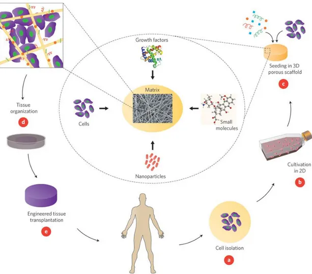
Organ transplantation is the ideal means of replacing organs or tissues lost due to trauma or other causes. However, a substantial number of people die annually without being included in waiting lists. Organ transplantations are limited by high costs, the lack of available donors and immune rejection of the organ [10]. Beyond these problems, even successful organ transplants face a series of problems. The field of regenerative medicine and tissue engineering has emerged largely with the intent to eliminate the problems associated with organ transplants, and utilizes the tenets of biology, engineering and materials sciences to maintain and renew damaged tissues and organs [11]. The three-dimensional (3D) composition of tissue microenvironments can be imitated by seeding patient- or donor-derived cells into the porous scaffold. The scaffold can be enriched with growth factors and small molecules according to the specific needs of the tissue of interest and subsequently transplanted into the patient (Figure 1.2) [10].
8
Figure 1.2 Basics of tissue engineering. Cells are initally isolated from the
patient or a donor (A) and expanded in cell culture (B). Expanded cells are seeded into a porous scaffold, which is decorated with the necessary signals for tissue repair (C) and incubated in a bioreactor to provide the optimal growth conditions (D). The constructed tissue is later transplanted to the damaged site
(E) (Reproduced from Ref. [10] with permission from Nature Publishing
Group.).
Tissue engineering and regenerative medicine approaches have been developed to provide cells with a facsimile of their natural microenviroment, which supports them and allows the formation of replacement tissues by
9
triggering the differentiation process. The main elements of this approach are cells, scaffold and signal molecules; and different combinations of these elements can be altered depending on the needs of the injured or diseased tissue [12].
1.2.1 Cell Types
The choice of cell type is vital to ensure that a sufficient number of cells can be generated to meet the needs of the damaged tissue. Three types of cell sources can be utilized for tissue engineering: Autologous, allogeneic and xenogeneic. Autologous cells are the patient’s own cells, which are isolated and subsequently expanded in cell culture for regenerative applications. The use of these cells is typical for tissue engineering, because there is no risk of immunogenity and no need for immunosuppressive treatment. In addition, they can be utilized for the repair many organs, such as the liver, blood vessels, bone, cartilage and skin. However, autologous cells have their own set of limitations. First of all, the tissue of interest should be reachable by biopsy, as the cells must be a derived from the patient [13]. Harvesting cells can also be a problem; e.g. if there is myocardial infarction in the patient’s medical history, it is not possible to obtain cardiac cells in sufficient numbers. The age of the patient also affects the amount of harvested cells, and even if the harvesting process is successful, contamination in culture environment is another potential risk. In addition to issues with the culture equipment, it is known that sera used for cell culture may lead to viral contamination. Allogeneic (from different individuals of the same species) or xenogeneic (cells from different species) sources are also utilized for tissue repair. However, immunogenicity
10
is the most common problem in both approaches. In addition, zoonoses can also pose a problem, as Patience et al. reported that porcine endogenous retrovirus can transfer from donor to acceptor [14]. Despite these limitations, allogeneic cells have been used as effective wound covers [15] and in the treatment of diabetes [16] and liver disease [17] (Figure 1.1), while feeder cells from xenogeneic sources have been evaluated for the formation of epidermal tissue[18].
Stem cells are another major source for regeneration of tissues. Stem cells have the exclusive capacity to proliferate in an undifferentiated state and differentiate under special conditions (which may be delivered through cell culture media) into various cell types [19]. These cells can be derived from both embryonic and adult tissues. Embryonic stem cells (ESC) have the potential to differentiate into all three germ lines: ectoderm [20], mesoderm [21] and endoderm [22]. However, controlling the fate of these cells is not easy and teratomas can form under in vivo conditions [23]. In addition to this, there are some ethical issues about the usage of ESCs, which is still forbidden in many countries – although the United States lifted the ban in 2009. Nevertheless, limitations associated with ESCs can be overcome with the use of induced pluripotent cells (iPSC), which closely resemble their embryonic counterparts but are derived from somatic cells that are transformed through Yamanaka factors [24-27].
Because of the tumorigenic potentials of ESCs and iPSCs, adult stem cells such as hematopoietic (HSC), mesenchymal (MSC) or adipose derived stem cells (ADSC) are generally preferred for regenerative medicine. These
11
cells are easily accessible, can be obtained in large amounts and pose fewer safety issues compared to ESCs and iPSCs. Somatic cells generally reach their fully differentiated forms soon after birth [28]. However, HSCs have the potential to facilitate tissue regeneration throughout the entire life span. HSCs have a high tendency to maintain myogenesis and cardiomyogenesis [29-31], and can also trigger the formation of liver cells [31]. Unlike HSCs, MSCs are found not only in bone marrow but also in bone [32], skeletal muscle [33], skin [34], pericytes [35], peripheral blood [36] and teeth [37]. They have attracted great attention for tissue engineering approaches because of their capability to differentiate into muscle, blood vessels, bone, tendon, cartilage, skin, nerve and adipocytes [18, 38]. In addition to HSCs and MSCs, ADSCs are popular for tissue engineering. Since adipose tissue is pervasive and easily accessible, they can be obtained in large amounts with less morbidity. The differentiation capacity of ADSCs is high and encompasses chondrogenic [39], myogenic [40], adipogenic [41], osteogenic [42], angiogenic [43] and neuronal lineages [44].
Stem cells are highly promising for the clinical applications of tissue regeneration. However, they still suffer from certain limitations; in particular, their quantity is typically insufficient to facilitate tissue repair. Although stem cells can be expanded in two dimensional cell culture environments, dedifferentiation eventually takes place before the desired number of cells is reached. In addition, the proliferation capacity of stem cells is quite low in 3D cultures. Another problem is the necessity of xenogeneic serum, which provides growth supplements to the cells but can also lead to contamination
12
[45]. While synthetic sera can be used instead of fetal calf serum, the proliferative capacity of cells decreases under these conditions.
1.2.2 Soluble macromolecules in ECM
Tissue engineering is a complex process that requires acts through several signal cascade events and ultimately culminates in the initiation of the repair process at the tissue of interest. Under natural conditions, repair-associated signals are activated through proteins called growth factors (GFs). GFs may be secreted endogenously to stimulate the cell itself (autocrine signaling) or to communicate with other cells (paracrine signaling). Although the effects of many growth factors are not fully described, stem cell research in particular led to a greater understanding of the remarkable capacity of growth factors to initiate and regulate processes such as development, differentiation and regeneration.
The delivery of growth factors into tissues of interest is a difficult prospect. While soluble GFs may be injected, their systemic dispersion prevents them from reaching the concentrations necessary for initiating local tissue repair. As such, three methods have been developed to selectively deliver GFs to a specific organ or tissue. The first is to use a DNA plasmid that codes for a given GF, allowing its continued in-site production. The second is to transfect the GF-coding gene to a certain cell type, and subsequently inject these cells to localize to the tissue of interest [18]. Lastly, GFs can be delivered without genetic engineering through the assistance of a carrier matrix called a scaffold. This is the most popular method for GF delivery, as it can readily overcome
13
the short half-lives associated with the injection of soluble GFs. However, the degradation kinetics of the scaffold should also be determined to ensure optimal release of GFs in tissue environments.
Bone morphogenic proteins (BMPs), fibroblast growth factor-2 (FGF-2), transforming growth factor-β (TGF- β) and vascular endothelial growth factor (VEGF) are the most commonly used GFs in tissue engineering. Some of these GFs are already available in commercial tissue engineered products (Figure 1). Recombinant BMP-2, for example, has been embedded into a collagen sponge and used for spinal fusion, tibial shaft fractures and oral-maxillofacial procedures [46]. In addition to ensuring direct delivery, the controlled release of GFs may also enhance their activity: for instance, the extracellular matrix production of vascular smooth muscle cells was found to increase in the presence of TGF-β1 immobilized in polyethylene glycol (PEG) hydrogel compared to the soluble TGF- β1[47].
Carrier systems can be decorated with specific structures to enhance their ability to bind to growth factors. It is known that glycosaminoglycan molecules can bind to and protect growth factors from degradation [48-49]. In addition, some growth factors such as FGF-2 have heparin-binding domains which establish an electrostatic interaction between the GF and the molecule through sulfate and carboxylic acid groups [50-51]. It has been shown that this binding interaction increases GF stability compared to the unbound form [52]. As such, the heparin molecule has been studied extensively for slow release applications.
14
1.2.3 Scaffolds
As a definition, a scaffold is a temporary platform used for the cultivation of cells and tissues [53]. The efficiency of tissue engineering approaches depends heavily on the ability of the scaffolds to provide cells with conditions closely resembling their native environment. Chemical and physical cues inherent to in vivo tissue environments are often mimicked for this purpose, and scaffolds are typically designed to facilitate the recruitment, attachment and proliferation of cells, imitate the mechanical properties of the tissue of interest and allow the exchange of oxygen, nutrients and waste products across the scaffold matrix. In addition, biochemical signals should be presented or retained in the scaffold structure to ensure the optimal growth and differentiation of the resident cell population [54].
As many of these requirements are met by the extracellular matrix (ECM) in the native tissue microenvironment, the investigation of ECM structure and function is a major aspect of tissue engineering.
1.2.3.1 The extracellular matrix and the importance of its interactions with cells
The ECM is the non-cellular part of tissues and contains proteins, proteoglycans, water and minerals. The formation, function, homeostasis and regeneration of tissues are orchestrated by the spatial and temporary organizations of signals arising from the ECM (Figure 1.3). The ECM provides not only a mechanical support to cells but also takes part in determining cellular fates, facilitating the replication, migration, differentiation, apoptosis or
15
proliferation of cells through molecules such as growth factors and pH adjustments [55-56]. The ECM is a dynamic 3D microenvironment and consists of different combinations of at least 100 types of molecules, which depend on the organ or tissue, age and physiological condition. Fibrillar proteins (collagens and non-collagenous glycoproteins), proteoglycans and glycosaminoglycans are the main components of the ECM, and serve as a reserve for soluble macromolecules such as growth factors, cytokines and chemokines [57].
16
Figure 1.3 Interactions between cells and their surrounding extracellular
matrix organize the biological dynamic. Specific binding of physical and soluble signals to cell surface receptors or cell-cell interactions initiate the signaling cascade inside the cells and fate of cells is determined by genetic regulation. PLC, phospholipase C; GAGs, glycosaminoglycans; PGs, proteoglycans; CAMs, cell adhesion molecules (Reproduced from Ref. [57] with permission from Nature Publishing Group.).
17
Collagen is the most common protein type in the ECM, and exhibits a triple helix (homo or heterotrimer) organization due to its α-chain structure [58]. There are 28 known members of the collagen family, which are derived from different combinations of 48 α-chains [59]. Other proteins may also bear collagen-like domains that allow them to exhibit similar properties [60]. Collagen fibers can form large-scale bundles, which play a major role in providing tensile strength to tissues and exhibit a variety of structures depending on the collagen types involved in network formation. In addition to its mechanical properties, collagen is also responsible for regulating cellular behaviors such as differentiation, adhesion and migration; thereby controlling the tissue development process [61-62]. In addition to collagen, elastin and fibrilin are the other fibrillar proteins in ECM, and largely exhibit similar properties [55].
Fibronectin is another key molecular player in the ECM. It can bind to GAGs, cell surface receptors such as integrins and other ECM proteins, and may also self-assemble by binding to other fibronectin molecules [63]. One of the most important roles of fibronectin is cell adhesion. The ‘RGD’ sequence found in fibronectin shows a strong affinity for integrins, including α5β1, αvβ3 and αIIbβ3 [64-65]. Recent research also suggests that this sequence is responsible not only for cell attachment, but also for cellular migration during the development and self-renewal of stem cells [66-68].
Laminin is another major protein of the ECM, and consists of epidermal growth factor-like repeats and globular and coiled domains [56]. This molecule commonly occurs in the basal lamina of tissues [69]. The modular domains in
18
the laminin molecule can interact with cell surface receptors, such as integrins, and other components of ECM, such as entactins, perlecan and collagens [70]. Like fibronectin, a variety of peptide epitopes have been derived from the laminin sequence; including “YIGSR” and “IKVAV”. The YIGSR peptide is associated with cell attachment and migration, metastasis and the inhibition of angiogenesis [71-72]; while IKVAV plays a role in neurite outgrowth, angiogenesis and tumor and cell growth [73-75].
Bornstein identified another class of ECM proteins, which are called matricellular proteins and include the tenascin family, thrombospondins, osteopontin and SPARC/osteonectin. These proteins also interact with cell surface receptors, other proteins and molecules such as cytokines, but are not involved in structural tasks [55, 76].
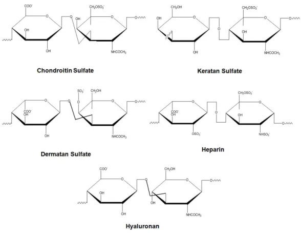
Proteoglycans are another important class of ECM proteins and typically consist of glycosaminoglycan (GAG) chains that are bound to a core protein [77-78]. Because of their structures, proteoglycans can bind to growth factors and cytokines [79]. They are categorized according to their GAG chains, core proteins and location, as cell-surface proteoglycans, modular proteoglycans and small leucine-rich proteoglycans (SLRPs) [78]. GAGs are long and charged molecules that consist of repeating disaccharide units [D-glucuronic or L-iduronic acid, sulfated N-aceltylglucosamine or N-acetylgalactosamine, and galactose (4 N-acetylglucosamine-β1,3-galactose-β1)]. These molecules can be further classified as sulfated and non-sulfated GAGs. Sulfated GAGs include chondroitin and dermatan sulfate (which are composed of galactosamine and either glucuronic acid or iduronic acid) heparin and
19
heparan sulfate (which are composed of glucosamine and either glucuronic acid or iduronic acid), and keratin sulfate (which is composed of glucosamine and galactose). Hyaluronan is the only non-sulfated GAG and does not bind to any core protein (Figure 1.4) Because of their hydrophilic structures, GAGs can interact with water molecules and form highly hydrated structures, thereby providing compressive strength to the tissues in which they are abundant [57].
Figure 1.4 Glycosaminoglycans and their structural units.
GAGs are one of the most studied polysaccharides and have substantial roles in various biological processes, including angiogenesis [80], cancer pathogenesis [81], neural development [82], embryogenesis [83] and anticoagulation [84]. These molecules modulate cells and their microenvironments through their interactions with ECM proteins, growth
20
factors, enzymes and cytokines. In addition, they can direct signaling by allowing the formation of enzyme-substrate and growth factor-receptor complexes.
1.2.3.2 Requirements of scaffolds
Numerous scaffolds have been developed to regenerate different tissues and organs in the literature, using a great variety of materials and fabrication techniques. Even though we do not have clear-cut rules of thumb to define an ideal scaffold, these materials must nevertheless meet certain general criteria to facilitate the regeneration of the tissue or cell of interest.
The decisive criterion for any scaffold for any tissue is biocompatibility. Neither the scaffold itself nor its degradation byproducts should raise any immune or inflammatory response in the body. Immune responses can delay the healing process or affect the acceptance of the scaffold [85]. Moreover, cells should function normally within the scaffold, which should be free of external contaminants and exhibit no toxic or mutagenic activity towards any organ or tissue.
The main aim of tissue engineering and regenerative medicine is to provide support to injured tissue until the recovery is completed. Therefore, the scaffold should be designed such that the regenerating tissue can gradually replace it. Tissue repair and scaffold degradation should be synchronous to ensure that this process occurs smoothly.
The optimization of mechanical properties is another other key element in tissue engineering. During the implantation or application process, the
21
structure should not be damaged as a result of surgical handling. Scaffolds intended to replace hard and ductile tissues, such as bone, cartilage and cardiovascular muscle, should be particularly resistant to mechanical stress and support the tissue during the healing process. Since the repair of injuries is slower in older patients, the degradation-resorption kinetics of scaffold should also be considered on a patient-dependent basis [86]. Nonetheless, the production of materials with ideal mechanical properties may also indirectly affect other aspects of tissue regeneration, possibly resulting in insufficient vascularization and cell infiltration [87].
The porous architecture of scaffolds is another crucial point. Scaffolds are typically intended to facilitate tissue regeneration at the site of implantation, and the infiltration of the scaffold by surrounding cells is essential for this process. Porous materials allow the exchange of nutrients and gases, which is necessary for the continuity of biological activities. Furthermore, the byproducts of the scaffold (and, if any, its resident cells) should be eliminated without damaging the newly formed tissue [88]. As cells communicate with the ECM through specific chemical groups and are heavily reliant on their microenvironment to establish these connections, pore size is also important for scaffold design. While pores should be large enough to allow cell movements, they should also be small enough for efficient cell binding [89].
Finally, it is important to mention the processes involved in scaffold production. Efforts in regenerative medicine generally aim for the clinical use of the scaffold. As such, the scaffold should be reproducible, cost effective and commercially available. Scaffolds that do not require any extra steps prior to
22
implantation (such as in vitro culture for cell growth) are preferred for this purpose [89].
1.2.3.3 Biomaterials utilized for tissue engineering and regenerative medicine
Providing an optimized milieu to regenerating tissue by using biomaterials is the primary aim of tissue engineering and regenerative medicine applications. The materials that have been chosen for this purpose can be divided into two categories called natural and synthetic. Decellularized biological materials will be discussed in naturally derived biomaterials part. Figure 1 has detailed examples of all materials.
1.2.3.3.1 Naturally derived biomaterials
The first materials used in clinical applications are naturally derived materials [89]. They carry the biological cues and other physical features to provide a better environment for cells to attach, proliferate, migrate and differentiate. Since they are extracted from natural sources such as skeleton and blood plasma, they do not have any toxicity [12]. The degradation is achieved by natural enzymatic system in the host. However, there are several disadvantages of these materials. Their degradation kinetics can be rapid and some chemical modifications such as cross-linking, which can cause toxicity, can be needed [90]. The other problem can be, albeit small, the inflammatory response.
Collagen is the most studied natural molecule for tissue engineering applications. It can be obtained from both animal and human sources.
23
According to used density, the degradation kinetics and the pore size can be controlled [91]. In addition, its combination with other synthetic materials increases the mechanical properties of collagen [92]. Development of organisms is based on cell-ECM interactions which are the communications between cell surface receptors and specific epitopes on molecules. These interactions can be direct like integrin family and discoidin domain 1 and 2 which bind GFO (Gly-Phe-Hyp) motif [93-94] and indirect like RGD (Arg-Gly-Asp) sequence in fibronectin [95]. In the indirect cell-collagen interaction, proteins containing RGD motif bind to both integrin and collagen. These binding profiles can lead the cellular behaviors like adhesion and proliferation and increase the importance of collagen scaffolds [96]. Collagen scaffolds have been widely investigated in neural system disorders [97], bone [98] and cartilage [99] disorders, cardiovascular system disorders [100], blood vessels [101], skin [102-104], cornea [105-107] and urogenital system regeneration [108].
Hyaluronan (hyaluronic acid or hyaluronate) is a GAG which iscomposed of β-(1,4) or β-(1,3) linked D-glucuronic acid and D-N-acetylglucosamine (Figure 1.4). Due to its water holding capacity, it has great viscoelastic properties. Hyaluronan can be obtained via bacterial production. It is found in wound sites, vitreous chamber of eye and synovial fluid in joints [109]. It has been also reported that hyaluronan has important roles in cell migration, inflammation, fertilization, mitosis and angiogenesis [110]. There are several applications of hyaluronan based scaffolds. It is extensively studied for cartilage repair due to its effect on chondrocyte proliferation and production of
24
GAGs and type II collagen [111-112]. In addition, it was shown that hyaluronan improves cartilage histogenesis and promote cellular condensation in chondrogenesis [113]. Hyaluronan-polyethylene glycol complex has been investigated for controlled release of BMP-2 and this complex contributed to the bone regeneration [114]. Apart from these applications, it was used for nerve and brain repair [115]; wound healing [116]; and as space filling material in otolaryngology [117] and reconstructive surgery [118].
As a polysaccharide, alginate is the main component of seaweeds, and is composed of β-(1,4) D-mannuronic acid and α-(1,4) Lguluronic acid [119]. This polysaccharide forms ionotropic and elastic gel with divalent cations including calcium and magnesium [120]. CaCO3 and CaSO4 salts affect the mechanical properties of alginate gels [121]. Alginate gels are biocompatible and injectable systems that form at physiological pH and temperature [122]. One of the limitations of the system is its slow degradation kinetics [123]. Alginate gels were used in pancreatic islet delivery [124], cell mobilization [125], wound dressing [126] and cartilage tissue engineering [120].
Chitosan is another biomolecule that has been widely investigated for biomedical applications. It is a soluble derivative of chitin, and has great properties for tissue engineering applications including biocompatibility, biodegradability, non-toxicity,hemostatic potential [127], antimicrobial activity and easy process ability. It can be processed as different forms such as sponges, hydrogels, membranes and fibers [128]. Due to its remarkable features, it has been investigated for bone and cartilage [129] regeneration, wound dressing [130] and nerve conduits [131].
25
Decellularized materials are prepared by removing all cell and remaining proteins and other biological elements. The source of material can be allogenic or xenogenic. They are also biodegradable and biocompatible materials, however, the cross-linking methods for improving the mechanical properties can evoke immune response [79]. The most common applications of decellularized matrices are in wound healing area [132]. Although studies on decellularized matrices increase the potential use of xenogenic sources in tissue engineering applications, there are still some issues that should be overcome including size differences between human and animal organs and difficulties in complex systems like heart and liver [13].
Except above mentioned materials, fibrinogen [133], fibrin [134], silk[135], agarose [136], dextran [137] etc have also been reported for tissue engineering and regenerative medicine applications.
1.2.3.3.2 Synthetic biomaterials
Naturally derived scaffolds have many advantages for supporting cells and creating the natural environment. However, concerns about immunological response due to xenogenic viral infections of natural materials have prompted researchers to develop synthetic materials with no immunogenicity responses, controllable properties and processing flexibility [138]. These materials can be fabricated according to the tissue needs. Their degradation can be controlled by material itself or its composites [139]. As biodegradable polyesters, poly (lactic acid) (PLA), poly (glycolic acid) (PGA), and poly (lactic-co-glycolic acid) (PLGA) have FDA approval in clinical
26
applications. In addition, their degradation byproducts are not toxic to body [140]. The degradation kinetics can be controlled from weeks to years. These polymers are biocompatible except from PGA. However, CO2 is the degradation products of PLA and PGA, which decrease the pH of environment and cause necrosis [88]. They are suitable for mass production with long shelf lives and are cheaper than biological polymers [53]. Other polymers which are used in tissue engineering applications include polyurethanes (for vascular valves and grafts, prostheses and catheters [141]); polyanhydrides (for drug-delivery applications [142]); and polyphosphazenes (for soft tissue regeneration [143]).
Despite their mechanical, physical and porous structures, one of the common drawbacks of these materials is lacking of bioactive signals for cell attachment, migration, differentiation and so on. However, they can be conjugated with essential peptide sequences to help cell-integrin recognition [144]. Hydrogel scaffolds made by these materials are biodegradable and have many similarities with ECM. Their applications are less invasive and site specific due to the injectable properties [145].
Hydrogels are hygroscopic materials that can harbor large amounts of water, up to thousands times than their own dry weight. Biocompatibility of hydrogels increases because of their high water content [146]. One of the first examples of polymeric hydrogels is PEG which is a well known polymer as food additive. Forming hydrogel from PEG is done by cross-linking that determine the mechanical properties and degradation kinetic of gel [142]. Photocross-linking approach for making PEG hydrogels has been used for
27
regenerative approaches including encapsulation of pancreatic islets [147], chondrocytes [148], osteoblasts [149], and mesenchymal stem cells and for delivery of nitric oxide to eliminate thrombosis and restenosis [150].
Unlike cross-linking, self-assembly mechanisms do not use any covalent interactions [151]. Self-assembly is used in biological system for macromolecular assembly without any external forces such as adjusting lipid content of cell membrane or correct folding of proteins [152]. During self-assembly, building blocks come together through non-covalent interactions such as electrostatic interactions, hydrogen bonding, π–π stacking, van der Waals interactions, and hydrophobic-hydrophilic forces [153]. Niece et al. described a new method by combining two oppositely charged peptide amphiphile (PA) molecules to form a hydrogel at aqueous and physiological environment [154]. Nanofibrous structure of the mixture is provided by hydrophobic collapse of molecules and intermolecular noncovalent interactions in aqueous solution (Figure 1.5). PA molecules are composed of amino acid sequences which can be degraded by natural enzymatic system in organisms [155]. They can also be designed to bear biological signals to create a system which is suitable for in vivo conditions. They have ability to be decorated with biological signals as well as present ligands or carry small molecules or GFs. Self-assembling peptide amphiphiles have been investigated in preclinical applications including cartilage regeneration [156], nerve regeneration [157], cardiac repair [158], bone regeneration [159], drug delivery systems [160] and stem cell differentiation [161] etc.
28
Figure 1.5 Schematic representations of peptide amphiphile molecules and
nanofibers. (A) Chemical structure, (B) different regions of structure, (C) nanofibrous structure of peptide amphiphiles (Reproduced from Ref. [162] with permission from Royal Society of Chemistry).
Self-assembled peptide gels are extensively studied in tissue engineering and regenerative medicine applications. Their porous and nanofibrous structures mimic the natural environment of ECM. In addition, decoration of these molecules with important biological signals can lead the regenerating tissues.
29
In this thesis, three different studies about applications of bioactive peptide amphiphile hydrogels on corneal stroma regeneration (Chapter 2), pancreatic islet culture and transplantation (Chapter 3) and wound healing (Chapter 4).
30
C
HAPTER
2
BIOACTIVE SELF-ASSEMBLED PEPTIDE NANOFIBERS FOR
CORNEAL STROMA REGENERATION
Part of this chapter of thesis is published in the following article [163] ;
Reprinted from Bioactive self-assembled peptide nanofibers for corneal stroma regeneration; Uzunalli, G., Soran, Z., Erkal, T.S., Dinc, E., Hondur, A.
M., Guler, M.O., and Tekinay, Acta Biomaterialia, 10 (2014),1156–1166, with permission from Elsevier
31
2.1 INTRODUCTION
The cornea acts as a barrier to protect the eye from external effects and has a major role in the refractive nature of the eye [164]. It is a dome-shaped, transparent, avascular and immune privileged tissue. Packed and well-ordered collagen fibrils provide its transparency. In addition, the fibril density and diameter are arranged by the interactions between keratan sulfate proteoglycans and collagen [165]. Cornea contains five major layers: the epithelium layer; Bowman’s layer; the stroma; Descment’s membrane and endothelium layer [166]. Epithelial layer of the cornea protects the inner layers from the external effects and provides gas exchange and nutrient supply to the cornea. Bowman’s layer, which is an acellular zone, places beneath the epithelium. Bowman’s layer and stroma are responsible for tensile strength of cornea due to their collagen content [167]. Descemet's membrane has a potential to extend, and because of its low stiffness, it protects endothelial layer from physical pressure [168]. Innermost layer of cornea is endothelium which regulates fluid balance inside the cornea, preserving the transparency of the tissue. The destruction of this layer causes corneal edema and blindness.
The structure and thickness of these layers are crucial for the cornea’s transparency. Corneal opacification due to inflammation, trauma or corneal dystrophies such as keratoconus or keratoglobus results in vision loss affecting ten million people worldwide annually, who are generally treated by cornea transplantation [169-170]. Even though the need for cornea donation increases each year, the number of suitable donors decreases owing to
32
widespread use of laser vision corrective surgery, which makes the cornea unusable for transplantation.
The stroma is the thickest and transparent part of the cornea. It plays an important role as a refractive layer and transmits the light to the retina. It is formed by quiescent corneal fibroblasts, keratocytes, which are sandwiched between collagen lamellae [171]. Uniform spacing between collagen fibers and their parallel organization are thought to have roles in corneal transparency. There are 200-250 lammellas of collagen bundles which primarily consist of collagen type I and lesser amount of collagen type V and VI in the stroma [172-173]. Apart from collagenous fibers, stroma hosts some extrafibrillar materials called proteoglycans including chondroitin sulfate/dermatan sulfate and keratin sulfate. Decorin is one of the chondroitin sulfate/dermatan sulfate proteoglycans which binds to type VI collagen [174]. Lumican, keratocan and mimecan are the other water-retentive proteoglycans in stroma which create a sulfated environment for hydration.
Damage to the ultrastructural organization of the corneal stroma can lead to irreversible loss of transparency. Stroma thinning, which is observed in keratoconus, also prevents the cornea from proper functioning. To address problems associated with corneal stroma, many different corneal substitutes, such as keratoprosthesis [175] or tissue engineered corneal equivalents [176], have been constructed. Keratoprosthesis has been employed for more than a century, but even the most promising commercial keratoprostheses [175, 177] were reported to cause progressive stroma melting and epithelial defects [178]. Stromal healing consists of collagen and proteoglycan synthesis and