Gunshot injuries are more frequently reported as the civil-ian (nongovernmental) armaments increase throughout the world.1) According to data obtained by the Small Arms Survey 2007 at the Graduate Institute in Geneva, Turkey is the 53rd country with a rate of 12.5 privately owned guns per 100 citizens.
Damage from a bullet depends on its ballistic fea-tures such as caliber, shape, and velocity. Since a tempo-rary cavity is created by the bullet as it passes through soft tissue, unexpected neurovascular injuries and contami-nation may occur. Missile penetration to the knee joint without neurovascular damage or a comminuted fracture
is very rare. Arthroscopic irrigation and debridement of the joint is the definitive treatment for this entity.1) Even with the excellent results of arthroscopic treatment of low-velocity gunshot wounds involving the knee joint, there may be some complications related to the injury properties and surgical technique. In this case presentation we aimed to highlight an important complication of arthroscopic removal of a bullet in the knee. We would like to report a case of leg compartment syndrome, after arthroscopic re-moval of a bullet in the knee joint with a noncomminuted medial femoral condyle fracture and intact neurovascular structures.
CASE REPORT
A 42-year-old male patient presented to our emergency room of Istanbul Medipol University Hospital with a low-velocity gunshot injury. We detected one entry point on the posterior aspect of the thigh, just superior to the
Compartment Syndrome Following Arthroscopic
Removal of a Bullet in the Knee Joint after a
Low-Velocity Gunshot Injury
Mert Keskinbora, MD, Sercan Yalçin, MD, İsmail Oltulu, MD,
Mehmet Emin Erdil, MD, Tuğrul Örmeci, MD*
Departments of Orthopaedics and Traumatology and *Radiology, İstanbul Medipol University, Istanbul, Turkey
Gunshot injuries are getting more frequently reported while the civilian (nongovernmental) armament increases in the world. A 42-year-old male patient presented to emergency room of Istanbul Medipol University Hospital due to a low-velocity gunshot in-jury. We detected one entry point on the posterior aspect of the thigh, just superior to the popliteal groove. No exit wound was de-tected on his physical examination. There was swelling around the knee and range of motion was limited due to pain and swelling. Neurological and vascular examinations were intact. Following the initial assessment, the vascular examination was confirmed by doppler ultrasonography of the related extremity. There were no signs of compartment syndrome in the preoperative physical examination. A bullet was detected in the knee joint on the initial X-rays. Immediately after releasing the tourniquet, swelling of the anterolateral compartment of the leg and pulse deficiency was detected on foot in the dorsalis pedis artery. Although the arthroscopic removal of intra-articular bullets following gunshot injuries seems to have low morbidity rates, it should always be considered that the articular capsule may have been ruptured and the fluids used during the operation may leak into surrounding tissues and result in compartment syndrome.
Keywords: Bullet, Knee, Joint, Arthroscopy
Copyright © 2016 by The Korean Orthopaedic Association
This is an Open Access article distributed under the terms of the Creative Commons Attribution Non-Commercial License (http://creativecommons.org/licenses/by-nc/4.0) which permits unrestricted non-commercial use, distribution, and reproduction in any medium, provided the original work is properly cited.
Clinics in Orthopedic Surgery • pISSN 2005-291X eISSN 2005-4408
Received November 8, 2014; Accepted March 31, 2015 Correspondence to: Mert Keskinbora, MD
Department of Orthopaedics and Traumatology, İstanbul Medipol University Hospital, Bağcılar, 34214, Istanbul, Turkey
Tel: +90-5532-714-1805, Fax: +90-212-460-7070 E-mail: [email protected]
116
Keskinbora et al. Compartment Syndrome Following Arthroscopic Removal of a Bullet in the Knee Joint
Clinics in Orthopedic Surgery • Vol. 8, No. 1, 2016 • www.ecios.org
popliteal groove. No exit wound was detected on physi-cal examination. There was swelling around the knee and range of motion was limited due to pain and swelling. Neurological and vascular examinations revealed intact. Following the initial assessment, the vascular examination was confirmed by Doppler ultrasonography of the related extremity. There were no signs of compartment syndrome in the preoperative physical examination. A bullet was de-tected in the knee joint on the initial X-rays (Fig. 1). Intra-venous antibiotics including cefazolin and ornidazole were started following the initial assessment.
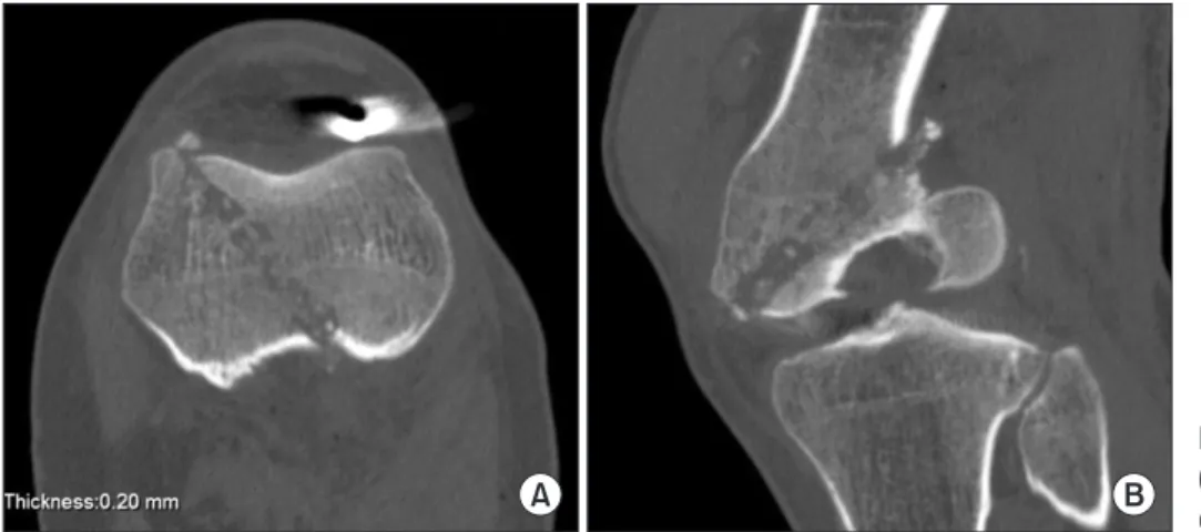
Preoperative computed tomography (CT) scans re-vealed an undeformed bullet just superior to the intercon-dylar eminence of the tibia in coronal plane and inferior to the patella in sagittal plane (Fig. 2). A knee arthroscopy was planned and performed under general anesthesia, 4 hours after the patient's admittance. Using standard
an-teromedial and anterolateral arthroscopic portals, the ante-rior cruciate ligament, and medial and lateral menisci were found to be intact. A non-displaced flap-like osteochon-dral defect, approximately 1 cm × 1 cm in size, was detect-ed on the mdetect-edial femoral condyle. A midline, 1 cm long mini-incision was performed through the patellar tendon as the Swedish portal is called.2) This extra portal allows direct access to the intercondylar notch and insertion of a large-sized grasper (Fig. 3). The bullet was removed using this portal. The knee joint was debrided and irrigated with 3,000 mL of saline solution. Immediately after releasing the tourniquet, swelling of the anterolateral compartment of the leg and pulse deficiency was detected in the dorsalis pedis artery on the foot. Thus, we performed an anterior and lateral compartment release through a single lateral incision fasciotomy.
Active knee flexion exercises up to 90° were started
A B
Fig. 1. Localization of the bullet on preoperative anteroposterior (A) and
sagittal (B) oblique radiographs.
A B
Fig. 2. Preoperative axial (A) and sagittal
(B) multiplanar reconstruction noncontrast computed tomography images.
Fig. 3. Arthroscopic view of the bullet in the knee joint in relation to the
117
Keskinbora et al. Compartment Syndrome Following Arthroscopic Removal of a Bullet in the Knee Joint
Clinics in Orthopedic Surgery • Vol. 8, No. 1, 2016 • www.ecios.org
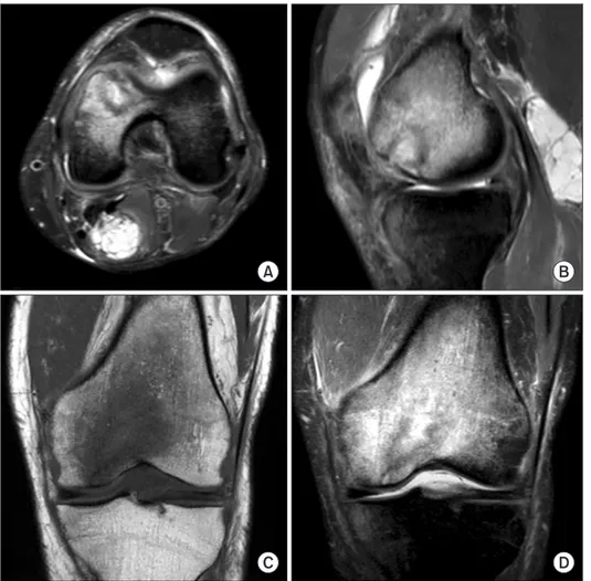
on the first postoperative day. However, weight bearing was not allowed on the surgical side due to the chondral lesion. On the third day after surgery, the incision of fas-ciotomy was primarily closed. No signs of infection were detected at follow-up visits. CT and magnetic resonance ımaging was performed in the 6th postoperative week in order to evaluate the bone and chondral healing (Fig. 4). Complete union was detected at the fracture line, and the chondral lesion seemed to be healed without an additional intervention.
DISCUSSION
In the present case, we preferred immediate arthroscopic debridement and removal of the bullet rather than an open surgical approach, because of the loose appearance of the intra-articular projectile and the absence of a comminuted fracture needing repair. Although arthroscopic removal seemed to be less morbid than an open surgical approach, we encountered compartment syndrome of the leg imme-diately after the tourniquet release.
An open procedure for removal of intra-articular bullet fragments has disadvantages such as increased blood loss, surgical site problems and prolonged recovery time.3) However, arthroscopical removal of lead projectiles in the knee reduces the postoperative complications like synovitis, arthropathy or systemic lead toxicity by debride-ment and lavage of the joint with minimal surgical mor-bidity.4) Additionally, intra-articular soft tissue injuries, especially including meniscal tears and free debris, would be safely and effectively treated by arthroscopic manage-ment simultaneously.5) In our patient, a small nondetached chondral lesion was observed in the lateral aspect of the medial femoral condyle. This lesion was healed in 6 weeks time without any further intervention.
Bullet fragments embedded in muscle or bone will frequently be encapsulated by a fibrous, avascular scar tissue, while fragments in a joint will be in subject to chemical degradation by hyaluronic acid and the pH of the synovial fluid.6,7) Thus, lead projectiles in joints are not considered to be physiologically inert and have to be removed when encountered.8) In addition, dissolution of
A B
C D
Fig. 4. Postoperative axial (A) and sagi ttal
(B) PD SPAIR, coronal T1 turbo spin echo (C), and coronal PD SPAIR (D) images. PD: proton density, SPAIR: spectral adiabatic inversion recovery.
118
Keskinbora et al. Compartment Syndrome Following Arthroscopic Removal of a Bullet in the Knee Joint
Clinics in Orthopedic Surgery • Vol. 8, No. 1, 2016 • www.ecios.org
the bullet by synovial fluid may yield to synovitis, lead arthropathy and increased levels of lead in the circula-tion.5,7,9) Either the direct mechanical effect of the intra-ar-ticular projectile, or the destructive chemical interactions between bullet-synovial fluid and articular cartilage makes the lead fragment removal necessary.5,6) An immediate arthroscopic intervention was preferred in our patient, be-cause of the mechanical joint motion restriction effect and direct contact of the bullet with the synovial fluid.
Arthroscopy is a relatively simple and less invasive procedure in the management of intra-articular knee problems. Although very rare, arthroscopic procedures may be complicated by instrument breakage or intra-articular damage,10) which would primarily result from the surgical practice of the surgeon. However, the most devastating complications are those related to neurovascu-lar injuries and compartment syndrome. In the literature, the infrequent cases of compartment syndrome after ar-throscopical interventions were mostly related to the use of mechanical infusion systems. In our patient, even though we used a manual Y-pump system, the extravasation of the fluid was associated with damage to the anterolateral knee capsule produced by the bullet at the end of its pathway. There were some reported cases of compartment syn-drome caused by rupture of a Baker’s cyst and a cadaveric study where extravasation was demonstrated by rupture
of the knee capsule from overflow of the irrigation fluid. The rare incidence of this complication, makes it difficult to reach a consensus. In contrast to some authors who rec-ommend waiting for spontaneous resorption of the fluid, we prefer an immediate single incision fasciotomy of the related compartment. Compartment pressure measure-ment is a useful diagnostic method for detecting compart-ment syndrome. However, in a patient like the one in our case under general anesthesia, a distended and pulseless leg following an arthroscopical intervention and a history of gunshot injury raises considerable suspicion of a com-partment syndrome. That’s why we preferred a minimally invasive fasciotomy and releasing of the compartment rather than compartment pressure measurement.
Although, the arthroscopic removal of intra-articu-lar bullets in gunshot injuries seems to have low morbidity rates, there is always the possibility that the articular cap-sule may rupture causing the fluids used during the opera-tion to leak into surrounding tissues resulting in compart-ment syndrome. In these patients, mini open surgery or dry arthroscopy may be better options.
CONFLICT OF INTEREST
No potential conflict of interest relevant to this article was reported.
REFERENCES
1. Dicpinigaitis PA, Koval KJ, Tejwani NC, Egol KA. Gunshot wounds to the extremities. Bull NYU Hosp Jt Dis. 2006; 64(3-4):139-55.
2. Mulhollan JS. Swedish arthroscopic system. Orthop Clin North Am. 1982;13(2):349-62.
3. Gupta RK, Aggarwal V. Late arthroscopic retrieval of a bul-let from hip joint. Indian J Orthop. 2009;43(4):416-9. 4. Diaz-Martin AA, Guerrero-Moyano N, Salinas-Sanchez P,
Guerado-Parra E. Arthroscopic removal of an intraarticular projectile from the knee. Acta Ortop Mex. 2011;25(4):223-6. 5. Tornetta P 3rd, Hui RC. Intraarticular findings after gunshot wounds through the knee. J Orthop Trauma. 1997;11(6): 422-4.
6. Rehman MA, Umer M, Sepah YJ, Wajid MA.
Bullet-in-duced synovitis as a cause of secondary osteoarthritis of the hip joint: a case report and review of literature. J Med Case Rep. 2007;1:171.
7. McQuirter JL, Rothenberg SJ, Dinkins GA, Kondrashov V, Manalo M, Todd AC. Change in blood lead concentration up to 1 year after a gunshot wound with a retained bullet. Am J Epidemiol. 2004;159(7):683-92.
8. Sclafani SJ, Vuletin JC, Twersky J. Lead arthropathy: ar-thritis caused by retained intra-articular bullets. Radiology. 1985;156(2):299-302.
9. Peh WC, Reinus WR. Lead arthropathy: a cause of delayed onset lead poisoning. Skeletal Radiol. 1995;24(5):357-60. 10. Pierzchała A, Kusz D, Widuchowski J. Complication of