An Unexpected Foreign Body Causing Twinkling Artifact On
Doppler Ultrasonography
Memede Doppler US'de “Twinkling” Artefaktına Yol Açan Yabancı Cisim
Ebru
Düșünceli Atman
1, Evren Üstüner
1, Hasan Özcan
11 Ankara Üniversitesi Tıp Fakültesi Radyoloji Anabilim Dalı
Bu bildiride ultrason incelemesi ile memesinde iğne saptanan olgu sunulmaktadır. Operasyon veya travma öyküsü olmayan olguda iğnenin metalik natüründen dolayı renkli Doppler ultrasonda “twinkling“ bulgusu saptanmıștır. Tanı mammografi ile doğrulanmıștır.
Anahtar Sözcükler: Yabancı Cisim, Meme, Twinkling Bulgusu, Renkli Doppler
We herein describe a patient with a needle in her breast which was initially shown on ultrasound examination. There was no operation or trauma history of the patient. Owing to the metallic nature of the needle, twinkling sign was detected on color Doppler ultrasound. Mammography confirmed the diagnosis.
Key Words: Foreign Body, Breast, “Twinkling” Sign, Color Doppler During routine practice,
mammographers occasionally encounter metallic foreign objects in the breast. Although they are easily recognized owing to the related history and high radiographic density on mammograms, they may cause diagnostic confusion with microcalcifications if they are small enough. However if the history is unknown and mammography is not the initial exam, clinical diagnosis may be problematic. In this case report we present the gray
scale and color Doppler ultrasound (US) and mammographic findings of a foreign body in the configuration of a needle in the breast which was first diagnosed by ultrasound examination. The patient did not report or recall any antecedent event related to the foreign body.
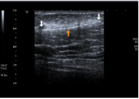
A 55-year-old woman presented with pain and erhythema involving the upper outer quadrant of the right breast for the last four days. A hyperechoic linear structure was detected within the breast parenchyma in the gray-scale US scan (Figure 1). Twinkling artifacts were noted posterior to this structure in the color mode and spectral analysis suggestive of a metallic foreign body (Figure 2A, 2B). The morphology of the structure resembled that of a needle but upon questioning, the patient did not recall any antecedent event related to a needle insertion, such as trauma or operation at all. Mammographic study was performed to confirm the sonographic findings and the metallic nature of the foreign body. The needle was clearly demonstrated in the oblique view (Figure 3).
Ankara Üniversitesi Tıp Fakültesi Mecmuası 2012, 65 (3) DOI: 10.1501/Tıpfak_000000827
DAHİLİ TIP BİLİMLERi/MEDICAL SCIENCES
Olgu Sunumu / Case Reports
Received: 15.11.2011 Accepted: 02.07.2013 Correspondig author
Uz.Dr.Ebru Düșünceli Atman, Phone: 0312 508 21 63 E-mail : [email protected]
Ankara Üniversitesi Tıp Fakültesi Mecmuası 2012, 65 (3)
An Unexpected Foreign Body Causing Twinkling Artifact On Doppler Ultrasonography 156
Figure 1: A hyperechoic linear object within
the right breast demonstrated on the gray scale US
Figure 2: Color mode US scan (A) and
spectral analysis (B) show twinkling artifacts posterior to the object indicating its metallic nature.
Figure 3: . On mediolateral oblique view the
metallic needle is seen in the superior aspect of the right breast.
Discussion
The foreign objects in the breast are usually related to diagnostic or therapeutic surgical or percutaneous interventions. Some of the foreign objects described so far are metallic suture materials, preoperative or postoperative localization needles or fragments of biopsy needles. An antecedent history is usually definitive for diagnosis when supported by the
imaging findings (1-3). In the literature migration of these materials outside the breast is also reported (2, 3). Presence of a metallic foreign body without history of a surgical or percutaneous intervention is very rare and described by a limited number of case reports in the literature (4). Of note, herbal Chinese treatment of breast abscesses with lead containing ointments may cause metallic densities in the breast (1). The distinguishing feature of our case report was the lack of a memorable surgical intervention or trauma in the patient with a reliable narration of antecedent events. The sonographic images and the additional color Doppler US findings including the twinkling artifact phenomenon strongly suggested the presence of a metallic foreign object in the form of a needle. Especially the twinkling artifact provided supportive evidence about the metallic nature of the object before the mammographic exam. The mammographic exam was confirmative because metallic foreign objects are easily recognizable due to their high radiographic density (1).
Twinkling artifact is a color Doppler ultrasound artifact which was first described by Rahmouni et al in 1996 (5). This phenomenon is most commonly encountered in structures which have strong reflective surfaces. Posterior acoustic attenuation is noted on gray-scale US. In the color mode, chaotic color bleeds coded in red and blue are observed posteriorly and in the spectral mode vertical lines are coded at the acoustic attenuation site (5-7).
Rahmouni et al (5), studied the mechanism of twinkling artifact
and they based this phenomena on multiple reflections on structures with irregular surfaces causing scattering of the sound beam from the transducer and delaying the time for the signal to return simulating movement (5). Other studies indicate that technical factors such as type, settings and the study parameters of the ultrasound equipment contribute to the phenomenon as well (8). If the twinkling artifact is not
recognized by the user, the studied structures may be misinterpreted as having blood flow within. The detection of this artifact is regarded as supportive sonographic evidence in the detection of urinary system stones, gall bladder and biliary pathologies, parenchymal and tumoral calcifications and in orbital pathologies such as metallic foreign objects and lens calcifications. This artifact may guide the management of urinary stones because may aid in the presumption of the biochemical components of the stones (6-8). No reports were found in the literature which mentioned the use of this artifact in the diagnosis of foreign objects in the breast. Presence of sewing needles within the breast tissue were described in the literature one of which resembled a tumor and the other was undetected despite several ultrasound examinations (4, 9). In our patient no significant tumor like soft tissue changes was present except a mild foreign body reaction.
We do not believe that the needle in this patient is from a surgical intervention as on the visual inspection and imaging, no evidence of a scar or structural distortion was noted. This needle could have been the part of a
Journal of Ankara University Faculty of Medicine 2012, 65 (3)
Ebru Düșünceli Atman, Evren Üstüner, Hasan Özcan 157
forgotten brooch or a simple needle pinned on her blouse which got dislodged and found way to her breast tissue while she was sleeping or moving.
In conclusion, foreign objects could be encountered in the breast with or without an antecedent history and especially during sonography twinkling artifact would be a
valuable additional diagnostic finding indicating the metallic nature of the foreign object.
REFERENCES
1. Moon WK, Park JM, Im JG, Noh DY,
Yeon KM, Han MC. Metallic punctate densities in the breast after Chinese herbal treatment: mammographic findings. Radiology. 2000;214:890-894.
2. Banitalebi H, Skaane P. Migration of
the breast biopsy localization wire to the pulmonary hilus. Acta Radiol. 2005;46:28-31.
3. Grassi R, Romano S, Massimo M, et al.
Unusual migration in abdomen of a wire for surgical localization of breastlesions. Acta Radiol. 2004;45:254-258.
4. Liszka G, Decker I. A foreign body
resembling a tumor (sewing needle) in the breast. Fortschr Geb Rontgenstr Nuklearmed. 1967;107:569-570.
5. Rahmouni A, Bargoin R, Herment A,
Bargoin N, Vasile N. Color Doppler “twinkling” artifact in hyperechoic regions. Radiology. 1996;199:269-271.
6. Conkbayır I, Yanık B, Şenyücel Ç,
Hekimoğlu B. “Twinkling” artifact in color Doppler ultrasonography (pictorial essay). Turk J Diagn Intervent Radiol. 2003;9:407-410.
7. Aytaç SK, Özcan H. Effect of color
Doppler system on the twinkling sign associated with urinary tract calculi. J Clin Ultrasound. 1999;27:433-439.
8. Kamaya A, Tuthill T, Rubin JM.
“Twinkling” artifact on color Doppler sonography: dependence on machine parameters and underlying cause. Am J Roentgenol 2003;180:215-222.
9. Solbach C, Diebold T, Louwen F,
Kaufmann M. Case report -- needle in the breast. Zentralbl Gynakol. 2006;128:229-23
Ankara Üniversitesi Tıp Fakültesi Mecmuası 2012, 65 (3)
An Unexpected Foreign Body Causing Twinkling Artifact On Doppler Ultrasonography 158