Ankara Üniv Vet Fak Derg, 61, 73-77, 2014
Short Communication / Kısa Bilimsel Çalışma
Cytological evaluation of pleural effusion with cell block technique
in a dog
Latife ÇAKIR¹, Ekrem Çağatay ÇOLAKOĞLU², Arda Selin TUNdz, Osman KUTSAL³
1 Erciyes University, Faculty of Veterinary Medicine, Department of Pathology, Kayseri; Ankara University, Faculty of Veterinary
Medicine, ²Department of Internal Medicine; ³Department of Pathology, Ankara, Turkey.
Summary: In this case, the use of the Cell Block technique in the diagnosis of pleural effusion collected from a 3.5 year old Setter Spaniel breed dog was investigated. The dog was presented to Ankara University Small Animal Hospital with the history of acute dyspnea, weakness, exercise intolerance and tachypnea for a week. Routine clinical examinations including blood testing were performed. Abdominal distension, pleural effusion and cyanotic cranial mucous membranes were remarkable during physical examination. The pleural fluid collected by thoracocentesis was brownish-colored, cloudy and has contained granular materials. The specimen collected from the dog was separated into three aliquots, one part was processed in a routine manner for cytological examination by cytocentrifugation, and the other parts of the sample were used for a cell block and bacteriological analysis. In microbiological analysis, Escherichia coli and Klebsiella spp. were isolated. The cytocentrifuged preparations and cell block sections revealed septic effusion. The cell block technique demonstrated morphological details better than cytocentrifugation. In conclusion, in this case, cytology and bacteriology of effusion, routine blood tests, abdominal radiography and echocardiography showed that the pleural effusion was resulted from pyothorax due to severe tricuspid dysplasia.
Key words: Cell block, cytocentrifugation, dog, pleural effusion, tricuspid dysplasia.
Bir köpekte hücre bloğu tekniği ile plevral efüzyonun sitolojik olarak değerlendirilmesi
Özet: Bu olguda, 3,5 yaşlı, dişi Setter İspanyol ırkı bir köpekte plevral efüzyonun tanısında hücre bloğu tekniğinin kullanım olanağı araştırıldı. Bir haftadan beri akut solunum güçlüğü, halsizlik, hızlı solunum öyküsü olan bir köpek Ankara Üniversitesi Veteriner Fakültesi Küçük Hayvan Hastanesine kabul edildi. Kan testleri dahil rutin klinik incelemeleri yapıldı. Yapılan fiziksel incelemede karın şişkinliği, plevral efüzyon ve kranial müköz membranlarda siyanoz dikkati çekti. Torakosentez ile alınan plevra sıvısı kahverengimsi renkte, bulanık ve granüler materyal içermekteydi. Alınan plevra sıvısı örnekleri üç kısıma ayrıldı. Bir kısmı santrifuj teknikleriyle çalışıldı, diğer kısımlar ise hücre bloğu ve bakteriyolojik izolasyon için kullanıldı. Mikrobiyolojik yoklamada
Escherichia coli ve Klebsiella spp. izole edildi. Plevral sıvının sitolojisinde septik efüzyona rastlandı. Hücre bloğunda sitosantrifüj
tekniğine göre hücrelerin morfolojik detayları daha belirgin olarak görüldü. Sonuç olarak, bu olguda plevral efüzyonlu bir köpeğe sitolojik, bakteriyolojik, abdominal radyografi, ekokardiyografi, rutin kan testleriyle şiddetli triküspid yetmezliğine bağlı pyotoraks tanısı konuldu.
Anahtar sözcükler: Hücre bloğu, köpek, plevral efüzyon, sitosantrifüj, triküspid displazisi
Pleural effusion, which is a common manifestation of several diseases, is the abnormal accumulation of fluid in the pleural space (5, 11). Inflammatory conditions of the pleura and pleural space changes capillary permeability and lymphatic function (4). Pulmonary and cardiac problems as well as malignancy, bacterial pleurisy and tuberculose (5) frequently result in pleural effusion in human. The aetiology of the pleural effusion
in dogs and cats are often unknown. Pyothorax, a purulent pleural effusion, is generally accepted as an uncommon but important disease in dogs (4, 7, 10, 12). It has been documented that the main cause for the development of the pyothorax is the presence of bacteriological agents in cat and dogs (4, 6, 7, 16).
Although clinical signs, radiography, thoracocentesis, cytology and microbiological culture are used for the
∗ This paper was presented as a poster in 37th European Congress of Cytology, 30 September-3 October 2012, Dubrovnik-Cavtat,
examination diagnosis of cytological e has been a cl the examinat smear has lo details of cel loss and ch methods (14 technique, w cellularity an common met in humans currently no technique fo pyothorax. T technique, w field, and cyt routine exam performed fo collected from Figure 1. Elec mm/sec. Şekil 1. Elektr 50 mm/sn. Figure 2. A) E elongated ante adhered to the Şekil 2. A) Ek anterior triküs bitişik olarak b of pleural fl f pyothorax evaluation and lassic cytologi tion of fluids ower sensitivi lls, overcrowd hanges in di 4). Although which is a se nd morpholog thods for eval (9), to the study determ for cytology Therefore, in which is used tocentrifugati minations and or the discrim m a dog. ctrocardiogram rokardiyogram. Echocardiograp erior leaflet. Ma e right side of th kokardiyografi spid kapak. Bel bulunan septal t luid in dogs can be con d culture (7, 12 ical method co in dogs (3, 4 ity due to bla ding or overlap ifferent labor h the cell b ensitive metho gical details, ( luation of the authors kno mining the uti in the diag this case stud for the first t on technique d microbiolog mination of th
(ECG). Deep . Derin S dalga
phy. Right para arked large righ he interventricu . Sağ parastern lirgin olarak ge triküspid kapak and cats (4), nfirmed only 2). A direct sm ommonly use 4, 7, 10, 12). and morpholog pping of cells ratory proces block (minibl od increasing (14) is one of body cavity fl owledge ther ility of minib gnosis of ca dy, the cell b time in veteri as well as clin gical culture w he pleural effu
S waves, right aları, sağ aks sa
asternal long ax ht atrial chamb lar septum. TV nal uzun eksen
enişlemiş sağ a k. TV: Triküspi , the y by mear d for The gical , cell ssing lock) g the f the fluids re is block anine block inary nical were usion weig Ani loss into thor brea feve reco jugu muf noti and rout Hea reve Tric pleu cyto fluid
t axis shift and apması ve ortal
xis view (PLAX er. B) Right pa V: Tricuspid valv görünüm.Septu atrium. B) Sağ id kapak. A 3.5 year ghing 20 kg, w mal Clinic w s, orthopnea, olerance for a racocentesis h athing in a priv Abdominal er and cyano orded during ular pulsation ffled heart and ceable. After n negative bloo tine blood an art Score: 12), e ealed that p cuspid dysplasi ural fluid w ological and d had exudativ d decreased ove ama QRS kom X). Septal tricu arasternal long ve. uma yapışık se parasternal uz -old female was referred t with the comp
, dyspnea, a month. The had been perfo vate clinic. distention, co otic cranial m physical exa n and capilla d lung sounds negative quick od smear for nalysis, radiog electrocardiogr pleural effusi ia (Figure 1, 2 was collected bacteriologic ve characteris erall QRS com mpleks amplitütü
uspid leaflet adh axis view (PLA eptal triküspid un eksen görün Setter Spanie to Ankara Uni plaint of ano weakness a e owner also ormed for two
ough test posit mucous mem amination. L ary refill tim s on auscultat k test results for
intraerythrocy graphic studi raphy and echo ion was ass
). During echo d by thoraco cal examinati stic.
mplex amplitude ünde azalma I-I
hered to septum AX). The septal
kapak ve tipik nüm İnterventr el breed dog, iversity Small rexia, weight and exercise reported that times to ease tivity, 38.6 oC mbranes were eft-right side me elevation, tion were also r Dirofilariasis ytic parasites; ies (Vertebral ocardiography ociated with ocardiography, ocentesis for ions and the
e Lead I-II. 50 II. Derivasyon. m and typically l leaflet is seen olarak uzamış riküler septuma , l t e t e C e e , o s ; l y h , r e 0 . y n ş a
Ankara Üniv Vet Fak Derg, 61, 2014 75
Figure 4.A) Cytocentrifugation of pleural fluid shows a mixed cell population including, macrophages, leucocytes and mesothelial cells (PAP). B) A cell block section of pleural fluid. Reactive mesothelial cells (arrowhead) showing a binuclear (thin arrows) throught the speciemen, which also contains lymhocytes, neutrophil, (thick arrows), and red blood cells, (HE). C) A cell block section. Cell population including erythrocytes neutrophils, macrophages and mesothelial cells. Inset shows a reactive mesothelial cells (arrowhead) (HE). D) Classic centrifuge smear of the pleural fluid. The fluid contains degenerative neutrophils (arrowheads), vacuolar cytoplasm of macrophages (thin arrow), lymphocytes, normal mesothelial cells (thick arrows), clump of bacteria (yellow arrow) (MGG).
Şekil 4. A) Plevral sıvının sitosantrifüjü nötrofil, makrofaj ve mezotel hücreleri göstermektedir (PAP). B) Plevral sıvıdan hücre bloğu kesiti. İki çekirdeklilik gösteren (ince oklar) reaktif mezotel hücreleri (ok başı), lenfosit, nötrofil lökositler (kalın oklar) ve eritrosit, (HE). C) Hücre blok kesiti. Eritrosit, nötrofil, makrofaj ve mezotel hücreleri içeren hücre bloğu. Ekli küçük resim, reaktif değişiklikler gösteren mezotel hücreleri, (HE). D) Plevral sıvının klasik santrifüj yayması. Sıvı; dejenere nötrofil lökositler (okbaşları), vakuoler sitoplazmalı makrofaj (ince ok), lenfosit ve normal mezotel hücreler (kalın oklar), bakteri kümelerini içermektedir (sarı ok) (MGG).
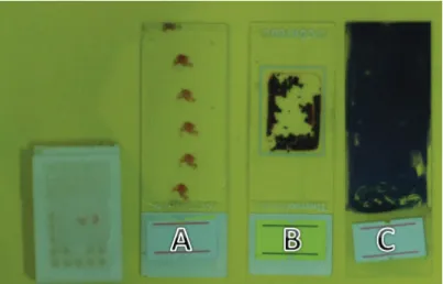
Figure 3. Preperation of centrifugation and paraffin block. (A: cell block section, B: cytospin preperation, C:classic centrifuge smear Şekil 3. Santrifüj tekniğiyle yayma / preparat ve parafin bloğu göstermektedir. (A: Hücre bloğu kesiti B: sitospin preparat C: santrifüj yayma)
In routine blood analysis, leukocytosis (Leukocyte index: WBC 17.89 10^9/l, GRA 16.33 10^9/l), anemia (Erythrocyte index: RBC 4.56 10^12/l, HGB 9.4 g/dl, HCT % 31.32) and the blood chemistry indicate liver congestion due to the Tricuspid regurgitation (ALP 915.4 IU/L, ALT 124.3 IU/L, AST 121.6 IU/L, CK 220 IU/L, Total Bilirubin 1.37 mg/dl).
The dog was placed in oxygen cage and treated initially with Furosemide, Spironolactone and Lisinopril. Lavage of chest cavity with sterile saline solution (containing heparin) was performed, and the animal was treated with intravenous Ampicillin-sulbactam. Two months later, the dog was died suddenly according to the owner's information.
Half of the pleural fluid was cytocentrifuged at 1500 rpm for 15 minutes (Cytospint; Shandon Southern Ltd., Cheshire, England) and the smears were prepared then stained with Papanicolaou (PAP) and haematoxylin-eosin (HE) for cytopathologic examinations Cytological examination of direct smears of fluid was performed after staining with May-Grünwald-Giemsa (MGG) method. For the cell block preperation, the other half of the fluid speciemen was fixed with 10% alcohol-formalin. After fixation, the speciemen was centrifuged at 2500 rpm for 15 minutes. The supernatant was poured off and the sediment was placed on cytoblock casset. After addition of reagent I and II supplied with the kit (Thermoshandon Cell Block Preperation System, USA) it was fixed in formalin and proscessed in histokinette as part of routine paraffin section for histopathology. Sections were stained with PAP and HE (Figure 3).
For microbiological examination, the fluid from the pleural cavity was inoculated onto 7% sheep blood agar and MacConcey agar. The media were then cultured in a humid atmosphere at 37ºC under aerobic and microaerophilic (candle jar) conditions.
A pleural fluid was 400 ml and cloudy, haemorrhagic to overtly purulent in appearance, and contained granular materials. Bacteriological culture revealed aerobic bacteria including Escherichia coli and Klebsiella spp.
Cytological examination of the fluid revealed large amounts of necrotic debris, massive numbers of mixed bacteria with degenerated and non-degenerated neutrophils, macrophages, lymphocytes, red blood cells and normal/reactive mesothelial cells. Reactive mesothelial cells showing irregular nuclei, different nucleus size, binucleated and increased chromatin have been observed. The smear was filled with degenerative neutrophils as well as a few lymphocytes and macrophages (Figure 4).
Tricuspid valva dysplasia (TVD) is a congenital malformation of tricuspid valve leaflets, chordae or papillary muscles that results in regurgitation. The septal leaflet of tricuspid valve is typically adhered to right side
of the interventricular septum and having little movement (2, 8).
Definitive diagnosis of tricuspid valve dysplasia can be made by echocardiography that confirmed the features include right atrial dilatation, septal leaflet adhesion and elongated anterior leaflet of tricuspid valve (2, 13). In the presented case, the smilar diagnostic features were seen, and especially adhered septal leaflet to interventricular septum was marked.
The dogs with tricuspid valve dysplasia may be initially asymptomatic but the symptoms of fatigue, ascites and dyspnea from pleural effusion often develop. Volume overload results from tricuspid valve dysplasia causes the effusion by changing the capillary permeability and lymphatic function (17). Pyothorax often results from penetrating thoracic wounds and bacterial contamination spreads the pleura with haematogenous and lymphogenous extension (1). Chest trauma, intrathoracic foreign body and esophageal perforations could not be determined in this case. Although the case has no anamnestic data of trauma, performing the intermittent nonsterile thoracocentesis or alterations in capillary permeability and lymphatic function associated volume overload led us to think them as the reasons of the septic contamination.
In this case, the presence of bacteria and the numerous degenerated neutrophils show a septic effusion. This hypothesis is supported also by the physico-chemical characteristics of the fluid. This result is consistent with the previous studies (4, 12). In the centrifugation method, presence of reactive mesothelial cells, an abundance of inflammatory cells and scarcity of typical cells cause difficulties in diagnosis (15). The cytodiagnosis with conventional smear has a lower sensitivity due to the overcrowding of the cells, cell loss as well as different laboratory processes (14). The paraffin block provide a concentrated material in smaller fields (9, 14). In this report, there was no difference between the types of obtained cells according to the sampling techniques. Centrifugation of effusion revealed scanty cellularity and inflammation with polymorphs. Cytologic examination of both centrifugation and cell block from pleural fluid revealed pyothorax. Although the number of the cells was less and the cells were unevenly distributed, the preservation of the cells was much better with cell block. In this case, cell block provided better diagnostic accuracy than cytocentrifuge and classic centrifuge smear, which was consistent with the results of previous studies (9, 14).
Therefore, the cell block technique can be recommend as a useful diagnostic tool in evaluating purulent, viscous fluids with intensive granules along with the routine centrifugation methods in veterinary application
Ankara Üniv Vet Fak Derg, 61, 2014 77 References
1. Bauer T, Woodfield JA (1995): Mediastinal, pleural, and
extrapleural diseases. 812-842. In: Ettinger SJ and Feldman
FJ (Eds). Textbook of Veterinary Internal Medicine. Fourth edition, WB Saunders Company, Philadelphia. 2. Boon JA (2011): Congenital Shunts and AV valve
dysplasia. 468-469.In: Boon, J.A. Veterinary
Echocardiography. Wiley-Blackwell, Ames, Iowa.
3. Boothe HW, Howe LM, Boothe DM, Reynolds LA, Carpenter M (2010): Evaluation of outcomes in dogs
treated for pyothorax: 46 cases (1983-2001). J Am Vet
Med Assoc, 15, 236, 657-63.
4. Demetriou J L, Foale RD, Ladlow J, McGrotty Y, Faulkner J, Kirby BM (2002): Canine and feline
pyothorax: a retrospective study of 50 cases in the UK and Ireland. J Small Anim Pract, 43, 388-394.
5. Gaur D D, Chauhan N, Kusum A, Harsh m, telekar M, Kishore S, Pathak V P (2007): Pleural fluid analysis-role
in diagnosing pleural malignancy. J Cytol, 24, 183-188.
6. Gülbahar MY, Gürtürk K (2002): Pyothorax associated
with a Mycoplasma sp and Arcanobacterium pyogenes in a kitten. Aust Vet J, 80, 344-345.
7. Johnson MS, Marti MWS (2007): Successful medical
treatment of 15 dogs with pyothorax. J Small Anim Pract,
48, 12-16.
8. Kittleson MD, Kienle RD (1998): Congenital abnormalities
of the atrioventricular valves. 273-281. In: Kittleson, MD,
Kienle, R D. Small animal cardiovascular medicine. Saint Louis, Mosby.
9. Kushwaha R, Shashikala P, Hiremath S, Basavaraj HG (2008): The cells in the pleural fluid and their value in the
differential diagnosis. J Cytol, 25, 138-43.
10. Piek CJ, Robben JH (2000): Pyothorax in nine dogs. Vet Q, 22, 107-11.
11. Rahman NM, Chapman SJ, Davies RJ (2005): Pleural
effusion: a structured approach to care. Br Med Bull, 72,
31-47.
12. Scott JA, Macintire DK (2003): Canine pyothorax:
Clinical Presentation, Diagnosis, and Treatment. Comp
Cont Educ Pract, 25, 180-194.
13. Shaughhnessy R (2004): Ebstein’s anomaly. 1335–1342. In: Crawford MH, DiMarco JP, Paulus WJ (Eds), Cardiology, 2nd edition, Tokyo, Mosby.
14. Thapar M, Mishra RK, Sharma A, Goyal V, Goyal V (2009): Critical analysis of cell block versus smear
examinations on effusions. J Cytol, 26, 60-64.
15. Udasimath S, Arakeril SU, Mahesh, Karigowdar MH, Yelikar BR( 2012): The role of the cell block method in
the diagnosis of malignant ascitic fluid effusions. JCDR, 6,
1280 – 1283.
16. Walker AL, Jang SS, Hirsh DC (2000): Bacteria
associated with pyothorax of dogs and cats: 98 cases (1989-1998). J Am Vet Med Assoc, 216, 359-363.
17. Ware WA (2007): Congenital cardiovascular diseases. 245. In: Ware, W.A. Cardiovascular disease in small animal medicine. Manson publishing, London.
Geliş tarihi: 02.08.2013 / Kabul tarihi: 09.10.2013
Address for correspondence:
Latife ÇAKIR, DVM, PhD Associate Professor Department of Pathology Faculty of Veterinary Medicine
University of Erciyes, 38039, Kayseri, Turkey e-mail: [email protected]