Diagnostic Value Of Mammography And Ultrasonography For
Differentiation Of Benign And Malignant Breast Masses
Meme Kitlelerinde Benign-Malign Ayrımında Mammografi Ve Ultrasonografinin Tanı Değeri
Eriz Özden
1 Ankara University, Faculty of Medicine, Department of Urology
Aim: The aim of this study is to determine the diagnostic value of mammography and ultrasonography for differentiating benign and malignant breast masses.
Material and Methods: Fifty-one patients who had palpable breast masses were evaluated with both mammography and US. The detected lesions were classified as benign or malignant according to their imaging features and these results were evaluated according to the histopathological reports.
Results: Histopathologically, 28 of the 51 breast masses (54.9%) were reported as benign and 23 (45.1%) as malignant. The sensitivity and specificity of mammography were 81.4% and 90.4% for benign masses. The sensitivity and specificity of US for benign masses were 85.7% and 82.6%. The sensitivity and specificity of mammography for malign masses were 90.9% and 81.4%. The sensitivity and specificity of US for malignant masses were 78.2% and 85.7%. Conclusion: Although ultrasonography is a valuable imaging method for the differentiation of solid – cystic breast masses, mammography is superior to ultrasonography for benign-malignant differentiation.
Key Words: Mammography, Ultrasonography, Breast mass
Amaç: Mammografi ve ultrasonografinin, meme kitlelerinde benign- malign ayrımı yapmaktaki değerini belirlemektir.
Gereç ve Yöntem: Palpabl meme kitlesi olan ve bu nedenle ultrasonografi ve mammografi incelemesi yapılan 51 olgu çalıșmaya dahil edilmiștir. İzlenen kitleler özelliklerine göre benign-malign olarak ayrılmıș, bu sonuçlar histopatoloji raporları ile karșılaștırılmıștır.
Bulgular: Histopatoloji sonuçlarında 51 meme kitlesinin 28’i (%54.9) benign, 23’ü (%45.1) malign olarak raporlanmıștır. Benign kitlelerde mamografinin sensitivite ve spesifisitesi %81.4 ve %90.4 olarak belirlenmiștir. Benign kitlelerde US’nin sensitivite ve spesifisitesi ise %85.7 ve %82.6 olarak hesaplanmıștır. Malign kitlelerde mamografinin sensitivite ve spesifisitesi %90.9 ve %81.4 olarak belirlenmiștir. Malign kitlelerde US’nin sensitivite ve spesifisitesi ise %78.2 ve %85.7 olarak hesaplanmıștır.
Sonuç: Meme kitlelerinde, kistik –solid ayrımında ultrasonografi değerli bir görüntüleme yöntemidir. Ancak benign-malign ayrımında mammografi ultrasonografiden üstündür. Anahtar Sözcükler: Mammografi, Ultrasonografi, Meme Kitlesi
Breast cancer is one of the leading causes of cancer deaths among women. Each year, nearly 40.000 deaths in the United States are attributed to this disease (1). The mortality of breast cancer is reported to be 25-35 in 100.000 people (2). The prognosis of breast cancer depends on the histological subtype and the dimension of the tumor, and spread at the time of the diagnosis. In an attempt to reduce this mortality, breast
self-examinations and periodic screening is recommended. Periodic screening is performed by mammographic examinations. If a lesion is detected at
mammography; ultrasonography (US), spot mammography, core needle biopsy or magnetic resonance imaging (MRI) can be used for the differential diagnosis. In this study, we have evaluated breast masses. With
Ankara Üniversitesi Tıp Fakültesi Mecmuası 2013, 66 (1)
DOI: 10.1501/Tıpfak_000000835 DAHİLİ BİLİMLER/MEDICAL SCIENCES
Araștırma Makale / Research Article
Received: Jan 30,2014 Accepted: Feb 14,2014 Correspondig Author Eriz Özden GSM : 0 532 730 96 72 Phone : + 90 - 312- 508 20 85 Fax : + 90-312- 311 21 67 E-mail : [email protected]
Eriz Özden, M.D., Specialist of Radiology, Ankara University, Faculty of Medicine, Department of Urology
Ankara Üniversitesi Tıp Fakültesi Mecmuası 2013, 66 (1)
Diagnostic Value Of Mammography And Ultrasonography For Differentiation Of Benign And Malignant Breast Masses
16
mammography and US in a series of patients with palpable breast masses, and sought to establish the diagnostic accuracy of mammography and US for differentiating benign and malignant breast masses.
MATERIALS AND METHODS
Fifty-one patients who palpable breast masses and were evaluated with both mammography and US, were retrospectively included to the study. After a physical examination and a finding of a palpable breast mass, the patients were referred by a clinician to the radiology department for mammography and US examinations.
Mammography was performed with a 800 Miliamper Siemens-Mammomat (Siemens- Germany) and, craniocaudal and mediolateral radiographs of the both breasts were taken. When needed, spot radiographs with magnification technique were also taken. When a mass lesion was detected by mammography; the dimensions,
margin characteristics (circumscribed or irregular),
spiculations and density of the lesion were evaluated and any associated microcalcifications were noted. Any lesion with contour microlobulation, ill defined margins, spiculation, ill defined posterior margin and intratumoral microcalcifications were defined as potentially malignant. Other lesions were defined as potentially benign.
Breast US was performed with a General Electric RT-X 400 (GE- Milwakue- USA) ultrasonogaphy equipment, using 7.5 Mhz linear transducer. When a lesion was detected by US; the dimensions, margins, echogenicity, internal echo pattern (homogenicity), posterior acoustic attenuation and
enhancement of the lesion were evaluated. Any lesion with contour microlobulation, ill defined margins, spiculated margins, posterior acoustic attenuation were defined as potentially malignant. Other solid lesions and anechoic cysts with none or thin lineer septations were defined as potentially benign. All US examinations were performed by a single radiologist (E.Ö).
All patients with solid or suspicious complicated cystic masses had undergone core needle biopsy or excisional biopsy, and the mammography and US results were compared with histopathological diagnosis. The sensitivity and specificity of mammography and US were calculated for benign and malignant breast tumours. Groups were statistically compared for proportions using chi-square tests, for means using one sample t test and one way Anova where appropriate. p<0.05 was considered as statistically significant.
RESULTS
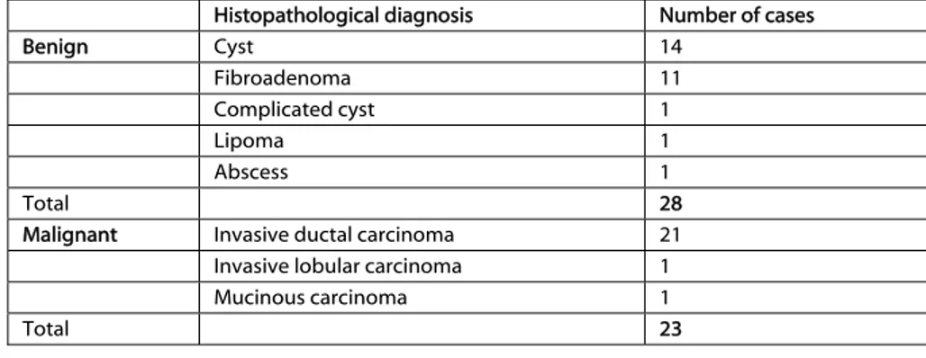
Our results show that; of the 51 breast masses 28 masses (54.9%) were benign and 23 masses (45.1%) were malignant histopathologically. The histopathology of benign and malignant masses listed in Table 1. Patient ages ranged between 19 and 70 years. The mean age was 45.3.
The patients with malignant diagnosis (mean age 51.2 years) were significantly (p<0.001) older than those with benign diagnosis (mean age 38.6 years). The size of the benign masses varied between 4 mm and 3 cm (mean 1.5 cm) and malignant tumors varied in size between 6 mm and 1.8 cm and there was not a statistically significant difference (p=0.53) between the mean dimensions. Mammographic evaluation revealed
that, 22 (78.5%) of the benign masses were diagnosed as benign and six (21.5%) of the benign masses were diagnosed falsely as malignant. Sonographically, 24 (85.7%) of the benign masses were diagnosed as benign and four (14.3%) of the benign masses were diagnosed falsely as malignant. As for malignant tumors; with
mammography, 20 (87%) were diagnosed as malignant and three (13%) were diagnosed falsely as benign. Sonographically, 18 (78%) of the malignant masses were diagnosed as malignant and five (22%) of the malignant masses were diagnosed falsely as benign. The sensitivity and specificity of
mammography and US for benign masses were 81.4%, 90.4%, 85.7% and 82.6, respectively. The sensitivity and specificity of mammography and US for malignant masses were 90.9%, 81.4%, 78.2% and 85.7, respectively.
Table 1: Histopathological diagnosis of breast masses
Histopathological diagnosis Number of cases
Benign Cyst 14 Fibroadenoma 11 Complicated cyst 1 Lipoma 1 Abscess 1 Total 28
Malignant Invasive ductal carcinoma 21
Invasive lobular carcinoma 1
Mucinous carcinoma 1
Journal of Ankara University Faculty of Medicine 2013, 66 (1)
Eriz Özden 17
DISCUSSION
Mammography and US are the most frequently used imaging methods for evaluating palpable breast masses. Mammography is accepted as the primary screening method, and it gives valuable information for differentiating benign and malignant lesions. When a mass lesion is detected at mammography, the lesion is first evaluated for the regularity of its margins. High density, irregular margins and spiculations are important findings for a malignant lesion (3, 4). Also, microcalcifications in a mass lesion should be evaluated carefully. But, some solid breast lesions, especially mucinous and medullary carcinomas are reported to have regular margins (3, 5). These lesions may be diagnosed as benign with mammography. In our series, 17 of the 23 malignant masses had irregular margins and spiculations. The only tumor in our series which was histopathologically diagnosed as mucinous carcinoma had regular margins at mammography and simulated a benign lesion. This finding is in accordance with the literature (3, 5, 6). The detection of microcalcifications in a mass lesion is another suspicious finding for maligancy (3, 7). In the literature, intratumoral microcalcifications have been reported in 25-60% of the malignant breast tumors (3, 6, 7). In our study group, microcalcifications were detected in six (26%) of the malignant masses. In our series, three tumors with regular margins were diagnosed as potentially malignant because of the existence of intratumoral microcalcifications. None of the benign lesions in this series had intratumoral microcalcifications. These findings show the significance of presence
of intratumoral microcalcifications. Based on the
demonstration of irregular margins, spiculated contours and intratumoral microcalcifications,
we determined a 90.9% sensitivity for the malignant masses using mammography. In our study, we had six false positive malignant diagnosis with mammography. Three of these cases were microcysts or complicated cysts with irregular margins and 3 of them were fibroadenomas with slight contour lobulation and irregularity. Some authors have also reported that some fibroadenomas may have irregular margins at mammography (8, 9). In our series, three of the 11 fibroadenomas (27%) were diagnosed as potentially malignant because of these features. This can be secondary to dense fibrous paranchyma surrounding the tumor which effects the evaluation of the margins. We determined a 81.4% sensitivity for the diagnosis of benign tumors with mammography.
Although mammography is a very effective imaging method for detecting breast tumors, US is more valuable in screening patients with mammographically dense breasts (4, 10). When a potentially benign lesion is detected with mammography, US is the best method for solid-cystic differentiation (11). Authors have reported a 95-100% accuracy for the differentiation of solid and cystic lesions with US (12-14). Anechoic lesions with regular margins, smooth walls, spheric or ovoid shape and posterior acoustic enhancement are regarded as simple cysts (3, 12). In some cysts with internal echogenicity, it may be difficult to differentiate the lesion from benign solid lesions such as fibroadenomas (15). In our study population, all cysts were thin walled and anechoic and US had a 100% sensitivity for differentiating cysts from solid lesions. This rate is statistically higher (p<0.001) than that of mammography (81.4%). The smallest cyst we have detected was 3 mm. Although US is very effective for differentiating cysts
from solid lesions, it is not such effective for the differentiation of benign-malign solid masses (16, 17). When a solid lesion is detected at US; the dimensions, shape,
margins, echogenicity, homogenicity and posterior
acoustic properties of the lesion must be evaluated. The most important diagnostic criteria for determining a malignant lesion by US is contour irregularity. Homogenous internal echogenicity and posterior acoustic shadowing are also valuable for differentiating benign and malign solid masses (18, 19). However, it has been reported that, 10-15% of malignant breast masses may also have regular margins and homogenous internal echogenicity at US (13). In addition, it is stated that 25-50% of fibroadenomas may have irregular margins (8). The other important criterion, acoustic attenuation caused by the dens internal structure of carcinomas, is also not very sensitive. Some authors have reported that the acoustic shadowing noted at the posterior aspect of the malignant masses is only detected at 20% of the malignant lesions, and added that, nearly 9-30% of benign solid breast masses can have posterior attenuation sign (8, 20). In our series, we had the correct benign diagnosis by US for nine of the 11 fibroadenomas. The other two lesions were reported as malignant because of the existence of irregular margins and posterior acoustic attenuation. US also could not make the correct diagnosis for one abscess and one complicated cyst in our series because of their heterogenous internal echogenicity and irregular margins, and these lesions were reported as suspicious for malignancy. In our study, we calculated a sensitivity rate of 85.7% by US for benign masses. On the other hand, 18 of the 23 malignant masses were diagnosed correctly by US. Other lesions were reported as benign because of their regular margins and homogenous echo texture. One of
Ankara Üniversitesi Tıp Fakültesi Mecmuası 2013, 66 (1)
Diagnostic Value Of Mammography And Ultrasonography For Differentiation Of Benign And Malignant Breast Masses
18
these masses was diagnosed as mucinous carcinoma, and the others were reported as invasive
ductal carcinoma histopathologically. In accordance
with the literature, the sensitivity of US for the diagnosis of malignant masses is low (78.2%) in our series.
In conclusion, US is a very effective imaging method for the differentiation of solid breast masses from simple cysts. But its diagnostic value is not satisfactory for the differentiation of benign- malignant solid masses. Mammography, which is a effective imaging method for
screening breast cancers, can not differentiate cysts from solid lesions appropriately but has a higher sensitivity than US for the differentiation of benign and malignant breast masses.
REFERENCES
1- Kopans DB. Breast imaging and the
standart of care for the symptomatic patient. Radiology 1993;187:608-611.
2- Sherman C, Colman K. Klinik
Onkoloji, Türk Kanser ve Savaş Kurumu 1990;162:174.
3- Feig AS. Breast masses. Radiol Clin
North Am. 1992;30:67-92.
4- Berg WA, Gutierrez L, NessAiver MS,
et al. Diagnostic accuracy of mammography, clinical examination, US , and MR imaging in preoperative assessment of breast cancer. Radiology 2004;223:830-849.
5- Conant EF, Dilian RL, Palazzo J.
Imaging findings in mucin-containing carcinomas of the breast: correlation with pathologic features. American Journal of Roentgenology 1994;163:821-824.
6- Yu PC, Lee YW, Chou FF, et al.
Clustered microcalcifications of intermediate concern detected on digital mammography: ultrasound assessment. Breast 2011;20:495-500.
7- Basset LW. Mammographic analysis of
calcifications. Radiol Clin North Am 1992;30:95-105.
8- Baert AL, Grenier PG, Willi UV, et al.
Imaging of the breast, An update. European Congress of Radiology, Vienna. European Society of Radiology. 1993; 78-83
9- Kopans DB. Breast imaging in: Kopans
DB, editor. Atlas of Breast Imaging. 1 st ed. Phil: Lippincott Williams & Wilkins; 1989:50-100.
10- Hooley RJ, Greenberg KL, Stackhouse
RM, et al. Screening US in patients with mammographically dense breasts: initial experience with Connecticut Public act 09-41. Radiology 2012;265:56-69.
11- Berg WA, Sechtin AG, Marques H, et
al. Cystic breast masses and the ACRIN 6666 experience: Radiol Clin North Am 2010;48:931-987.
12- Mc Sweeney MB, Murphy CH. Whole
breast sonography, Radiol Clin North Am 1985; 23 : 157-167.
13- Feig AS. The role of ultrasound in a
breast imaging center, Semin Ultrasound CT MR 1989;10:90-105.
14- Hooley RJ, Scoutt LM, Pilpotts LE.
Breast ultrasonography, state of the art. Radiology 2013;268:642-659.
15- Chang YW, Kwon KH, Goo DE, et al.
Sonographic differentiation of benign and malign cystic lesions of the breast. J Ultrasound Med 2007;26:47-53.
16- Linda A, Zuiani C, Lorenzon M, et al.
Hyperechoic lesions of the breast: not always benign American Journal of Roentgenology 2011;196:1219-1224.
17- Kim JM, Kim JY, Yoon JH, et al. How
to find an isoechoic lesion with breast US. Radiographics 2011;31:663-676.
18- Paulinelli RR, Freitas-Junior R,
Morrreira MAR, et al. Risk of malignancy in solid breast nodules according to their sonographic features. J Ultrasound Med 2005;24:635-641.
19- Wang LC, Sullivan M, Hongyan D, et
al. US appearance of ductal carcinoma in situ. Radiographics 2013;33:213-228.
20- Basset LW, Kime-Smith C. Breast
Sonography. American Journal of Roentgenology1991;156:449-455.