Filename: CMR468984
Article-No: 468984, Fig.: 2, Tab.: 3; Pag.: 7 <Issueid>000</Issueid>
<BezNr>418120</BezNr><I>E.</I><F>Esin</F><S>Sakalli Çetin</S><F16_Sup>a</F16_Sup> <BezNr>418121</BezNr><I>H.</I><F>Hasan</F><S>Tetiker</S><F16_Sup>b</F16_Sup> <BezNr>418122</BezNr><I>Ö.</I><F>Özgür</F><S>İlhan Çelik</S><F16_Sup>c</F16_Sup> <BezNr>418123</BezNr><I>N.</I><F>Nigar</F><S>Yılmaz</S><F16_Sup>d</F16_Sup>
<BezNr>418124</BezNr><I>I.H.</I><F>İbrahim Hakkı</F><S>Ciğerci</S><F16_Sup>e</F16_Sup>
Original Article · Originalarbeit
Complement Med Res DOI: 10.1159/000468984
Methotrexate-Induced Nephrotoxicity in Rats:
Protective Effect of Mistletoe (Viscum album L.) Extract
Esin Sakalli Çetin
aHasan Tetiker
bÖzgür ˙Ilhan Çelik
cNigar Yılmaz
d˙Ibrahim Hakkı Ci˘gerci
ea Department of Medical Biology, Faculty of Medicine, Mu˘gla Sıtkı Kocman University, Mu˘gla, Turkey;
b Department of Anatomy, Faculty of Medicine, Mu˘gla Sıtkı Kocman University, Mu˘gla, Turkey;
c Department of Pathology, Faculty of Medicine, Mu˘gla Sıtkı Kocman University, Mu˘gla, Turkey;
d Department of Biochemistry, Faculty of Medicine, Mu˘gla Sıtkı Kocman University, Mu˘gla, Turkey;
e Department of Molecular Biology and Genetics, Faculty of Science, Afyon Kocatepe University, Afyon, Turkey
Schlüsselwörter
Mistel · Methotrexat · Oxidativer Stress · Nephrotoxizität · Comet-Assay
Zusammenfassung
Hintergrund: Der Schutzeffekt von Mistelextrakt (Helixor®, HLX)
gegen Methotrexat (MTX)-induzierten akuten oxidativen Stress und Nephrotoxizität bei Ratten wurde mit histologischen und biochemischen Methoden sowie dem Comet-Assay evaluiert.
Material und Methoden: 32 weibliche Wistar-Albino-Ratten
wurden in 4 Gruppen eingeteilt: Kontrollgruppe, HLX-Gruppe (5 mg/kg Körpergewicht (bw), Tag 1–10, intraperitoneal (i.p.)), MTX-Gruppe (10 mg/kg bw, Tag 7, 8, und 9, i.p.), und MTX + HLX-Gruppe (10 mg/kg bw, Tag 7, 8, und 9, i.p. + 5 mg/kg bw, Tag 1–10, i.p.). Am Ende des Experiments wurden die Gluta-thion-Peroxidase (GSH-Px)-, Superoxid-Dismutase (SOD)-, Stickoxid (NO)- und Myeloperoxidase (MPO)-Spiegel gemes-sen; eine histopathologische Analyse und ein Comet-Assay wurden durchgeführt. Ergebnisse: MTX führte bei den Ratten zu oxidativem Stress in der Niere und zu Nephrotoxizität. Eine Vorbehandlung mit HLX führte bei der MTX + HLX-Gruppe im Vergleich zur MTX-Gruppe zu einer signifikanten Verbesserung der renalen GSH-Px- und SOD-Aktivitäten. Der Abfall der NO- und MPO-Spiegel in den mit HLX vorbehandelten Rattengrup-pen war nicht signifikant. Die histochemische Untersuchung ergab, dass HLX in der MTX + HLX-Gruppe im Vergleich zur MTX-Verabreichungsgruppe eine signifikante Verbesserung der MTX-induzierten degenerativen Nierenveränderungen her-vorrief, einschließlich der Tubulierweiterung, der interstitiellen Entzündung, der perirenalen Entzündung, der glomerulären Blutstauung, der glomerulären Degeneration und der parenchy-malen Blutung. Basierend auf dem Comet-Assay erniedrigt eine Vorbehandlung mit HLX die MTX-induzierten DNA-Schäden in den endogenen Lymphozyten, allerdings nicht signifikant.
Schlussfolgerung: Diese Studie zeigt, dass die Gabe von HLX
aufgrund seiner antioxidativen und antientzündlichen Eigen-schaften den MTX-induzierten akuten oxidativen Stress und die Nephrotoxizität bei Ratten deutlich reduziert.
Keywords
Mistletoe · Methotrexate · Oxidative stress · Nephrotoxicity · Comet assay
Summary
Background: The protective effect of mistletoe extract
(He-lixor®, HLX) against methotrexate (MTX)-induced acute
oxida-tive stress and nephrotoxicity in rats was evaluated by histo-logical and biochemical methods as well as the comet assay.
Material and Methods: 32 female Wistar albino rats were
di-vided into 4 groups: control group, HLX group (5 mg/kg body weight (bw), days 1–10, intraperitoneally (i.p.)), MTX group (10 mg/kg bw, days 7, 8, and 9, i.p.), and MTX + HLX group (10 mg/kg bw, days 7, 8, and 9, i.p. + 5 mg/kg bw, days 1–10, i.p.). At the end of the experiment, the glutathione peroxidase (GSH-Px), superoxide dismutase (SOD), nitric oxide (NO), and myeloperoxidase (MPO) levels were measured, and a histo-pathological analysis and comet assay were carried out.
Results: MTX induced renal oxidative stress and nephrotoxicity
in the rats. Pretreatment with HLX significantly improved the renal GSH-Px and SOD activities in the MTX + HLX group com-pared to the MTX group. The decrease in the NO and MPO lev-els in the rat groups pretreated with HLX was not significant. The histochemical evaluation revealed that HLX provided sig-nificant improvement in the MTX-induced renal degenerative changes, including tubule distension, interstitial inflammation, perirenal inflammation, glomerular congestion, glomerular de-generation, and parenchymal hemorrhage, in the MTX + HLX group compared to the MTX-administered group. According to the comet assay, pretreatment with HLX lowered the MTX- induced DNA damage in endogenous lymphocytes, although not significantly. Conclusion: This study demonstrated that HLX administration markedly reduced the MTX-induced acute oxi-dative stress and nephrotoxicity in rats through its antioxidant and anti-inflammatory properties.
© 2017 S. Karger GmbH, Freiburg
Published online: May 4, 2017
Schlüsselwörter
Mistel
Methotrexat
Oxidativer Stress
Nephrotoxizität
Comet-Assay
Zusammenfassung
Hintergrund: D
Complementary
Medicine Research
Practice I Methods I Perspectives
© Free Author Copy - for personal use only
ANY DISTRIBUTION OF THIS ARTICLE WITHOUT WRITTEN CONSENT FROM S. KARGER AG, BASEL IS A VIOLATION OF THE COPYRIGHT. Written permission to distribute the PDF will be granted against payment of a permission fee, which is based on the number of accesses required. Please contact [email protected]
Introduction
Methotrexate (MTX) is a well-known chemotherapeutic agent
and it is used in the treatment of autoimmune diseases due to its
anti-inflammatory and immunosuppressive effects [1]. Despite the
spectrum of clinical use, the efficacy of MTX is often limited by
se-vere adverse effects, mainly nephrotoxicity and hepatotoxicity; it
also has other side effects such as intestinal injury and
myelosup-pression [2]. As MTX is primarily cleared by the kidneys, both the
precipitation of MTX in the kidney tubules and glomerular
filtra-tion rate decreases cause kidney injury at high doses of MTX. The
risk of kidney toxicity is 2% in patients with MTX treatment [3].
Although the exact pathogenesis of MTX-induced nephrotoxicity is
not understood, enhancement of the formation of reactive oxygen
species (ROS), neutrophil infiltration, inhibition of DNA synthesis,
and release of inflammatory mediators including interleukin
(IL)-1β and tumor necrosis factor (TNF)-α are reported to play
im-portant roles [2, 4–6]. Increased expression of TNF-α, a key
modu-lator in liver and kidney homeostasis, was reported in a model of
MTX-induced hepatic, renal, and intestinal damage [6, 7]. The
pro-inflammatory effects of TNF-α are mediated through nuclear factor
kappa B (NF-κB)-regulated proteins, such as inducible nitric oxide
synthase (iNOS) and cyclooxygenase-2 (COX-2) [8].
In the present study, a mistletoe preparation was selected due to
its anti-inflammatory effects, which inhibit cytokine-induced
se-cretion of prostaglandin E2, an important molecular mediator of
inflammatory reactions, by selectively inhibiting COX-2 [9, 10]. Its
selection was also based on its antioxidant properties, which have
been previously reported to prevent oxidative damage [11–17].
Mistletoe (Viscum album L.), a semiparasitic plant, grows on
different host trees. V. album preparations, including Helixor
®,
Is-cador
®, Isorel
®, Plenosol
®, and Iscucin
®, are standardized
aque-ous extracts of mistletoe [18–22]. They are composed mainly of
mistletoe lectins and viscotoxins and other molecules, such as
poly-saccharides, flavonoids, thiols, cyclitols, phytosterols, and
triterpe-nes, depending on the harvesting time and host tree [18–22]. V
.
album preparations have been used as complementary therapies in
cancer, in addition to conventional treatments. When utilized with
standard chemotherapy or radiotherapy, V. album preparations
contribute to a significant improvement in the patient’s quality of
life [23]. Reported effects of V. album preparations on tumors
in-clude not only the induction of tumor cell apoptosis and inhibition
of angiogenesis but also the modulation of the immune system,
ex-erting a potent anti-inflammatory effect, and protection of the
DNA of healthy cells against damage caused by cytostatic drugs
[24–26]. As a direct result, the side effects of chemotherapy and
radiotherapy are reduced [24–26].
Several studies demonstrated that various agents, including
caf-feic acid phenethyl ester [5], melatonin [27], curcumin [28],
thy-moquinone [29], pentoxifylline [2, 30], and alpha-lipoic acid [30],
had beneficial effects helping to reduce MTX-induced tissue
dam-age. Considering the nephrotoxic and genotoxic effects of MTX,
we hypothesized that a V. album preparation (Helixor
®(HLX))
may improve MTX-induced oxidative stress and nephrotoxicity.
To date, the effects of HLX on MTX-induced nephrotoxicity have
not been studied. Thus, the aim of this study was to investigate the
effect of HLX on MTX-induced oxidative stress and nephrotoxicity
by using biochemical methods, histological examinations, and
mo-lecular methods.
Materials and Methods
Chemicals
MTX was purchased from Koçak Pharma Drug and Chemical Industry Co. Ltd. Helixor M (lot 4112505) was purchased from Helixor Heilmittel GmbH & Co KG, Rosenfeld, Germany. The vial of Helixor M contained 50 mg total plant extract of V. album L. in 1 ml water.
Animals
The experimental procedures and the protocols for animal use were ap-proved by the Animal Ethics Committee of the Süleyman Demirel University, Isparta, Turkey (No. 28.08.2012/05). 32 female Wistar albino rats, each weigh-ing 200–220 g, were purc hased and maintained in accordance with the Animal Welfare Act and the Guide for the Care and Use of Laboratory Animals set up by the Süleyman Demirel University, Isparta, Turkey.
Experimental Protocol
The dose of HLX was selected as 5 mg/kg because previous studies [31, 32] had demonstrated that complementary treatment with HLX can beneficially re-duce the side effects of chemotherapy and improve the quality of life in cancer patients at doses of 1–500 mg/kg body weight (bw). We performed a prelimi-nary experiment with different doses determined on the basis of dose transla-tion from human dosage to animal dosage [33].
The experimental rats were further randomly divided into 4 subgroups with 8 rats in each group;
– Group I. Control group: The rats were intraperitoneally (i.p.) injected with isotonic saline solution.
– Group II. HLX group: The rats were i.p. injected with Helixor M (5 mg/kg bw, on days 1–10).
– Group III. MTX group: The rats were i.p. injected with MTX (10 mg/kg bw, on days 7, 8, and 9) [34].
– Group IV. MTX + HLX group: The rats were i.p. injected with Helixor M (5 mg/kg bw, on days 1–10) and MTX (10 mg/kg bw, on days 7, 8, and 9).
Specimen Collection
At the end of the experiment, the rats were anesthetized with intramuscular ketamine hydrochloride (Ketalar, 50 mg/kg; Eczacibasi, Istanbul, Turkey), ve-nous blood samples were taken, and the sera were separated after centrifugation
at 4,000 rpm for 5 min at 4 ° C. Then, both kidneys were rapidly excised and the
left kidneys were equally divided into 2 longitudinal pieces. One half of the left kidney was placed in formaldehyde solution for routine histopathological ex-amination, and the entire right kidney and the other half of the left kidney were washed with physiological saline for biochemical analyses. The kidney tissue
samples were stored at –80 ° C until analysis.
Biochemical Analysis
Measurement of Renal SOD Activity
Tissue samples were homogenized with ice-cold buffer containing 20 mM
HEPES buffer (pH 7.4), 1 mM EGTA, 210 mM mannitol, and 70 mM sucrose
per gram tissue for 2 min at 16,000 rpm and 4 ° C. Then, the homogenate was
centrifuged at 1500 × g for 5 min at 4 ° C to remove the debris. The clear
super-natant fluid was collected to carry out the SOD activity assays. Analysis of the SOD activity was performed with Cayman’s Superoxide Dismutase Assay Kit (Cayman Chemical Co., Ann Arbor, MI, USA) and read out in a Bio-Tek ELx-800 (Winooski, VT, USA) absorbance reader. The SOD activity was expressed
as units per milligram protein for tissue and units per milliliter for serum. 1 U of SOD was described as the amount of enzyme causing 50% inhibition in the nitro blue tetrazolium (NBT) reduction rate by the xanthine-xanthine oxidase system as a superoxide generator.
Measurement of Renal GSH-Px Activity
Tissue samples were homogenized with ice-cold buffer (50 mM Tris-HCl
(pH 7.5), 5 mM EDTA, and 1 mM dithiothreitol (DTT)) per gram tissue at 4 ° C.
Then, the homogenate was centrifuged at 10,000 × g for 15 min at 4 ° C to
re-move the debris. The clear supernatant fluid was collected to determine the GSH-Px activities. The GSH-Px activity analysis was performed with Cayman’s GSH-Px Assay Kit (Cayman Chemical Co.) and read out in a Bio-Tek ELx-800 absorbance reader. The principle of the method relies on the detection of nico-tinamide adenine dinucleotide phosphate (NADPH) oxidation by hydrogen peroxide, at 340 nm. 1 U of GSH-Px activity is defined as the amount of enzyme needed to oxidize 1 nmol of NADPH per minute.
Measurement of the Renal NO Levels
The tissue was homogenized in phosphate-buffered saline (PBS; pH = 7.4)
and centrifuged at 10,000 × g for 15 min at 4 ° C. The principle of the method is
based, briefly, on measuring the total nitrite by spectrophotometry at 545 nm in a Bio-Tek ELx-800, after conversion of nitrate into nitrite, by using a nitrate/ nitrite colorimetric assay kit (Cayman Chemical Co.). A standard curve was es-tablished from nitrite standards to analyze unknown sample concentrations, and the NO level was expressed as µM/g protein.
Measurement of the Renal MPO Levels
Tissue-associated MPO activity was determined by an enzyme-linked im-munosorbent assay kit (MPO Instant ELISA; eBioscience, Vienna, Austria) and measured in a Bio-Tek ELx-800. 1 U of enzyme activity is expressed as ng/ml protein.
Histological Evaluation
For the light-microscopic evaluation, renal tissues were fixed in 10% for-maldehyde and processed routinely for embedding in paraffin. Tissues were sectioned into 4-µm-thick slices, stained with hematoxylin and eosin (H&E) and examined under an Olympus BX51 (Tokyo, Japan) photomicroscope.
Renal injury was evaluated based on 10 sections per rat kidney at 100–400 × magnification (assessed by an examiner who did not know the treatment group) according to the following criteria: (1) distension of the tubules, (2) interstitial inflammation, (3) perirenal inflammation, (4) glomerular congestion, (5) glo-merular degeneration, (6) parenchymal hemorrhage, and (7) perirenal eosino-phil infiltration. Each criterion was scored (1 score per rat) using a semiquanti-tative scale as follows: 0 = none, 1 = mild, 2 = moderate, 3 = severe [35–37].
Fig. 1. Renal (A) GSH-Px activity, (B) SOD
ac-tivity, (C) NO levels, (D) MPO levels in the
con-trol, HLX, MTX, and MTX + HLX groups. Each group consists of 8 animals. Different characters (a, b, c) above the columns represent significance at p < 0.05; ‘x’ represents significance at p < 0.001. GSH-Px = glutathione peroxidase, SOD = superox-ide dismutase, NO = nitric oxsuperox-ide, MPO = myelo-peroxidase, HLX = Helixor, MTX = methotrexate.
To date, the effects of HLX on MTX-induced nephrotoxicity have
not been studied. Thus, the aim of this study was to investigate the
effect of HLX on MTX-induced oxidative stress and nephrotoxicity
by using biochemical methods, histological examinations, and
mo-lecular methods.
Materials and Methods
Chemicals
MTX was purchased from Koçak Pharma Drug and Chemical Industry Co. Ltd. Helixor M (lot 4112505) was purchased from Helixor Heilmittel GmbH & Co KG, Rosenfeld, Germany. The vial of Helixor M contained 50 mg total plant extract of V. album L. in 1 ml water.
Animals
The experimental procedures and the protocols for animal use were ap-proved by the Animal Ethics Committee of the Süleyman Demirel University, Isparta, Turkey (No. 28.08.2012/05). 32 female Wistar albino rats, each weigh-ing 200–220 g, were purc hased and maintained in accordance with the Animal Welfare Act and the Guide for the Care and Use of Laboratory Animals set up by the Süleyman Demirel University, Isparta, Turkey.
Experimental Protocol
The dose of HLX was selected as 5 mg/kg because previous studies [31, 32] had demonstrated that complementary treatment with HLX can beneficially re-duce the side effects of chemotherapy and improve the quality of life in cancer patients at doses of 1–500 mg/kg body weight (bw). We performed a prelimi-nary experiment with different doses determined on the basis of dose transla-tion from human dosage to animal dosage [33].
The experimental rats were further randomly divided into 4 subgroups with 8 rats in each group;
– Group I. Control group: The rats were intraperitoneally (i.p.) injected with isotonic saline solution.
– Group II. HLX group: The rats were i.p. injected with Helixor M (5 mg/kg bw, on days 1–10).
– Group III. MTX group: The rats were i.p. injected with MTX (10 mg/kg bw, on days 7, 8, and 9) [34].
– Group IV. MTX + HLX group: The rats were i.p. injected with Helixor M (5 mg/kg bw, on days 1–10) and MTX (10 mg/kg bw, on days 7, 8, and 9).
Specimen Collection
At the end of the experiment, the rats were anesthetized with intramuscular ketamine hydrochloride (Ketalar, 50 mg/kg; Eczacibasi, Istanbul, Turkey), ve-nous blood samples were taken, and the sera were separated after centrifugation
at 4,000 rpm for 5 min at 4 ° C. Then, both kidneys were rapidly excised and the
left kidneys were equally divided into 2 longitudinal pieces. One half of the left kidney was placed in formaldehyde solution for routine histopathological ex-amination, and the entire right kidney and the other half of the left kidney were washed with physiological saline for biochemical analyses. The kidney tissue
samples were stored at –80 ° C until analysis.
Biochemical Analysis
Measurement of Renal SOD Activity
Tissue samples were homogenized with ice-cold buffer containing 20 mM
HEPES buffer (pH 7.4), 1 mM EGTA, 210 mM mannitol, and 70 mM sucrose
per gram tissue for 2 min at 16,000 rpm and 4 ° C. Then, the homogenate was
centrifuged at 1500 × g for 5 min at 4 ° C to remove the debris. The clear
super-natant fluid was collected to carry out the SOD activity assays. Analysis of the SOD activity was performed with Cayman’s Superoxide Dismutase Assay Kit (Cayman Chemical Co., Ann Arbor, MI, USA) and read out in a Bio-Tek ELx-800 (Winooski, VT, USA) absorbance reader. The SOD activity was expressed
as units per milligram protein for tissue and units per milliliter for serum. 1 U of SOD was described as the amount of enzyme causing 50% inhibition in the nitro blue tetrazolium (NBT) reduction rate by the xanthine-xanthine oxidase system as a superoxide generator.
Measurement of Renal GSH-Px Activity
Tissue samples were homogenized with ice-cold buffer (50 mM Tris-HCl
(pH 7.5), 5 mM EDTA, and 1 mM dithiothreitol (DTT)) per gram tissue at 4 ° C.
Then, the homogenate was centrifuged at 10,000 × g for 15 min at 4 ° C to
re-move the debris. The clear supernatant fluid was collected to determine the GSH-Px activities. The GSH-Px activity analysis was performed with Cayman’s GSH-Px Assay Kit (Cayman Chemical Co.) and read out in a Bio-Tek ELx-800 absorbance reader. The principle of the method relies on the detection of nico-tinamide adenine dinucleotide phosphate (NADPH) oxidation by hydrogen peroxide, at 340 nm. 1 U of GSH-Px activity is defined as the amount of enzyme needed to oxidize 1 nmol of NADPH per minute.
Measurement of the Renal NO Levels
The tissue was homogenized in phosphate-buffered saline (PBS; pH = 7.4)
and centrifuged at 10,000 × g for 15 min at 4 ° C. The principle of the method is
based, briefly, on measuring the total nitrite by spectrophotometry at 545 nm in a Bio-Tek ELx-800, after conversion of nitrate into nitrite, by using a nitrate/ nitrite colorimetric assay kit (Cayman Chemical Co.). A standard curve was es-tablished from nitrite standards to analyze unknown sample concentrations, and the NO level was expressed as µM/g protein.
Measurement of the Renal MPO Levels
Tissue-associated MPO activity was determined by an enzyme-linked im-munosorbent assay kit (MPO Instant ELISA; eBioscience, Vienna, Austria) and measured in a Bio-Tek ELx-800. 1 U of enzyme activity is expressed as ng/ml protein.
Histological Evaluation
For the light-microscopic evaluation, renal tissues were fixed in 10% for-maldehyde and processed routinely for embedding in paraffin. Tissues were sectioned into 4-µm-thick slices, stained with hematoxylin and eosin (H&E) and examined under an Olympus BX51 (Tokyo, Japan) photomicroscope.
Renal injury was evaluated based on 10 sections per rat kidney at 100–400 × magnification (assessed by an examiner who did not know the treatment group) according to the following criteria: (1) distension of the tubules, (2) interstitial inflammation, (3) perirenal inflammation, (4) glomerular congestion, (5) glo-merular degeneration, (6) parenchymal hemorrhage, and (7) perirenal eosino-phil infiltration. Each criterion was scored (1 score per rat) using a semiquanti-tative scale as follows: 0 = none, 1 = mild, 2 = moderate, 3 = severe [35–37].
Comet Assay
The comet assay (alkaline single-cell gel electrophoresis assay) was used to determine the endogenous lymphocyte DNA damage occurring as single-strand breaks, by measuring the migration of DNA fragments from the nucleoid, visu-ally resembling a comet. 100 randomly chosen nuclei per rat (50 cells analyzed in each slide) were examined at 400 × magnification using a fluorescence micro-scope (Olympus, Japan). Each image was classified according to the intensity of the fluorescence in the comet tail and was given a value of 0, 1, 2, 3, or 4, so that the total scores of the slide amounted to between 0 and 400 arbitrary units (AU).
Statistical Analysis
Statistical evaluations were performed using the program SPSS 20.0 for Windows. In general, any significant differences between these groups were evaluated using the Kruskal-Wallis test. The Mann-Whitney U test was used to compare the groups with each other. Results are presented as mean + standard deviation (SD); p < 0.05 was regarded as statistically significant; p < 0.01 was regarded as highly statistically significant.
Results
NO, MPO Levels and GSH-Px, SOD Activities in the Kidney
Both table 1 and figure 1 summarize the results. All rats
sur-vived without major complications.
The renal GSH-Px activity was found to be significantly higher in
the MTX + HLX group compared to both the control group (p <
0.05) and the MTX group (p < 0.001). However, it was found to be
increased in the HLX group and decreased in the MTX group
com-pared to the control group, albeit not significantly (p > 0.05) (fig. 1A).
The renal SOD activity was significantly increased in the HLX +
MTX group compared with the control group (p < 0.05) and the
MTX group (p < 0.05). The SOD values of the HLX group
com-pared to the control group were increased, and the values in the
MTX group were decreased, but these values were not significantly
different when compared to the control group (p > 0.05) (fig. 1B).
The renal NO values in the MTX group compared to the control
group were found to be increased, and pretreatment with HLX
de-creased the values in the MTX + HLX group, but not significantly
(p > 0.05). However, no significant difference was found between
the MTX group and the MTX + HLX group (p > 0.05) (fig. 1C).
Fig. 1. Renal (A) GSH-Px activity, (B) SOD
ac-tivity, (C) NO levels, (D) MPO levels in the
con-trol, HLX, MTX, and MTX + HLX groups. Each group consists of 8 animals. Different characters (a, b, c) above the columns represent significance at p < 0.05; ‘x’ represents significance at p < 0.001. GSH-Px = glutathione peroxidase, SOD = superox-ide dismutase, NO = nitric oxsuperox-ide, MPO = myelo-peroxidase, HLX = Helixor, MTX = methotrexate.
A C 2,5 2,0 1,5 1,0 0,5 Control GSH-Px
(U/mg)
HLI< MTX MTX+HLX NO(µm/g)
o,o -J.-L__...L_-,---J'-J__r----1--'---,--"""""-L--., I Control HLI< MTX MTX+HLX B 0,25 0,2 0,15 -0,1 0,05 aSOD (U/mg)
C b a 0 + -l__J__----"'-"-~----'--'---'-"---'----,----"""""---, D~
]
1,5 1,0 0,5 0,0 Control Control MTX MTX+HLXMPO(ng/ml)
HLI< MTX MTX+~LXThe renal MPO values were found to be increased in the MTX
group and decreased in the HLX group compared to the control
group, but not significantly (p > 0.05). Pretreatment with HLX
de-creased the MPO values in the MTX + HLX group; however, no
significant difference was found between the MTX group and the
MTX + HLX group (p > 0.05) (fig. 1D).
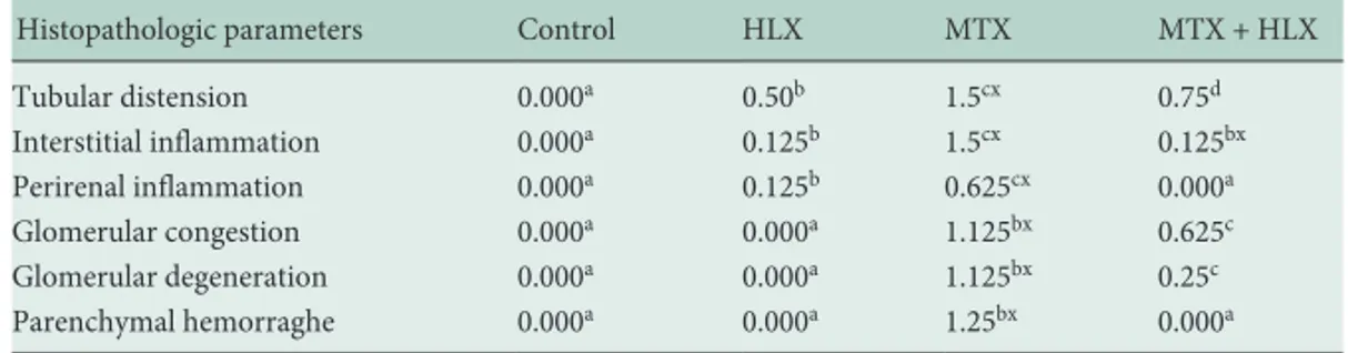
Kidney Histopathology
Table 2 summarizes the kidney histopathology results of all
groups. In the histologic examination, the kidney tissues of the
control and HLX groups showed normal kidney morphology
(fig. 2A and 2B). MTX significantly caused tubular distension (p =
0.000) (fig. 2C), interstitial inflammation (p = 0.003) (fig. 2D),
per-irenal inflammation (p = 0.009), glomerular congestion (p = 0.000),
glomerular degeneration (p = 0.001) (fig. 2E), and parenchymal
hemorrhage (p = 0.004) (fig. 2F).
Moreover, administration of HLX plus MTX provided a great
improvement regarding the tubule distension (p = 0.015) (fig. 2G),
the interstitial inflammation (p = 0.009), the perirenal
inflamma-tion (p = 0.009), the glomerular congesinflamma-tion (p = 0.044) (fig. 2G),
the glomerular degeneration (p = 0.011), and the parenchymal
hemorrhage (p = 0.004) (fig. 2H); these values were found to be
statistically significant compared to the MTX group.
Comet Assay Results
The comet assay results showed that the DNA damage was
higher in the MTX group compared to the control group. While
the highest genotoxic activity was observed in the MTX group
(31.00 ± 7.00), the lowest one was observed in the control group
(15.00 ± 3.00). The decrease in DNA damage in the MTX + HLX
group is statistically non-significant compared to that of the MTX
group. The DNA damage in the MTX group is significantly higher
than that in the control and HLX group, but no significant
differ-ences could be seen between MTX + HLX or MTX alone (table 3).
Discussion
In the present study, mistletoe extract clearly exerted a
protec-tive effect against MTX-induced oxidaprotec-tive stress, inflammatory cell
infiltration, and nephrotoxicity in rats due to its powerful
antioxi-dant and anti-inflammatory properties. The results showed that
MTX caused oxidative renal tissue damage, as evidenced by renal
histopathological findings in the form of tubular distension,
inter-stitial inflammation, perirenal inflammation, glomerular
conges-tion, glomerular degeneraconges-tion, parenchymal hemorrhage, and
per-irenal eosinophil infiltration, which is in agreement with the
find-ings of previous studies [35–37]. Pretreatment with HLX before the
administration of MTX ameliorated the MTX-induced damage of
the kidneys. The exact mechanism of MTX-induced nephrotoxicity
remains obscure. However, some studies demonstrated that the
main factor in MTX-associated tissue injury was oxidative damage,
with subsequent free-radical generation [4–6]. The role of
oxida-tive stress has been documented in MTX-induced nephrotoxicity
[2, 5, 6, 27–30, 38] and hepatotoxicity [6, 29, 30]. Here, we showed
for the first time that HLX ameliorated MTX-induced oxidative
stress and nephrotoxicity. The mechanism included reversing the
MTX-induced renal oxidative stress, as indicated by the significant
increase in GSH-Px and SOD activities. The decrease in the NO
Control HLX MTX MTX + HLX
GSH-Px, U/mg 0.35713 ± 0.08a 0.38250 ± 0.04a 0.30037 ± 0.06a 0.45538 ± 0.05bx
SOD, U/mg 0.10023 ± 0.05a 0.11246 ± 0.04b 0.09363 ± 0.02a 0.16344 ± 0.06c
NO, µm/g 1.18576 ± 0.39 1.45688 ± 0.81 1.47944 ± 0.32 1.32408 ± 0.05
MPO, ng/ml 1.26298 ± 0.53 1.21612 ± 0.39 1.52706 ± 0.24 1.27288 ± 0.07
Results are presented as mean ± SD. Groups of data were compared with the Kruskal-Wallis test followed by the Mann-Whitney U test. GSH-Px = Glutathione peroxidase, SOD = superoxide dismutase, NO = nitric oxide, MPO = myeloperoxidase, SD = standard deviation. Values followed by different characters (a, b, c) in the columns are significantly different at p < 0.05.
Values followed by the ‘x’ character in the columns are significantly different at p < 0.001.
Table 1. GSH-Px, SOD, NO, and MPO values in the kidneys of the 4 groups of rats (n = 8 each)
Histopathologic parameters Control HLX MTX MTX + HLX
Tubular distension 0.000a 0.50b 1.5cx 0.75d Interstitial inflammation 0.000a 0.125b 1.5cx 0.125bx Perirenal inflammation 0.000a 0.125b 0.625cx 0.000a Glomerular congestion 0.000a 0.000a 1.125bx 0.625c Glomerular degeneration 0.000a 0.000a 1.125bx 0.25c Parenchymal hemorraghe 0.000a 0.000a 1.25bx 0.000a
Results are presented as the median of the scores. Groups of data were compared with the Kruskal-Wallis test followed by the Mann-Whitney U test.
HLX = Helixor, MTX = methotrexate.
Values followed by different characters (a, b, c, d) in the columns are significantly different at p < 0.05. Values followed by the ‘x’ character in the columns are significantly different at p < 0.001.
Table 2. Histopathological findings in the kid-neys of the 4 groups of rats (n = 8 each)
Fig. 2. (A) Control group (H&E, × 40): normal renal morphology, (B) HLX group (H&E, × 100): normal renal morphology, (C) MTX group (H&E, × 40):
disten-sion of tubules (arrows), (D) MTX group (H&E, × 40): interstitial in flammation (stars), (E) MTX group (H&E, × 200): glomerular degeneration (arrows), (F) MTX
group (H&E, × 200): parenchymal hemorrhage (arrows), (G) MTX + HLX group (H&E, × 200): mild interstitial inflammation (stars) and minimal glomerular
congestion (arrows), (H) MTX + HLX group (H&E, × 200): mild parenchymal hemorrhage (stars). H&E = hematoxylin and eosin, HLX = Helixor, MTX =
metho-trexate.
Table 3. Comet assay values of the 4 groups of rats
Kidney Histopathology
Table 2 summarizes the kidney histopathology results of all
groups. In the histologic examination, the kidney tissues of the
control and HLX groups showed normal kidney morphology
(fig. 2A and 2B). MTX significantly caused tubular distension (p =
0.000) (fig. 2C), interstitial inflammation (p = 0.003) (fig. 2D),
per-irenal inflammation (p = 0.009), glomerular congestion (p = 0.000),
glomerular degeneration (p = 0.001) (fig. 2E), and parenchymal
hemorrhage (p = 0.004) (fig. 2F).
Moreover, administration of HLX plus MTX provided a great
improvement regarding the tubule distension (p = 0.015) (fig. 2G),
the interstitial inflammation (p = 0.009), the perirenal
inflamma-tion (p = 0.009), the glomerular congesinflamma-tion (p = 0.044) (fig. 2G),
the glomerular degeneration (p = 0.011), and the parenchymal
hemorrhage (p = 0.004) (fig. 2H); these values were found to be
statistically significant compared to the MTX group.
Comet Assay Results
The comet assay results showed that the DNA damage was
higher in the MTX group compared to the control group. While
the highest genotoxic activity was observed in the MTX group
(31.00 ± 7.00), the lowest one was observed in the control group
(15.00 ± 3.00). The decrease in DNA damage in the MTX + HLX
group is statistically non-significant compared to that of the MTX
group. The DNA damage in the MTX group is significantly higher
than that in the control and HLX group, but no significant
differ-ences could be seen between MTX + HLX or MTX alone (table 3).
Discussion
In the present study, mistletoe extract clearly exerted a
protec-tive effect against MTX-induced oxidaprotec-tive stress, inflammatory cell
infiltration, and nephrotoxicity in rats due to its powerful
antioxi-dant and anti-inflammatory properties. The results showed that
MTX caused oxidative renal tissue damage, as evidenced by renal
histopathological findings in the form of tubular distension,
inter-stitial inflammation, perirenal inflammation, glomerular
conges-tion, glomerular degeneraconges-tion, parenchymal hemorrhage, and
per-irenal eosinophil infiltration, which is in agreement with the
find-ings of previous studies [35–37]. Pretreatment with HLX before the
administration of MTX ameliorated the MTX-induced damage of
the kidneys. The exact mechanism of MTX-induced nephrotoxicity
remains obscure. However, some studies demonstrated that the
main factor in MTX-associated tissue injury was oxidative damage,
with subsequent free-radical generation [4–6]. The role of
oxida-tive stress has been documented in MTX-induced nephrotoxicity
[2, 5, 6, 27–30, 38] and hepatotoxicity [6, 29, 30]. Here, we showed
for the first time that HLX ameliorated MTX-induced oxidative
stress and nephrotoxicity. The mechanism included reversing the
MTX-induced renal oxidative stress, as indicated by the significant
increase in GSH-Px and SOD activities. The decrease in the NO
and MPO levels in the rat groups pretreated with HLX was not
sig-nificant. HLX lowered, although not significantly, the
MTX-in-duced DNA damage in endogenous lymphocytes.
NO is a free radical formed from
L-arginine by NOS. The
over-production of NO, which reacts with superoxide anions, leads to
the formation of peroxynitrite (ONOO
–). Peroxynitrite oxidizes
cellular structures and causes lipid peroxidation and ROS
forma-tion, resulting in cellular injury. It has been reported that increased
peroxynitrite caused renal injury and damage to arteries and
tu-bules [39]. Previous studies showed that an MTX overdose led to
nephrotoxicity due to lipid peroxidation, which resulted in
in-creased levels of malondialdehyde (MDA), NO release, and ROS
formation [2, 5, 6, 27–30]. Similarly, in the present study, the level
of NO was increased in the kidney tissues of the MTX group. It was
also elevated in the HLX group. V. album extract exerts a positive
effect on cardiac tissue via its vasodilatory activity, which is
medi-ated by increases in NO production. NO formation in vascular
en-dothelial cells modulates the vasodilator tone, and it is necessary
for the regulation of blood flow and pressure. Tenorio et al. [40]
and Tenorio-Lopez et al. [41] reported that a V. album-induced
in-crease in cardiac NO levels had hypotensive and vasodilatory
ef-fects in an isolated and perfused heart model. Similarly, elevated
NO levels following V. album administration in rats were
demon-strated in heart tissue [42]. However, in the present study, HLX
ad-ministration decreased the MTX-induced NO levels, although not
significantly. Korean mistletoe (
V. album coloratum) lectin was
re-ported to exert an immunomodulatory effect by blocking
lipopoly-saccharide-induced NO production in macrophage-like cells [43].
The protective effects of Korean mistletoe lectin against oxidative
stress were reported to be linked to the down-regulation of mRNA
and protein expression of iNOS and COX-2 through NF-κB
regu-lation and inhibition of NO production [17]. Overexpression of
pro-inflammatory mediators including TNF-α, NF-κB, COX-2,
and iNOS was shown to play an important role in the direct
ne-phrotoxicity effects of MTX [6, 28, 29, 35, 44]. Therefore, we
sug-gest that HLX treatment might prevent the MTX-induced increases
in iNOS levels. This suggestion is in agreement with the
histologi-cal findings in the present study, which demonstrated that the
ad-ministration of HLX greatly ameliorated the MTX-induced
inflam-mation in renal tissues.
MPO is a heme peroxidase enzyme found in neutrophil primary
granules and monocyte lysosomes that leads to tissue damage in
acute and chronic inflammation [45]. Thus, inhibiting the
enzy-matic activity of MPO may be beneficial in the treatment of
inflam-mation-related diseases [45]. In the present study, MTX elevated
the MPO activity, pointing to an accumulation of inflammatory
Control HLX MTX MTX + HLX
GSH-Px, U/mg 0.35713 ± 0.08a 0.38250 ± 0.04a 0.30037 ± 0.06a 0.45538 ± 0.05bx
SOD, U/mg 0.10023 ± 0.05a 0.11246 ± 0.04b 0.09363 ± 0.02a 0.16344 ± 0.06c
NO, µm/g 1.18576 ± 0.39 1.45688 ± 0.81 1.47944 ± 0.32 1.32408 ± 0.05
MPO, ng/ml 1.26298 ± 0.53 1.21612 ± 0.39 1.52706 ± 0.24 1.27288 ± 0.07
Results are presented as mean ± SD. Groups of data were compared with the Kruskal-Wallis test followed by the Mann-Whitney U test. GSH-Px = Glutathione peroxidase, SOD = superoxide dismutase, NO = nitric oxide, MPO = myeloperoxidase, SD = standard deviation. Values followed by different characters (a, b, c) in the columns are significantly different at p < 0.05.
Values followed by the ‘x’ character in the columns are significantly different at p < 0.001.
Histopathologic parameters Control HLX MTX MTX + HLX
Tubular distension 0.000a 0.50b 1.5cx 0.75d Interstitial inflammation 0.000a 0.125b 1.5cx 0.125bx Perirenal inflammation 0.000a 0.125b 0.625cx 0.000a Glomerular congestion 0.000a 0.000a 1.125bx 0.625c Glomerular degeneration 0.000a 0.000a 1.125bx 0.25c Parenchymal hemorraghe 0.000a 0.000a 1.25bx 0.000a
Results are presented as the median of the scores. Groups of data were compared with the Kruskal-Wallis test followed by the Mann-Whitney U test.
HLX = Helixor, MTX = methotrexate.
Values followed by different characters (a, b, c, d) in the columns are significantly different at p < 0.05. Values followed by the ‘x’ character in the columns are significantly different at p < 0.001.
Fig. 2. (A) Control group (H&E, × 40): normal renal morphology, (B) HLX group (H&E, × 100): normal renal morphology, (C) MTX group (H&E, × 40):
disten-sion of tubules (arrows), (D) MTX group (H&E, × 40): interstitial in flammation (stars), (E) MTX group (H&E, × 200): glomerular degeneration (arrows), (F) MTX
group (H&E, × 200): parenchymal hemorrhage (arrows), (G) MTX + HLX group (H&E, × 200): mild interstitial inflammation (stars) and minimal glomerular
congestion (arrows), (H) MTX + HLX group (H&E, × 200): mild parenchymal hemorrhage (stars). H&E = hematoxylin and eosin, HLX = Helixor, MTX =
metho-trexate.
Control HLX MTX MTX + HLX
DNA damage, AU ± SD* 15.00 ± 3.00a 19.00 ± 3.60ab 31.00 ± 7.00c 26.66 ± 2.30bc
*Mean ± SD.
HLX = Helixor, MTX = methotrexate, AU = arbitrary unit, SD = standard deviation.
Values followed by different characters (a, b, c, d) are significantly different at p < 0.05 (Duncan test).
Table 3. Comet assay values of the 4 groups of rats
cells (neutrophils and monocytes) in the kidney tissue. This
obser-vation is in agreement with the histological findings, which revealed
interstitial and perirenal inflammation in the renal tissue of the
MTX group. The MPO activity elevation following MTX
adminis-tration in rats has already been demonstrated earlier, in the kidneys
[27, 46] and the liver [46]. The HLX-induced decrease in the MPO
activity in this study, although not significant, suggests that
inflam-matory cell infiltration might be restricted. This protective
mecha-nism appears to be related to the increased NO levels, which inhibit
platelet and neutrophil aggregation and therefore mitigate the
ef-fects of elevated MPO activity [47]. A previous study demonstrated
that V. album extract attenuated cyclophosphamide (CP)-induced
increases in MPO activity in both heart and bladder tissues [38].
The production of free radicals is prevented by the endogenous
antioxidative defense system. SOD and GSH-Px are the main
anti-oxidative enzymes in the cytosol of living cells that protect against
ROS-induced oxidative damage. The release of free radicals results
in extensive cellular damage when the levels surpass the
antioxida-tive capacity of the biological system. An increase in the activity of
antioxidative enzymes has been shown to prevent oxidative
stress-associated tissue injury [48]. In the present study, the activities of
SOD and GSH-Px decreased in the kidney tissue of the MTX-only
group, which is consistent with the findings of recent studies [2, 6,
28, 30]. HLX, a powerful antioxidant, confers protection against
MTX-induced toxicity by inhibiting the initiation of oxidative
stress. Following the mistletoe administration, the activities of
GSH-Px and SOD significantly increased in the HLX + MTX
group. The increases in antioxidant enzyme activities may reflect
an improved antioxidant status of the rats pretreated with HLX, as
indicated by the elevation of the GSH-Px and SOD levels. This
ob-servation is in agreement with the findings of an earlier study,
which reported that pretreatment with a methanolic extract of
Eu-ropean mistletoe (V. album L.) increased the antioxidant enzyme
activities of catalase, SOD, GSH-Px, and glutathione S-transferases
in the heart of a CP + V. album group as compared to a
CP-only-treated group [34]. Moreover, animal studies reported protective
effects of V. album extract against oxidative stress in the liver,
kid-ney, brain, and heart of rats [15, 34, 49]. This antioxidant activity
of V. album extract is associated with its pharmacologically active
constituents, mostly flavonoids and lectins, which act as
free-radi-cal scavengers, reducing agents, singlet oxygen quenchers,
hydro-gen donors, and metal chelators [50–52]. Similar results were
ob-served in different studies in which other parasitic plant extracts
(mistletoe-like plants) were used. Treatment with a mistletoe alkali,
which is a lipid-soluble antioxidant isolated from Chinese
mistle-toe extract (V. coloratum (Komar) Nakai), elevated the GSH-Px
and SOD activities in the liver and kidney tissue and in the plasma
of rats treated with carbon tetrachloride (CCI
4) [52]. Similarly, the
high phenol content of Eastern Nigerian mistletoe (Loranthus
mi-cranthus Linn.) was responsible for its high antioxidant potential
observed in diabetic rats [11]. Furthermore, the antioxidant and
hepatoprotective activity of African mistletoe Tapinanthus
bang-wensis (Engl. & K. Krause) in rats was reported to be due to the
presence of flavonoids [53]. In a study of Korean mistletoe (V.
album coloratum) lectin, the authors suggested that it showed
radi-cal-scavenging activity and protective effects against oxidative
stress induced by free radicals, NO, superoxide anions (O
2–), and
peroxynitrite in vitro [17].
MTX, a folate antagonist, competitively binds to the folate-
dependent enzyme dihydrofolate reductase, inhibiting
thymi-dylate synthesis and, hence, DNA synthesis. MTX also causes
folate deficiency, which leads to genotoxic damage [1]. In the
pre-sent study, DNA damage caused by MTX was demonstrated by a
comet assay. Previous studies used comet assays to evaluate
MTX-induced germ cell toxicity and MTX-MTX-induced DNA damage of
in-testinal cells [1, 54].
In addition to the antitumor activities and chemopreventive
ef-fects of V. album, antigenotoxic efef-fects of V. album have been
dem-onstrated [13, 34, 49]. In the present study, pretreatment of the rats
with HLX lowered, although not significantly, the MTX-induced
DNA damage in endogenous lymphocytes, as determined by
de-creased DNA damage values in the comet assay. Similar results
were obtained in another study, which reported that
V. album
ex-tract attenuated the cytogenotoxic effects of MTX by reducing the
number of chromosomal aberrations and significantly increasing
the mitotic index in mouse bone marrow cells [49]. Another study
demonstrated similar results in CP-induced mouse bone marrow
cells [34]. V. album was also reported to protect against H
2O
2-in-duced oxidative nuclear and mitochondrial DNA damage in vitro,
due to its high quercetin content [13].
In conclusion, the present study demonstrated that
pretreat-ment with HLX alleviated the MTX-induced nephrotoxicity in rats
via its antioxidant and anti-inflammatory properties, as evident
from histopathological improvements and significant increases in
the activities of the antioxidative enzymes SOD and GSH-Px. The
decrease in the NO and MPO levels in the rat groups pretreated
with HLX was not significant. In addition, pretreatment with HLX
lowered, albeit not significantly, the MTX-induced DNA damage
in endogenous lymphocytes. The improvement in animals
pre-treated with V. album may suggest that further investigations
should be performed to explore the beneficial effects of V. album to
overcome one of the most serious problems in chemotherapy.
Disclosure Statement
The authors declare that there are no conflicts of interest.
Acknowledgement
The present study was supported by the Research Fund of Mugla Sıtkı Koç-man University (Project Number 13/11).
References
1 Padmanabhan S, Tripathi DN, Vikram A, Ramarao P, Jena GB: Methotrexate-induced cytotoxicity and geno-toxicity in germ cells of mice: intervention of folic and folinic acid. Mutat Res 2009;673:43–52.
2 Asvadi I, Hajipour B, Asvadi A, Asl NA, Roshangar L, Khodadadi A: Protective effect of pentoxyfilline in renal toxicity after methotrexate administration. Eur Rev Med Pharmacol Sci 2011;15:1003–1009.
3 Widemann BC, Adamson PC: Understanding and managing methotrexate nephrotoxicity. Oncologist 2006;11:694–703.
4 Leitão RF, Brito GA, Oriá RB, Braga-Neto MB, Bel-laguarda EA, Silva JV, et al: Role of inducible nitric oxide synthase pathway on methotrexate-induced in-testinal mucositis in rodents. BMC Gastroenterol 2011; 11:90.
5 Uz E, Oktem F, Yilmaz HR, Uzar E, Ozgüner F: The activities of purine-catabolizing enzymes and the level of nitric oxide in rat kidneys subjected to methotrexate: protective effect of caffeic acid phenethyl ester. Mol Cell Biochem 2005;277:165–170.
6 Hafez HM, Ibrahim MA, Ibrahim SA, Amin EF, Goma W, Abdelrahman AM: Potential protective effect of etanercept and aminoguanidine in methotrexate-in-duced hepatotoxicity and nephrotoxicity in rats. Eur J Pharmacol 2015;768:1–12.
7 Cetiner M, Sener G, Sehirli AO, Ekşioğlu-Demiralp E, Ercan F, Sirvanci S, et al: Taurine protects against methotrexate-induced toxicity and inhibits leukocyte death. Toxicol Appl Pharmacol 2005;209:39–50. 8 Aggarwal BB, Gupta SC, Kim JH: Historical
perspec-tives on tumor necrosis factor and its superfamily: 25 years later, a golden journey. Blood 2012;119:651–665. 9 Hegde P, Maddur MS, Friboulet A, Bayry J, Kaveri SV:
Viscum album exerts anti-inflammatory effect by
selec-tively inhibiting cytokine-induced expression of cy-clooxygenase-2. PLoS One 2011;6:e26312.
10 Saha C, Hegde P, Friboulet A, Bayry J, Kaveri SV:
Vis-cum album-mediated COX-2 inhibition implicates
destabilization of COX-2 mRNA. PLoS One 2015;10: e0114965.
11 Onunogbo C, Ohaeri OC, Eleazu CO, Eleazu KC: Chemical composition of mistletoe extract (Loranthus
micranthus) and its effect on the protein, lipid
metabo-lism and the antioxidant status of alloxan induced dia-betic rats. J Med Res 2012;1:57–62.
12 Önay-Uçar E, Karagöz A, Arda N: Antioxidant activity of
Viscum album ssp. album. Fitoterapia 2006;77:556–560. 13 Önay-Uçar E, Erol O, Kandemir B, Mertoğlu E,
Karagöz A, Arda N: Viscum album L. extract protects HeLa cells against nuclear and mitochondrial DNA damage. Evid Based Complement Alternat Med 2012; 2012:958740.
14 Oluwaseun AA, Ganiyu O: Antioxidant properties of methanolic extracts of mistletoes (Viscum album) from cocoa and cashew trees in Nigeria. Afr J Biotechnol 2008;7:3138–3142.
15 Orhan DD, Aslan M, Sendogdu N, Ergun F, Yesilada E: Evaluation of the hypoglycemic effect and antioxidant activity of three Viscum album subspecies (European mistletoe) in streptozotocin-diabetic rats. J Ethnophar-macol 2005;98:95–102.
16 Kim MS, Lee J, Lee KM, Yang SH, Choi S, Chung SY, et al: Involvement of hydrogen peroxide in mistletoe lectin-II-induced apoptosis of myeloleukemic U937 cells. Life Sci 2003;73:1231–1243.
17 Kim BK, Choi MJ, Park KY, Cho EJ: Protective effects of Korean mistletoe lectin on radical-induced oxidative stress. Biol Pharm Bull 2010;33:1152–1158.
18 Deliorman D, Calis I, Ergun F: A new acyclic monoter-pene glucoside from Viscum album ssp. album. Fito-terapia 2001;72:101–105.
hepatoprotective activity of African mistletoe Tapinanthus
bang-wensis (Engl. & K. Krause) in rats was reported to be due to the
presence of flavonoids [53]. In a study of Korean mistletoe (
V.
album coloratum) lectin, the authors suggested that it showed
radi-cal-scavenging activity and protective effects against oxidative
stress induced by free radicals, NO, superoxide anions (O
2–), and
peroxynitrite in vitro [17].
MTX, a folate antagonist, competitively binds to the folate-
dependent enzyme dihydrofolate reductase, inhibiting
thymi-dylate synthesis and, hence, DNA synthesis. MTX also causes
folate deficiency, which leads to genotoxic damage [1]. In the
pre-sent study, DNA damage caused by MTX was demonstrated by a
comet assay. Previous studies used comet assays to evaluate
MTX-induced germ cell toxicity and MTX-MTX-induced DNA damage of
in-testinal cells [1, 54].
In addition to the antitumor activities and chemopreventive
ef-fects of V. album, antigenotoxic efef-fects of V. album have been
dem-onstrated [13, 34, 49]. In the present study, pretreatment of the rats
with HLX lowered, although not significantly, the MTX-induced
DNA damage in endogenous lymphocytes, as determined by
de-creased DNA damage values in the comet assay. Similar results
were obtained in another study, which reported that V. album
ex-tract attenuated the cytogenotoxic effects of MTX by reducing the
number of chromosomal aberrations and significantly increasing
the mitotic index in mouse bone marrow cells [49]. Another study
demonstrated similar results in CP-induced mouse bone marrow
cells [34]. V. album was also reported to protect against H
2O
2-in-duced oxidative nuclear and mitochondrial DNA damage in vitro,
due to its high quercetin content [13].
In conclusion, the present study demonstrated that
pretreat-ment with HLX alleviated the MTX-induced nephrotoxicity in rats
via its antioxidant and anti-inflammatory properties, as evident
from histopathological improvements and significant increases in
the activities of the antioxidative enzymes SOD and GSH-Px. The
decrease in the NO and MPO levels in the rat groups pretreated
with HLX was not significant. In addition, pretreatment with HLX
lowered, albeit not significantly, the MTX-induced DNA damage
in endogenous lymphocytes. The improvement in animals
pre-treated with V. album may suggest that further investigations
should be performed to explore the beneficial effects of V. album to
overcome one of the most serious problems in chemotherapy.
Disclosure Statement
The authors declare that there are no conflicts of interest.
Acknowledgement
The present study was supported by the Research Fund of Mugla Sıtkı Koç-man University (Project Number 13/11).
References
1 Padmanabhan S, Tripathi DN, Vikram A, Ramarao P, Jena GB: Methotrexate-induced cytotoxicity and geno-toxicity in germ cells of mice: intervention of folic and folinic acid. Mutat Res 2009;673:43–52.
2 Asvadi I, Hajipour B, Asvadi A, Asl NA, Roshangar L, Khodadadi A: Protective effect of pentoxyfilline in renal toxicity after methotrexate administration. Eur Rev Med Pharmacol Sci 2011;15:1003–1009.
3 Widemann BC, Adamson PC: Understanding and managing methotrexate nephrotoxicity. Oncologist 2006;11:694–703.
4 Leitão RF, Brito GA, Oriá RB, Braga-Neto MB, Bel-laguarda EA, Silva JV, et al: Role of inducible nitric oxide synthase pathway on methotrexate-induced in-testinal mucositis in rodents. BMC Gastroenterol 2011; 11:90.
5 Uz E, Oktem F, Yilmaz HR, Uzar E, Ozgüner F: The activities of purine-catabolizing enzymes and the level of nitric oxide in rat kidneys subjected to methotrexate: protective effect of caffeic acid phenethyl ester. Mol Cell Biochem 2005;277:165–170.
6 Hafez HM, Ibrahim MA, Ibrahim SA, Amin EF, Goma W, Abdelrahman AM: Potential protective effect of etanercept and aminoguanidine in methotrexate-in-duced hepatotoxicity and nephrotoxicity in rats. Eur J Pharmacol 2015;768:1–12.
7 Cetiner M, Sener G, Sehirli AO, Ekşioğlu-Demiralp E, Ercan F, Sirvanci S, et al: Taurine protects against methotrexate-induced toxicity and inhibits leukocyte death. Toxicol Appl Pharmacol 2005;209:39–50. 8 Aggarwal BB, Gupta SC, Kim JH: Historical
perspec-tives on tumor necrosis factor and its superfamily: 25 years later, a golden journey. Blood 2012;119:651–665. 9 Hegde P, Maddur MS, Friboulet A, Bayry J, Kaveri SV:
Viscum album exerts anti-inflammatory effect by
selec-tively inhibiting cytokine-induced expression of cy-clooxygenase-2. PLoS One 2011;6:e26312.
10 Saha C, Hegde P, Friboulet A, Bayry J, Kaveri SV:
Vis-cum album-mediated COX-2 inhibition implicates
destabilization of COX-2 mRNA. PLoS One 2015;10: e0114965.
11 Onunogbo C, Ohaeri OC, Eleazu CO, Eleazu KC: Chemical composition of mistletoe extract (Loranthus
micranthus) and its effect on the protein, lipid
metabo-lism and the antioxidant status of alloxan induced dia-betic rats. J Med Res 2012;1:57–62.
12 Önay-Uçar E, Karagöz A, Arda N: Antioxidant activity of
Viscum album ssp. album. Fitoterapia 2006;77:556–560. 13 Önay-Uçar E, Erol O, Kandemir B, Mertoğlu E,
Karagöz A, Arda N: Viscum album L. extract protects HeLa cells against nuclear and mitochondrial DNA damage. Evid Based Complement Alternat Med 2012; 2012:958740.
14 Oluwaseun AA, Ganiyu O: Antioxidant properties of methanolic extracts of mistletoes (Viscum album) from cocoa and cashew trees in Nigeria. Afr J Biotechnol 2008;7:3138–3142.
15 Orhan DD, Aslan M, Sendogdu N, Ergun F, Yesilada E: Evaluation of the hypoglycemic effect and antioxidant activity of three Viscum album subspecies (European
mistletoe) in streptozotocin-diabetic rats. J Ethnophar-macol 2005;98:95–102.
16 Kim MS, Lee J, Lee KM, Yang SH, Choi S, Chung SY, et al: Involvement of hydrogen peroxide in mistletoe lectin-II-induced apoptosis of myeloleukemic U937 cells. Life Sci 2003;73:1231–1243.
17 Kim BK, Choi MJ, Park KY, Cho EJ: Protective effects of Korean mistletoe lectin on radical-induced oxidative stress. Biol Pharm Bull 2010;33:1152–1158.
18 Deliorman D, Calis I, Ergun F: A new acyclic monoter-pene glucoside from Viscum album ssp. album. Fito-terapia 2001;72:101–105.
19 Deliorman D, Ergun F, Sener B, Palittapongarnpim P: Evaluation of antibacterial activity of Viscum album subspecies. Pharm Biol 2001;39:381–385.
20 Büssing A, Schietzel M: Apoptosis-inducing properties of Viscum album L. extracts from different host trees, correlate with their content of toxic mistletoe lectins. Anticancer Res 1999;19:23–28.
21 Radenkovic M, Ivetic V, Popovic M, Mimica-Dukic N, Veljkovic S: Neurophysiological effects of mistletoe (Viscum album L.) on isolated rat intestines. Phytother Res 2006;20:374–377.
22 Wollenweber E, Wieland A, Haas K: Epicuticular waxes and flavonol aglycones of the European mistletoe,
Vis-cum album L. Z Nat Forsch 2000;55:314–317. 23 Maier G, Fiebig HH: Absence of tumor growth
stimula-tion in a panel of 16 human tumor cell lines by mistletoe extracts in vitro. Anticancer Drugs 2002;13:373–379. 24 Kovacs E, Hajto T, Hostanska K: Improvement of DNA
repair in lymphocytes of breast cancer patients treated with Viscum album extract (Iscador). Eur J Cancer 1991;27:1672.
25 Bussing A, Regnery A, Schweizer K: Effects of Viscum
album L. on cyclophosphamide-treated peripheral
blood mononuclear cells in vitro: sister chromatid ex-changes and activation/proliferation marker expres-sion. Cancer Lett 1995;94:199.
26 Kovacs E: The in vitro effect of Viscum album (VA) ex-tract on DNA repair of peripheral blood mononuclear cells (PBMC) in cancer patients. Phytother Res 2002; 16:143–147.
27 Abraham P, Kolli VK, Rabi S: Melatonin attenuates methotrexate-induced oxidative stress and renal dam-age in rats. Cell Biochem Funct 2010;28:426–433. 28 Morsy MA, Ibrahim SA, Amin EF, Kamel MY, Rifaai
RA, Hassan MK: Curcumin ameliorates methotrexate-induced nephrotoxicity in rats. Adv Pharmacol Sci 2013;2013:387071.
29 El-Sheikh AA, Morsy MA, Abdalla AM, Hamouda AH, Alhaider IA: Mechanisms of thymoquinone hepatore-nal protection in methotrexate-induced toxicity in rats. Mediators Inflamm 2015;2015:859383.
30 Armagan I, Bayram D, Candan IA, Yigit A, Celik E, Ar-magan HH, et al: Effects of pentoxifylline and alpha lipoic acid on methotrexate-induced damage in liver and kidney of rats. Environ Toxicol Pharmacol 2015; 39:1122–1131.
31 Kim MH, Lee SH, Kim SC, Kim YK, Park SH: Com-parative study on the effects of a Viscum album (L.) ex-tract (mistletoe) and doxycycline for pleurodesis in pa-tients with malignant pleural effusion. Korean J Med 1999;57(suppl 2):121.
32 Piao BK, Wang YX, Xie GR, Mansmann U, Matthes H, Beuth J, Lin HS: Impact of complementary mistletoe extract treatment on quality of life in breast, ovarian and non-small cell lung cancer patients. A prospective randomized controlled clinical trial. Anticancer Res 2004;24:303–309.
33 Reagan-Shaw S, Nihal M, Ahmad N: Dose translation from animal to human studies revisited. FASEB J 2008; 22:659–661.
34 Sekeroğlu V, Aydin B, Sekeroğlu ZA: Viscum album L. extract and quercetin reduce cyclophosphamide-in-duced cardiotoxicity, urotoxicity and genotoxicity in mice. Asian Pac J Cancer Prev 2011;12:2925–2931. 35 Abdel-Raheem IT, Khedr NF: Renoprotective effects of
montelukast, a cysteinyl leukotriene receptor antago-nist, against methotrexate-induced kidney damage in rats. Naunyn Schmiedebergs Arch Pharmacol 2014; 387:341–353.
36 Grönroos M, Chen M, Jahnukainen T, Capitanio A, Aizman RI, Celsi G: Methotrexate induces cell swelling and necrosis in renal tubular cells. Pediatr Blood Can-cer 2006;46:624–629.
37 Chelab KG, Majeed SK: Methotrexate-induced histo-pathological changes in the kidneys of mice. Iraqi J Vet Sci 2009;23:219–222.
38 Çakır T, Polat C, Baştürk A, Gül M, Aslaner A, Durgut H, et al: The effect of alpha lipoic acid on rat kidneys in methotrexate induced oxidative injury. Eur Rev Med Pharmacol Sci 2015;19:2132–2139.
39 Ashtiyani SC, Najafi H, Firouzifar MR, Shafaat O: Grape seed extract for reduction of renal disturbances following reperfusion in rats. Iran J Kidney Dis 2013;7: 28–35.
40 Tenorio FA, del Valle L, González A, Pastelín G: Vaso-dilator activity of the aqueous extract of Viscum album. Fitoterapia 2005;76:204–209.
41 Tenorio-Lopez FA, Valle Mondragon LD, Olvera GZ, Torres Narvaez JC, Pastelin G: Viscum album aqueous
extract induces NOS-2 and NOS-3 overexpression in Guinea pig hearts. Nat Prod Res 2006;20:1176–1182. 42 Lee JY, Kim JY, Lee YG, Byeon SE, Kim BH, Rhee MH,
et al: In vitro immunoregulatory effects of Korean mis-tletoe lectin on functional activation of monocytic and macrophage-like cells. Biol Pharm Bull 2007;30:2043– 2051.
43 Ibrahim MA, El-Sheikh AA, Khalaf HM, Abdelrahman AM: Protective effect of peroxisome proliferator activa-tor recepactiva-tor (PPAR)-α and -γ ligands against metho-trexate-induced nephrotoxicity. Immunopharmacol Immunotoxicol 2014;36:130–137.
44 Kohnen S, Franck T, Van Antwerpen P, Boudjeltia KZ, Mouithys-Mickalad A, Deby C, et al: Resveratrol inhib-its the activity of equine neutrophil myeloperoxidase by a direct interaction with the enzyme. J Agric Food Chem 2007;55:8080–8087.
45 Jahovic N, Cevik H, Sehirli AO, Yeğen BC, Sener G: Melatonin prevents methotrexate-induced hepatorenal oxidative injury in rats. J Pineal Res 2003;34:282–287. 46 Mollace V, Salvemini D, Anggard E, Vane J: Nitric
oxide from vascular smooth cells: regulation of platelet reactivity and smooth muscle cell guanylate cyclase. Br J Pharmacol 1991;104:633–638.
47 Reiter RJ, Poeggeler B, Tan DX, Chen LD, Manchester LC, Guerrero JM: Antioxidant capacity of melatonin: a novel action not requiring a receptor. Neuroendocrinol Lett 1993;15:103–116.
48 Sekeroğlu ZA, Sekeroğlu V: Effects of Viscum album L. extract and quercetin on methotrexate-induced cyto-genotoxicity in mouse bone-marrow cells. Mutat Res 2012;746:56–59.
49 Amic D, Davidovic-Amic D, Beslo D, Trinasjstic N: Structure-radical scavenging activity relationship of flavonoids. Croatia Chem Acta 2003;76:55–61. 50 Oboh G, Rocha JBT: Polyphenols in red pepper
[Capsi-cum annuum var. aviculare (Tepin)] and their
protec-tive effect on some pro-oxidants induced lipid peroxi-dation in brain and liver. Eur Food Res Technol 2007; 225:239–247.
51 Ademiluyi AO, Oboh G: Antioxidant properties of methanolic extracts of mistletoes (Viscum album) from cocoa and cashew trees in Nigeria. Afr J Biotechnol 2008;7:3138–3142.
52 Shi ZM, Feng P, Jiang DQ, Wang XJ: Mistletoe alkali inhibits peroxidation in rat liver and kidney. World J Gastroenterol 2006;12:4052–4055.
53 Patrick-Iwuanyanwu KC, Onyeike EN, Wegwu MO: Anti-inflammatory effect of crude methanolic extract and fractions of African mistletoe Tapinanthus
bang-wensis (Engl. & K. Krause) on Wistar albino rats. Der
Pharmacia Lettre 2010;2:76–83.
54 Dadhania VP, Tripathi DN, Vikram A, Ramarao P, Jena GB: Intervention of alpha-lipoic acid ameliorates methotrexate-induced oxidative stress and genotoxic-ity: a study in rat intestine. Chem Biol Interact 2010; 183:85–97.
© Free Author Copy - for personal use only
ANY DISTRIBUTION OF THIS ARTICLE WITHOUT WRITTEN CONSENT FROM S. KARGER AG, BASEL IS A VIOLATION OF THE COPYRIGHT. Written permission to distribute the PDF will be granted against payment of a permission fee, which is based on the number of accesses required. Please contact [email protected]