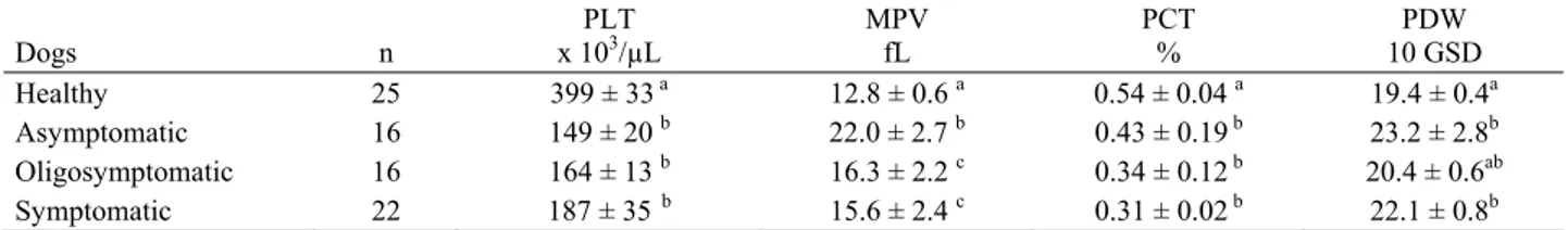
Başlık: Evaluation of erythrocyte and platelet indices in canine visceral leishmaniasisYazar(lar):TEMİZEL, Ethem Mutlu;CİHAN, Hüseyin;YILMAZ, Zeki;AYTUĞ, Nilüfer Cilt: 58 Sayı: 3 Sayfa: 185-188 DOI: 10.1501/Vetfak_0000002472 Yayın Tarihi: 2011 PDF
Tam metin
Şekil
Benzer Belgeler
Oyuncağın, çocuğun dünyasında çok önemli bir yeri olduğunu vurgulayan ve 20 yıldır Türk çocukla rına kaliteli sevimli oyun caklar yaratmak için uğra
The main reason why political values meet the requirements of the development of our society, the vital needs of the state independence of Uzbekistan, the interests of our people
The mean lymphocyte value was lower in severe COVID-19 cases compared to moderate COVID- 19 cases both on the day of hospital admission and on the third follow-up day (p <
We aimed to evaluate hemogram parameters (especially platelet indices) in diabetic patients with myocardial bridges.. Methods: We reviewed angiograms performed between May 2017
White blood cell and platelet count, MPV and PDW values, and serum LDH levels of the patients who had been diagnosed with reactive lymph node hyperplasia, patients
When the pre-operative and post-operative values of the patients in the study group were compared, a statistically significant difference was found between the two groups in terms
In terms of MPV, although there was no significant difference between the ARF patients in the acute stage and those in remission; the MPV/platelet ratio was significantly lower
Platelet parameters were noted from the hospital-based data system and compared with the severity of disease (Bleeding score, Severity score, Warning signs and Duration of
