ROLE OF FAM134B IN LIVER CANCER
A THESIS SUBMITTED TO
THE DEPARTMENT OF MOLECULAR BIOLOGY AND GENETICS AND THE GRADUATE SCHOOL OF ENGINEERING AND SCIENCE OF
BILKENT UNIVERSITY
IN PARTIAL FULFILLMENT OF THE REQUIREMENTS FOR THE DEGREE OF
MASTER OF SCIENCE
BY
AYŞE DERYA SONER
AUGUST 2013
ii
I certify that I have read this thesis and that in my opinion it is fully adequate, in scope and in quality, as a thesis for the degree of Master of Science.
Prof. Dr. Mehmet Öztürk
I certify that I have read this thesis and that in my opinion it is fully adequate, in scope and in quality, as a thesis for the degree of Master of Science.
Assoc. Prof. Dr. Rengül Çetin-Atalay
I certify that I have read this thesis and that in my opinion it is fully adequate, in scope and in quality, as a thesis for the degree of Master of Science.
Assist. Prof. Dr. Arzu Atalay
Approved for the Graduate School of Engineering and Science
Prof. Dr. Levent Onural
Director of the Graduate School of Engineering and Science
iii
ABSTRACT
ROLE OF FAM134B IN LIVER CANCER
Ayşe Derya Soner
M.Sc. in Molecular Biology and Genetics Supervisor: Prof. Dr. Mehmet Öztürk
August 2013, 74 Pages
The family with sequence similarity 134, member B (FAM134B) protein initially caught attention in our laboratory through demonstrating elevated levels possibly associated with senescent, cirrhotic, and mesenchymal-like states in hepatocellular carcinoma (HCC), and the aim of this thesis work was to identify the role of FAM134B in HCC. The work in this thesis initially demonstrated that the induction of endoplasmic reticulum (ER) stress in normal mice livers with 8 hours of tunicamycin treatment did not significantly alter the levels of FAM134B mRNA or protein. We then demonstrated that induction of ER stress in four HCC cell lines using several ER stress inducers such as thapsigargin, tunicamycin or DTT did not cause a significant increase in the levels of FAM134B mRNA or protein, with the exception of high dose DTT treatment of Snu449 cells which also demonstrated apoptosis. We also observed that FAM134B protein levels may be elevated during epithelial-to-mesenchymal transition (EMT) in PLC cells induced by TGF-β treatment. Snu449 cells in which FAM134B was silenced demonstrated an altered morphology in cell culture, and appeared to lose their migratory capabilities. Most significantly though, FAM134B-silenced Snu449 cells demonstrated a dramatic loss of resistance to treatment with thapsigargin and adriamycin, which are both known to inhibit the calcium pumps on the ER. The knockdown cells also demonstrated loss of resistance to serum starvation, TGF-β treatment, tunicamycin and alcohol treatment, but no significant difference was observed in resistance to 5-FU, camptothecin and hydrogen peroxide. These results implicated that FAM134B may play a role that is essential for survival under several forms of cytotoxic threat, especially those that disturb calcium homeostasis.
iv
ÖZET
FAM134B’NİN KARACİĞER KANSERİNDEKİ ROLÜ
Ayşe Derya Soner
Moleküler Biyoloji ve Genetik Yüksek Lisansı Tez Yöneticisi: Prof. Dr. Mehmet Öztürk
Ağustos 2013, 74 Sayfa
FAM134B (family with sequence similarity 134, member B) isimli protein laboratuarımızda ilk olarak seviyelerinin artışı hepatosellüler karsinomda hücre yaşlanması, siroz ve mezenkimal benzeri davranış durumları ile bağlantılı olabilecek bir protein olarak dikkat çekti, ve bu tez çalışmasının amacı FAM134B’nin hepatosellüler karsinomdaki rolünü tanımlamaktı. Bu tezdeki çalışmalar ilk olarak normal fare karaciğerinde 8 saatlik tünikamisin uygulamasi ile indüklenen endoplazmik reticulum (ER) stresinin, FAM134B mRNA ve protein seviyelerine önemli bir etkisi olmadığını gösterdi. Daha sonra da tapsigargin, tunikamisin ve DTT gibi indukleyiciler kullanılarak oluşturulan ER stresinin dört hepatosellüler karsinom hücre hattında da FAM134B mRNA ve protein seviyelerine önemli bir etkisi olmadığını gösterdik; tek istisnai durum yüksek doz DTT uygulanan ve hücre ölümü gösteren Snu449 hücreleri idi. Bunların yanısıra, FAM134B protein seviyelerinin PLC hücrelerinde TGF-β ile indüklenen epitel-mezenkimal geçiş sırasında da artıyor olabileceğini gözlemledik. FAM134B’nin susturulduğu Snu449 hücrelerinin kültürde farklı bir morfolojiye sahip olduklarını, ve hareket kabiliyetlerini yitirdiklerini gözlemledik. Fakat en önemlisi, FAM134B’nin susturulduğu Snu449 hücreleri, tapsigargin ve adriamisin uygulamasına olan dirençlerini çok ciddi bir şekilde kaybettiler, ve bu iki ilacın da ER membranındaki kalsiyum pompalarının çalışmasına engel oldukları biliniyor. FAM134B’nin susturulduğu hücreler aynı zamanda serum yetersizliği, TGF-β, tunikamisin ve alkol uygulamalarına olan dirençlerinde de kayıp gösterdiler, fakat 5-FU, camptotesin ve hidrojen peroksite olan dirençlerinde ciddi bir değişim gözlenmedi. Bu sonuçlar, FAM134B’nin hücre ölümüne neden olabilecek (özellikle de kalsiyum dengesinin bozulması gibi) birçok durumda hücrenin hayatta kalma mekanizmasında önemli bir rol oynayabileceğini gösteriyor.
v
vi
ACKNOWLEDGEMENTS
This thesis work has been made possible by the scientific guidance and emotional support of many people.
First and foremost, I would like to thank my thesis supervisor Prof. Dr. Mehmet Öztürk for his great scientific vision; the perspectives he preferred to pursue during this research work steered us towards very interesting findings. I have had the great fortune of working in Prof. Öztürk’s laboratory starting with my senior projects, and I have learned a lot from him throughout these past three years; I am very thankful for all his scientific guidance, fatherly advice, and his encouraging, positive attitude towards his students.
As an extension of our supervisor’s motivating attitude, the Öztürk lab has always been a place of hard work, as well as lots of laughs, support, and sharing. I am very grateful to have had the blessing of working with the past and present members of our group; Dr. Ceyhan Ceran, Dr. Pelin Telkoparan, Dr. Çiğdem Özen, Dr. Hani Alotaibi, Mustafa Yılmaz, Dilek Çevik, Ayşegül Örs, Gökhan Yıldız, Umur Keleş, Hande Topel, Alper Dağcan, Yusuf İsmail Ertuna, Emre Yurdusev, Engin Demirdizen, Merve Deniz Abdüsselamoğlu, Umar Raza and Dr. Ayaz Mustufa have all been great people to work with, and have provided lots of support.
I am also very grateful to all past and present Bilkent MBG faculty members; I was lucky to have the chance to meet them all and I learned a lot from them over the past six years. I have learned a lot from Assist. Prof. Dr. Ebru Erbay, especially during the endoplasmic reticulum stress-related section of this thesis work. Assoc. Prof. Işık Yuluğ, Assoc. Prof. Rengül Çetin-Atalay and Assist. Prof. Özlen Konu have not only taught me a lot about molecular biology, but have also always provided motherly support and advice. I was also lucky enough to receive the support and advice of Assoc. Prof. Uygar Tazebay, for which I am also very grateful. I have also learned a lot from Prof. Can Akçalı, Assoc. Prof. İhsan Gürsel, Assist. Prof. Ali Güre, Prof. Tamer Yağcı, and Prof. Tayfun Özçelik, both inside and outside the classroom. Further members of the MBG family have also been of great help throughout this
vii
thesis work; I am very thankful to Bilge Kılıç, Füsun Elvan, Sevim Baran, Yıldız Karabacak, Abdullah Ünnü, Turan bey and Ümmühan hanım for their patience. The MBG family has always felt like an actual family, and I am very lucky to have worked among such friends. I am especially grateful for having the friendship and support of Damla Gözen, Merve Çakır, Şahika Cıngır, Mehmet Şahin, Sıla Özdemir, Büşra Yağabasan and Özlem Tufanlı throughout this thesis work; they have been more like sisters and brothers to me than friends. I am also very grateful for the great support I received from my family, my parents, my sister, and Ömer Faruk Cavga, who have always helped me through the times of hard work.
Finally, I would like to thank the Scientific and Technological Research Council of Turkey (TÜBİTAK) for providing the grants that have made this research work possible, through funding project numbers 101S191 and 111T558.
viii TABLE OF CONTENTS SIGNATURE PAGE...II ABSTRACT...III ÖZET...IV DEDICATION...V ACKNOWLEDGEMENTS...VI TABLE OF CONTENTS...VIII LIST OF TABLES...XII LIST OF FIGURES...XIII 1. INTRODUCTION ... 1
1.1 Cancers of the Liver; Status Worldwide & in Turkey ... 1
1.2 Hepatocellular Carcinoma ... 3
1.3 Pathogenesis of Hepatocellular Carcinoma ... 3
1.3.1 Fibrosis as an Initial state of Hepatocarcinogenesis ... 4
1.3.2 Senescence and Hepatocarcinogenesis ... 7
1.3.3 Cirrhosis ... 9
1.4 Epithelial to Mesenchymal Transition and TGF-β ... 9
1.4.1 Epithelial to Mesenchymal Transition and TGF-β in Liver Fibrosis ... 10
1.4.2 Epithelial to Mesenchymal Transition and TGF-β in the progression of Hepatocellular Carcinoma ... 11
ix
1.5 The Endoplasmic Reticulum ... 12
1.5.1 The Abundance and Functions of Rough and Smooth ER in Hepatocytes ... 13
1.5.2 Endoplasmic Reticulum Stress and the Unfolded Protein Response ... 14
1.5.3 Endoplasmic Reticulum Stress, Obesity and Liver Disease ... 15
1.5.4 ER Stress, Epithelial to Mesenchymal Transition and Src Kinase ... 17
1.6 FAM134B in Literature ... 17
2. OBJECTIVES AND RATIONALE ... 20
3. MATERIALS AND METHODS ... 21
3.1 MATERIALS ... 21
3.1.1 General Laboratory Reagents and Kits ... 21
3.1.2 Cell Culture Solutions and Materials ... 21
3.1.3 Electrophoresis Apparatus ... 22
3.1.4 cDNA Synthesis and Polymerase Chain Reaction Reagents ... 22
3.1.5 Primers ... 22
3.1.6 Antibodies ... 23
3.2 SOLUTIONS AND MEDIA ... 24
3.2.1 General solutions ... 24
3.2.2 Tissue culture solutions ... 24
3.2.3 RIPA Lysis Buffer for Protein Extraction ... 25
3.2.4 Sodium Deodecyl Sulphate–Polyacrylamide Gel Electrophoresis (SDS-PAGE) Gels and Solutions for Western Blotting ... 26
3.2.5 Immunoperoxidase staining solutions ... 27
3.2.6 Sulforhodamine B (SRB) staining solutions ... 27
3.3 METHODS ... 27
x
3.3.2 Tissue culture methods ... 29
3.3.3 mRNA Expression Analyses ... 31
3.3.4 Protein Level Analyses ... 32
3.3.5 Stable Knockdown with shRNA ... 34
3.3.6 Wound Healing / Scratch Assay ... 35
3.3.7 Sulforhodamine B (SRB) Colorimetric Assay ... 35
4. RESULTS ... 36
4.1 FAM134B in Humans & Mouse ... 36
4.1.1 FAM134B Gene & Transcript Information ... 36
4.1.2 Protein Information on FAM134B ... 37
4.1.3 Initial Findings on FAM134B ... 37
4.2 FAM134B in Endoplasmic Reticulum Stress in vivo ... 38
4.2.1 Tunicamycin-induced ER stress causes a steatosis-like appearance in mice liver tissues ... 38
4.2.2 FAM134B expression is not affected in tunicamycin-induced ER stress in mice liver tissues ... 39
4.2.3 FAM134B protein level is not affected in tunicamycin-induced ER stress in mice liver tissues ... 39
4.3 FAM134B in vitro ... 40
4.3.1 Basal Levels of FAM134B in HCC cell lines ... 40
4.3.2 FAM134B and ER stress ... 42
4.3.3 Effect of 6 hours of ER stress induction on FAM134B protein levels in selected HCC cell lines ... 44
4.3.4 FAM134B and EMT ... 46
4.3.5 FAM134B knockdown experiments in Snu449 cells ... 48
4.3.6 Effects of FAM134B Knockdown on Cytotoxicity of Different Drugs ………...52
xi
4.4 Proteins predicted to phosphorylate FAM134B isoforms ... 59
5. DISCUSSION AND CONCLUSION ... 62
5.1 Domains and Motifs found on the FAM134B Protein ... 62
5.2 FAM134B and ER Stress ... 62
5.3 FAM134B and Epithelial-to-Mesenchymal Transition ... 63
5.4 FAM134B and Survival under Cytotoxic Stimuli ... 63
5.5 A Possible Relationship between the Src/PI3K and MEK1/ERK Pathways, the ER, FAM134B, and Calcium Homeostasis ... 65
6. FUTURE PERSPECTIVES ... 67
xii
LIST OF TABLES
Table 3-1: Primer sequences and Tm values... 23 Table 3-2: Antibodies, source companies and dilutions used. ... 23 Table 3-3: RIPA lysis buffer ingredients and recipe. ... 25
xiii
LIST OF FIGURES
Figure 1.1: Mortality-to-incidence ratios of cancers in Turkey and Worldwide ... 1 Figure 1.2 Histopathological progression and molecular features of HCC. [12]... 3 Figure 4.1: Genomic region and transcripts of FAM134B. ... 36 Figure 4.2: 8 hours of 1ug/g body weight tunicamycin treatment causes lipid
accumulation and a steatosis-like morphology in mice liver tissue. ... 38 Figure 4.3: ER stress induction by 8 hours of 1ug/g body weight tunicamycin
treatment does not affect the levels of FAM134B mRNA expression in liver tissues of mice. ... 39 Figure 4.4: ER stress induction by 8 hours of 1ug/g body weight tunicamycin
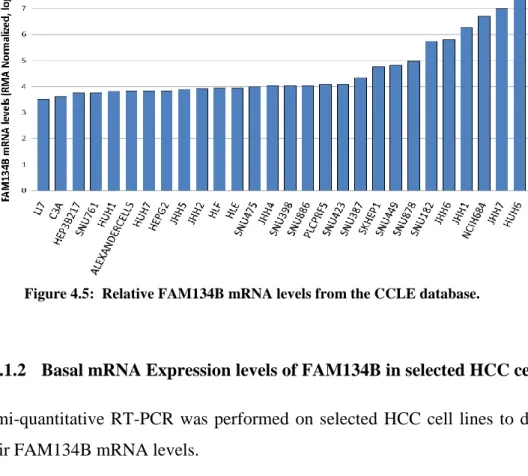
treatment does not affect the protein levels of FAM134B significantly in liver tissues of mice. ... 39 Figure 4.5: Relative FAM134B mRNA levels from the CCLE database. ... 40 Figure 4.6: Basal FAM134B mRNA levels of selected HCC cell lines. ... 40 Figure 4.7: Basal FAM134B protein levels were higher in the more
poorly-differentiated HCC cell lines compared to the well-poorly-differentiated HCC cell lines selected. ... 41 Figure 4.8: FAM134B demonstrates a perinuclear staining like that of Calnexin,
which indicates localization on the Endoplasmic Reticulum. ... 42 Figure 4.9: In Huh7, Snu387, Snu449 and Snu475 cell lines, FAM134B mRNA
expression levels are not significantly affected by ER stress induction with Tunicamycin, Thapsigargin and DTT for 1, 6 and 24 hours. ... 43 Figure 4.10: 6 hours of ER stress induction with tunicamycin, thapsigargin and DTT
does not significantly affect FAM134B protein levels in selected cell lines, with the exception of DTT treatment in Snu449 cells. ... 44 Figure 4.11: Cell morphology and cell death patterns under ER stress induction for 6 hours with different inducers. ... 45 Figure 4.12: The well-differentiated HCC cell lines Huh7, PLC and Hep40 assume a
more mesenchymal-like morphology after 72 hours of treatment with TGF-β1. ... 46 Figure 4.13: Western blot and graph of relative expression levels normalized to the
Ponceau staining demonstrate a dramatic increase in Vimentin levels in PLC cells, which is accompanied by an increase in FAM134B levels. ... 47
xiv
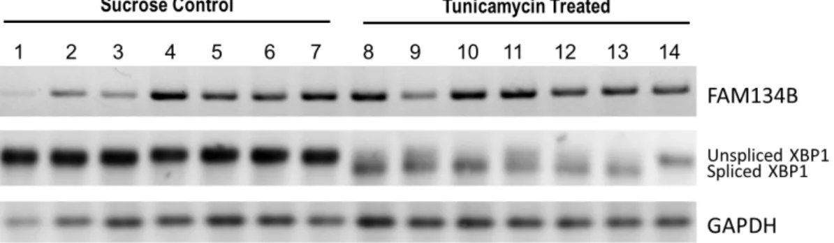
Figure 4.14 : shRNA knockdown of FAM134B is effective, especially in clones 59-11 and -12. ... 48 Figure 4.15: shRNA silencing of FAM134B is also visible at the mRNA level in
clones 59-11 and -12. ... 48 Figure 4.16: FAM134B-silenced Snu449 cells displayed an altered morphology in
culture. ... 49 Figure 4.17: Vimentin levels are not significantly increased in FAM134B
knocked-down Snu449 cells. ... 50 Figure 4.18: FAM134B-silenced Snu449 cells had poorer survival and migratory
capabilities under serum starved conditions that inhibited proliferation. ... 50 Figure 4.19: FAM134B-silenced Snu449 cells have poorer survival capabilities
under serum starvation conditions. ... 51 Figure 4.20: Snu449 cells have a significantly high resistance to thapsigargin. ... 52 Figure 4.21: Silencing of FAM134B causes a dramatic decrease in thapsigargin
resistance of Snu449 cells. ... 53 Figure 4.22: Silencing of FAM134B drastically reduces Adriamycin resistance in
Snu449 cells both in higher ug/ml (top) and lower ng/ml (bottom)
concentrations. ... 54 Figure 4.23: Tunicamycin cytotoxicity is not affected by the silencing of FAM134B.
... 55 Figure 4.24: FAM134B silencing enhances antitumor activity of TGF-β. ... 56 Figure 4.25: FAM134B silencing only slightly enhances EtOH-induced cell death. 56 Figure 4.26: FAM134B silencing does not affect resistance to 5-fluorouracil and
camptothecin induced cytotoxicity. ... 57 Figure 4.27: Silencing of FAM134B does not significantly affect survival rates
under oxidative stress induced by H2O2. ... 58
Figure 4.28: Protein candidates that are predicted to phosphorylate FAM134B isoform 1. ... 59 Figure 4.29: Protein candidates that are predicted to phosphorylate FAM134B
1
1. INTRODUCTION
1.1 Cancers of the Liver; Status Worldwide & in Turkey
Cancer is a leading cause of death worldwide, and although the most common incidences are cancers of the breast, prostate and lung, liver cancers draw attention with the second poorest survival rates, preceded only by cancers of the pancreas, as shown in Figure 1.1 [1] .
Figure 1.1: Mortality-to-incidence ratios of cancers in Turkey and Worldwide.
2
As demonstrated in Figure 1.1, 93% of liver cancer patients do not survive this disease, while this ratio is 86% for lung cancers, and 33 and 29% for cancers of the breast and prostate respectively. These high fatality rates make liver cancer the third most common cause of death from cancers worldwide, causing 600,000 deaths per year, although it is the seventh most common type of cancer [2] [3].
These poor survival rates associated with cancers of the liver are caused by difficulties in timely diagnoses and inoperability of the tumors; most cases are diagnosed when the tumor has passed the localized state and reached the distant stage, [4] and only 10-20% of the tumors can be removed completely by surgery [5]. For the time being, the best treatment for liver cancer is liver transplantation, but it can be offered only to patients with small tumors and is limited by the availability of organ donors [6]. The prognostic outcome is better in more developed countries where more advanced facilities are available for both diagnosis and treatment [3]. A significant ratio of liver cancers is attributable to infection by hepatitis B virus (HBV), for which vaccination is available; the ratio of liver cancers caused by HBV infection is 60% in developing countries and 23% in developed countries [3]. The World Health Organization is promoting a vaccination program, and 177 countries have integrated the HBV vaccine to their national infant immunization schedules as of 2008 [3]. Hepatitis C virus (HCV) infection, on the other hand, accounts for 33% of liver cancers in developing countries and for 20% in developed countries, however, vaccination is not possible thus far [3].
Importantly, despite the implementation of preventative measures such as vaccination programs, the incidence rates of liver cancers are still increasing in many regions, including Central Europe and the United States, and this increase is partly attributable to the increasing rates of obesity [3].
This worrying state draws attention to the mechanisms involved on the contribution of obesity and other non-viral factors on the onset and progression of cancers of the liver; obesity, metabolic syndrome, diabetes, exposure to aflatoxins, and alcohol consumption constitute an aspect of liver cancers for which the endoplasmic reticulum (ER) appears to play key roles, and these will be discussed in detail.
3
Figure 1.2 Histopathological progression and molecular features of HCC. [12]
1.2 Hepatocellular Carcinoma
Hepatocellular carcinoma (HCC) is the most common type of liver cancer, making up 70% to 85% of all primary liver cancers worldwide [3]. Rarer forms of liver cancer include hepatoblastoma, angiosarcoma or cholangiocarcinoma [7].
The liver is one of the major secretory organs of the human body, and hepatocytes constitute approximately 80% of the total mass of this organ; these cells are the main functional cells of the liver, and have essential roles in the synthesis and storage of many proteins, the metabolisms of carbohydrates and lipids, synthesis phospholipids, cholesterol and bile salts, and detoxification of drugs and toxins [8].
Both the dedifferentiation of mature hepatocytes and imperfect differentiation of hepatic progenitor cells (HPCs) appear to contribute to hepatocarcinogenesis [9] during the attempt of the liver to make up for hepatocytes lost by persistent damage; the mechanisms involved will be discussed in further detail.
1.3 Pathogenesis of Hepatocellular Carcinoma
Risk factors leading to hepatocarcinogenesis include viral infections such as Hepatitis B (HBV) and Hepatitis C (HCV) infections, metabolic conditions such as obesity, non-alcoholic fatty liver disease and diabetes, toxic factors such as alcohol
4
and aflatoxins, as well as other factors such as autoimmune hepatitis and hereditary haemochromatosis [10][11][12]. These factors can directly cause DNA damage, mutations and epigenetic irregularities involving oncogenes and tumor suppressor genes, telomerase activation and chromosomal abnormalities. Moreover, persistent damage to hepatocytes also contribute to carcinogenesis through causing hepatocyte death either via apoptosis or via necrosis, which, in turn, leads to the development of chronic liver disease, fibrosis, and cirrhosis [13]. It is important to note that 80% of HCC cases are observed in patients that initially experience chronic liver disease and cirrhosis [14].
As a way to make up for cell death, the liver cells start regenerating, and this involves high rates of cell division and proliferation. As demonstrated in Figure 1.2, this high rate of proliferation leads to telomere shortening and genomic instability [12]. The high rates of DNA replication may also make the cells more prone to errors and the accumulation of mutations.
Most frequently reported molecular causes of aberrant proliferation and dedifferentiation of hepatocytes include; inactivation of tumor suppressors such as p53, retinoblastoma protein (pRb) or the p14ARF and p16INK4a proteins encoded by the CDKN2A gene, overexpression of oncogenes such as CyclinD1/Cdk4, insulin-like growth factor-II or c-MET, and activation of Ras/mitogen activated protein (MAPK), transforming growth factor-β (TGF- β) or Wnt/ β-catenin signaling cascades [15].
1.3.1 Fibrosis as an Initial state of Hepatocarcinogenesis
Damage to hepatocytes may be caused by several factors such as elevated metabolic overload resulting in high levels of reactive oxygen species (ROS) and ER stress, chemical toxicity by drugs, alcohol or aflatoxins, or viral activity causing metabolic deregulation [16].
Steatosis, which involves the expanding of hepatocytes due to excessive lipid droplet storage, is one of the first signs of cellular stress, and is highly reversible. Irreversible or persistent damage, however, may cause hepatocyte death through apoptosis or necrosis. The death-mediated signals trigger the activation of Kupffer
5
and quiescent hepatic stellate cells which coordinate an inflammatory and wound healing response; Kupffer cells are specialized macrophages of the liver, and stellate cells are pericytes involved in formation of scar tissue. As illustrated in Figure 1.3, in an acute setting, this response might lead to tissue regeneration and repair, however, when the injury is chronic, the consequences are fibrogenesis, cirrhosis, and cancer [13].
Fibrosis is the scarring of a tissue due to excessive deposition of extracellular matrix (ECM) proteins as an attempt of repair. During chronic liver injury, the quiescent hepatic stellate cells become activated by signals described in Figure 1.4, such as inflammatory cytokines and give rise to myofibroblasts. Myofibroblasts are the cell type responsible for fibrosis in the liver [13].
As also shown in Figure 1.4, transforming growth factor-β (TGF-β) is considered one of the major pro-fibrogenic cytokines, as demonstrated by data obtained from animal models of liver damage [13]. In fact, TGF-β plays important roles in many stages of liver disease, which will be discussed in detail.
Figure 1.3: The progression of hepatocellular carcinoma & the role of senescence as a means of eliminating fibrotic cells.Adapted from Dooley & Ten Dijke, 2012 [13]
6
Figure 1.4: Molecular mechanisms involved in activation of hepatic stellate cells, fibrosis, and perpetuation into a tumorigenic form.
Friedman, 2010
Although majority of evidence suggests that hepatic stellate cells are the main contributors of the myofibroblast build that forms during fibrogenesis, there is evidence that other cell types such as portal fibroblasts of the liver, bone-marrow-derived fibrocytes, or circulating mesenchymal cells may also contribute to the fibrosis through deposition of extracellular matrix proteins [13] [17].
Interestingly, some research also indicates that hepatocytes and biliary epithelial cells may also contribute to myofibroblast formation during fibrosis of the liver. Hepatocytes and biliary epithelial cells of the liver may transdifferentiate into myofibroblasts through epithelial-to-mesenchymal transition (EMT, which will be explained later in detail) that may be mediated by TGF-β, Hedgehog and bone morphogenic protein 7 (BMP7), and contribute to ECM deposition and fibrosis [17]. Unlike the later stages of cirrhosis, fibrosis of the liver is a state that can be resolved, either through reversal of the fibroblastoid morphology by mesenchymal-to-epithelial transition (MET) [17], or through the elimination of fibrotic cells by senescence and apoptosis [13].
7
1.3.2 Senescence and Hepatocarcinogenesis
Senescence is a term that describes the process of becoming old, and cellular senescence is a phenomenon characterized by permanent cell cycle arrest [14]. Senescence can be identified by markers such as senescence-associated DNA-damage foci, senescence-asssociated heterochromatin foci, senescence-associated β-galactosidase (SABG) and p16INK4A [14].
Senescence can be inspected in three pathways; telomere-dependent senescence, oncogene-induced senescence, and ROS-induced senescence. All three of these pathways lead to senescence by triggering a DNA damage response in the cell (often induced by double strand breaks in the DNA), which activates ATM and ATR kinases, leading to the phosphorylation of p53 by CHK1 and CHK2 kinases. Phosphorylated p53 is released from MDM and stabilized, and can then induce senescence or apoptosis [14]. The form of senescence that is dependent on telomere shortening is also known as “replicative senescence” since telomere length acts as a cell-division counter in this case, as will be described in detail [14].
This cell cycle arrest, followed by apoptosis, may act as a barrier to the progression of HCC by acting as a means of eliminating fibrotic cells [13]. Senescent cells are observed at a ratio of 10% in normal liver, which is elevated to 84% in cirrhosis, and is reduced to 60% in HCC. These rates of senescence are mainly attributable to replicative senescence dependent on telomere shortening of hepatocytes. It was also observed that senescence-inducing proteins such as p16 INK4a and p21Cip1 accumulate in cirrhotic livers. Senescence-escape mechanisms such as telomerase reactivation, or mutations or epigenetic silencing events in senescence-inducing genes such as p53 and p16INK4a play important roles in the progression of HCC [14].
1.3.2.1 Telomere Shortening & Telomere-Dependent Senescence
Human chromosomes contain DNA-protein complexes named “telomeres” at the ends of each chromosome. Since DNA replication cannot initiate from the very ends of linear DNA, important genetic information would be lost by each replicative cycle without the availability of the TTAGGG telomere repeats at the ends of the
8
chromosomes. Therefore, the telomeres prevent genomic instability and the loss of essential genetic information by forming a cap at the ends of the chromosomes [14]. Majority of human somatic cells are incapable of synthesizing new telomeres due to the repression of telomerase reverse transcriptase (TERT) expression, therefore, telomeres become shorter with every cycle of cell division, acting as a “cell cycle counter” which helps determine when the cell should stop replicating. Shortened telomeres begin to lose their protected status, and such loss of telomere protection or any other form of telomere dysfunction leads to the formation of abnormal chromosomal end-to-end fusions through non-homologous end joining or homologous recombination pathways of DNA repair. The triggering of these DNA repair pathways indicate that open-ended telomere DNA is recognized as double strand breaks. This triggers a DNA damage response in the cell, which leads to senescence through the phosphorylation of p53, as previously mentioned, which activates p21cip1, which is an inhibitor of Cyclin-dependent kinase 2 (CDK2). These events keep the tumor suppressing pRb protein in the active form, inhibiting E2F function and the expression of growth-promoting genes [14].
1.3.2.2 ROS-induced and Oncogene-induced Senescence
Reactive oxygen species (ROS) are mostly produced during cellular respiration at the mitochondria. In vitro methods of ROS induction such as mild H2O2 treatment and
culturing under high O2 conditions were shown to induce or aggravate senescence
and cause reduced lifespans. ROS and oxidative stress may give rise to senesce through causing DNA damage or inducing changes in other signaling pathways [14]. Senescence has also been observed in response to the expression of well-studied oncogenes such as Ras, Raf, Myc, Cyclin E, Mek and Mos. In the case of Ras, expression of this oncogene was demonstrated to cause G1 arrest together with elevated levels of cell-cycle arrest associated proteins such as p53 and p16INK4a [14]. These two forms of senescence, like telomere-dependent replicative senescence, work primarily through DNA damage response pathways as well, but they can also induce other signals that lead to the activation of cyclin-dependent kinase inhibitors
9
such as p16INK4a and p15INK4b that inhibit CDK4 and CDK6 to keep the tumor suppressing pRb protein active [14]
1.3.3 Cirrhosis
As mentioned earlier, 80% of HCC cases originate as chronic liver disease and cirrhosis [14]. Although often mistakenly considered simply as an extended form of fibrosis, cirrhosis is, in fact, different from fibrosis in many ways. Cirrhosis is characterized by extensive inflammatory signaling, architectural disruption of the tissue due to excessive secretion of ECM proteins by myofibroblasts originating from stellate cells, aberrantly regenerating or senescent hepatocytes, nodule formation and vascular changes, and is often not reversible [13] [14].
1.4 Epithelial to Mesenchymal Transition and TGF-β
Epithelial cells of the adult human have a distinct apical-basal cell polarity, are often in layers of cells that are held together by cell-to-cell adhesion proteins such as E-cadherin, form a sheet on a basement membrane, and are immobile. Organs of the human body are often made up of organized, immobile epithelial cells that may mainly carry out secretory functions as in the cases of the liver and the pancreas. Mesenchymal cells, on the other hand, have very different phenotypes; they do not express E-cadherin, and are therefore not organized in layers having polarities, and they can migrate easily. Mesenchymal stem cells of the adult human reside in the bone marrow, and give rise to the highly mobile cells of the circulatory and lymphatic systems, as well as bone and cartilage tissues [18].
Epithelial to mesenchymal transition (EMT) is the reversible change of an epithelial cell into a mesenchymal or, in some cases, fibroblastoid phenotype. Epithelial cells that undergo EMT assume a more mesenchymal phenotype, losing their cell polarity and cell-to-cell adhesion characteristics and gaining the ability to transmigrate the
10
basement membrane and connective tissues to move into circulation, and becoming motile and invasive [15].
This switch from an epithelial to a mesenchymal phenotype is a normal part of embryonic development. It initially occurs during the gastrulation stage of the blastula, forming the mesoderm which gives rise to mesenchymal cells that contribute to extraembryonic tissues. Later in development, EMT contributes to the development of the circulatory, nervous and musculoskeletal systems [15].
In the adult, however, EMT usually occurs under pathophysiological circumstances such as chronic inflammation, wound healing response, and cancer progression. During wound healing, epithelial cells obtain reduced adhesiveness and increased migration capabilities during the process of producing new and intact epithelial layers. EMT is also observed during the non-regenerative repair attempt of an organ called fibrosis, which includes the dedifferentiation of epithelial cell types into a more fibroblastoid phenotype, contributing to the wound healing and inflammation response by secreting ECM proteins, and this event has been demonstrated to take part in liver fibrosis as well [15].
Transforming growth factor-β (TGF-β) is a cytokine that functions through Smad2/3 and Smad4 to control gene transcription and induce cellular effects such as growth inhibition, apoptosis, differentiation and extracellular matrix deposition [19]. The ultimate result of TGF-β signaling depends on the activities of other pathways in the cell, such as Ras signaling, and can have complex effects, as will be described.
1.4.1 Epithelial to Mesenchymal Transition and TGF-β in Liver Fibrosis
EMT has been demonstrated to participate significantly in liver fibrosis, with major contributions from the cytokine TGF-β. The first evidence that hepatocytes give rise to fibroblasts by going though EMT during liver fibrosis was provided by a study where EMT was induced in mouse hepatocytes by treatment with TGF-β1, and the hepatocytes were observed to change into fibroblast-specific-protein (FSP)-1 positive hepatic fibroblasts [20]. Another research study demonstrated that hepatocytes isolated from cirrhotic livers, which had been exposed to elevated levels of TGF-β in
11
vivo, had elongated, fibroblastoid-like phenotypes and expressed vimentin and
collagen I [21]. Further research demonstrated that loss of epithelial phenotype in primary mouse hepatocytes in response to TGF-β treatment was associated with upregulation of SNAIL and consequent loss of E-cadherin expression[22] and that this also involved the activation of Smad2/3 signaling and collagen I synthesis [23]. Also very interestingly, it was demonstrated that EMT in primary murine hepatocytes was associated with FAK/Src dependent activation of the PI3K/Akt signaling, which causes resistance to apoptosis, as well as Erk1/2 signaling, which causes induction of EMT, demonstrating that EMT confers resistance against TGF-β-induced apoptosis [24]. This observation may be of particular interest to this study, as the Akt/Erk pathway is known to play key roles in cell survival under ER stress [25], for which the protein studied here, FAM134B may also play key roles, as will be discussed later in detail.
1.4.2 Epithelial to Mesenchymal Transition and TGF-β in the progression of Hepatocellular Carcinoma
TGF-β and EMT appear to play dual roles during the progression of hepatocellular carcinoma; in the healthy liver and during tumor initiation through fibrosis, TGF-β appears to induce cell cycle arrest, apoptosis and the development of a fibroblastoid-like phenotype, and at the neoplastic stage, TGF-β controls dedifferentiation and spreading of the neoplastic hepatocytes, contributing to the development of a poorly-differentiated state and metastatic behavior. [15]
Murine tumor models have demonstrated that a synergy between oncogenic Ras and TGF-β1 signaling causes EMT, malignant progression and metastasis, and that these are associated with loss of E-cadherin and the tight junction protein ZO-1, and activation of Smad2/3 leading to autocrine TGF-β signaling. Further studies showed that the induction phase of EMT requires crosstalk between TGF-β and MAPK, and that the maintenance phase depends on the activation of PI3K/Akt. EMT was also shown to be associated with increased platelet-derived growth factor (PDGF) signaling, independent of the genetic background of EMT, which was demonstrated
12
to cause migration and tumor progression. Interleukin-like EMT inducer (ILE1), which is a downstream target of TGF-β signaling, was shown to collaborate with oncogenic Ras to trigger the activation of STAT3, as well as the nuclear accumulation of β-catenin, via PDGF/ PDGF-R. [15]
Human studies were also supportive of the previous findings; studies with HCC cell lines revealed that elevated expression of the ECM protein laminin (Ln)-5 leads to the activation of Erk1/2, which is required for cell proliferation. In patient studies, 58% of HCC cases demonstrated reduced levels of E-cadherin. Loss of E-cadherin in HCC patients was reported to be accompanied by loss of β-catenin from cell borders and partial translocation to nucleus, as well as elevated levels of the ECM protein Laminin-5. Loss of E-cadherin also significantly correlated with intrahepatic metastasis and poor survival of patients. Also, expression of Twist, a negative regulator of E-cadherin expression, in non-metastatic HCC cells induced EMT and increased invasion. Also interestingly, viral proteins of HCV were also observed to render hepatocytes less sensitive to anti-proliferative effects of TGF-β. [15]
1.5 The Endoplasmic Reticulum
The endoplasmic reticulum (ER) is an organelle made up of a membrane-enclosed network of sacs, with central roles in protein and lipid synthesis, membrane biogenesis, xenobiotic detoxification, cellular calcium storage and the proper folding and modifying of proteins [26]. The ER membrane makes up more than half of the total membrane area of an animal cell, and is the production site of all transmembrane proteins in a cell, including those of the membrane-bound organelles and the plasma membrane. And the ER lumen is where almost all of the secretory proteins and proteins of the ER lumen itself, the Golgi apparatus and lysosomes are delivered for processing, folding and modifications. [27]
13
1.5.1 The Abundance and Functions of Rough and Smooth ER in Hepatocytes
Hepatocytes are the major providers of glucose and lipids to the entire human body, and they can produce millions of proteins per minute, possessing one of the highest rates of protein synthesis in the human body, and the overwhelming majority of these proteins are processed in the ER. [28]
The rough ER, which contains ribosomes on the cytosolic face, is mainly concerned with protein synthesis, and is abundant especially in cells that secrete proteins, for example, in hepatocytes that secrete proteins such as serum albumin or coagulation factors, or pancreatic β-cells that are responsible for insulin secretion. Most proteins synthesized in the rough ER are N-glycosylated and are destined for transport to the plasma membrane, Golgi apparatus, lysosomes or extracellular space, while proteins destined for the cytosol are often not glycosylated. Ca2+ dependent chaperones such as calnexin retain incompletely folded proteins in the ER until they are properly folded, depending on their N-glycosylation status. [27]
The smooth ER, on the other hand, lacks ribosomes, and is mainly concerned with lipid metabolism, carbohydrate metabolism, and detoxification. Smooth ER is very abundant in hepatocytes since these cells play key roles in lipid and carbohydrate metabolism, and are responsible for producing lipoprotein particles and detoxifying lipid-soluble drugs and metabolic byproducts. During detoxification of drugs or toxins such as phenobarbital or ethanol, the smooth ER of hepatocytes can double its surface area within a few days in order to be able to synthesize the unusually large amounts of detoxification enzymes required. [27]
Another fundamental responsibility of the ER is to store Ca2+; the release and uptake of Ca2+ to and from the cytosol is involved in many rapid responses to extracellular signals, [27] and is involved in timing of the exocytosis of synthesized proteins, such as the release of insulin by pancreatic β-cells. The ER membrane contains an ATP-dependent channel that pumps Ca2+ from the cytosol into the ER lumen, generally named as sarco-endoplasmic reticulum Ca2+ ATPases (SERCA).
Calcium homeostasis is especially important for liver cells; Hotamisligil et al. have recently reported its implications in obesity by demonstrating that aberrant lipid
14
metabolism disrupts calcium homeostasis and causes liver endoplasmic reticulum stress in obesity [26]. Furthermore, recent research has shown that Ca2+ gradients are also important for the coating and uncoating of transport vesicles that are trafficked between the ER and the Golgi apparatus [29].
1.5.2 Endoplasmic Reticulum Stress and the Unfolded Protein Response
A sudden high demand of protein synthesis, inducers of oxidative stress, or some other disruption of ER homeostasis can cause the accumulation of misfolded proteins in the ER, and this triggers the unfolded protein response (UPR). The accumulation of unfolded proteins in the ER is sensed by the binding immunoglobulin protein/glucose-regulated protein 78 (BiP/GRP78). Upon being sequestered by misfolded proteins, BiP/GRP78 dissociates from the three known ER-membrane transducers, inositol requiring 1α (IRE1α), PKR-like ER kinase (PERK), and activating transcription factor 6α (ATF6α), allowing their activation. These three transducers initiate a response that aims to alleviate ER stress by reducing the load of new peptides entering the ER, increasing the folding capacity of the ER and degrading misfolded proteins. [30]
PERK phosphorylates eukaryotic initiation factor 2 alpha (eIF2α), which attenuates global mRNA translation, aiming to alleviate some of the peptide burden on the ER. PERK also selectively increases the translation of ATF4, which is a transcription factor that induces the expression of chaperones, amino acid transporters, antioxidant stress response genes, ER-associated degradation (ERAD) proteins and C/EBP homologous protein (CHOP), which can cause apoptosis if ER stress persists. [30] IRE1α has kinase and RNAse activities; its autophosphorylation activates the RNase activity to splice X-box binding protein 1 (XBP1) mRNA, allowing the production of the active transcription factor sXBP1. sXBP1 upregulates ER chaperones, ERAD proteins, and CHOP. IRE1a activation also facilitates mRNA degradation and translation attenuation, in collaboration with PERK. IRE1α also recruits and activates C-jun N-terminal kinase (JNK), which is a stress kinase that can mediate apoptosis under persistent ER stress. [30]
15
ATF6α released from BiP/GRP78 transports to the Golgi apparatus, where it is cleaved and activated by intramembrane proteolysis. The activated transcription factor ATF6α heterodimerizes with and activates sXBP1 to induce the transcription of chaperones, ERAD proteins and CHOP. [30]
Unresolved ER stress results in apoptosis through mechanisms involving CHOP, JNK, Bcl-2 family proteins, calcium and redox homeostasis, and caspase activation [30]. For instance, apoptosis induced by ER stress has been shown to involve disruption of calcium homeostasis and protein accumulation, which leads to the calpain-mediated activation of caspase-12, which is localized to the ER [31]. In fact, disruption of calcium homeostasis appears to be very important for the link between ER stress and apoptosis. Overexpression of the anti-apoptotic protein Bcl-2 was shown to reduce free ER calcium and cell death, while reduction of the pro-apoptotic proteins Bak and Bax resulted in lower free ER calcium and reduced sensitivity to apoptotic stimuli such as hydrogen peroxide treatment. [30]
1.5.3 Endoplasmic Reticulum Stress, Obesity and Liver Disease
Hepatocytes, pancreatic exocrine cells and adipoctyes constitute a triad of cells that have very important metabolic functions, have high protein synthesis rates, and are therefore very sensitive to ER stress brought on by disruptions of metabolic homeostasis, such as excess nutrients, insulin peaks, and obesity. [28]
Obesity is a risk factor for non-alcoholic fatty liver disease, type 2 diabetes, and hepatocellular carcinoma, which are all interconnected. In mouse models, obesity has been demonstrated to induce ER stress in liver and adipose tissues of mice. A high demand on adipocytes to store more lipids than they can handle causes production of reactive oxygen species (ROS), inflammation, ER stress, apoptosis, and increased fatty acid (FA) release. The excess circulating FAs are taken up by the liver, and cause a lipotoxicity that alters glucose utilization and insulin action, and results in steatosis of hepatocytes. In the livers of obese mice, ATF6α expression was observed to be reduced, and its reconstitution was observed to improve glucose homeostasis. [32]
16
The UPR influences metabolic processes in complex ways, as described in Figure 1.5. In hepatocytes, XBP1 induces increased FA synthesis, while activation of ATF6α inhibits the induction of lipogenic genes by sterol regulatory element-binding protein (SREBP), however, mouse models lacking hepatic ATF6α showed increased steatosis regardless of the reduced levels of lipogenesis, indicating that more complex mechanisms were involved. ATF6α also has an inhibitory effect on gluconeogenesis, modulating the activity of the key transcriptional regulator cAMP-response element binding protein (CREB). [32] Also interestingly, liver samples
from patients with non-alcoholic fatty liver disease have demonstrated elevated BiP expression and eIF2α phosphorylation. Genetic modulation studies on the pathways involving eIF2α phosphorylation, IRE1α/XBP1, and ER protein translocation have linked ER stress and hepatocyte steatosis. [30] Also, CHOP has been implicated in hepatocyte death as a mediator of decreased gene expression under severe ER stress [32]
Several damaging agents that disrupt ER homeostasis, such as drugs, alcohol, lipids, and HBV and HCV have been shown to cause ER dysfunction through mediators such as ROS that cause oxidative stress, altered membrane lipid composition, and Figure 1.5: Metabolic causes and consequences of ER stress and the UPR. Hotamisligil, 2010 [32]
17
formation of protein aggregates, which can either be resolved or have more serious consequences such as inflammation, steatosis, and apoptosis of hepatocytes [30].
1.5.4 ER Stress, Epithelial to Mesenchymal Transition and Src Kinase
It is also important to note that sustained ER stress and EMT appear to be closely related. Recent studies on thyroid and alveolar epithelial cells have demonstrated that induction of ER stress by chemical inducers such as thapsigargin and tunicamycin resulted in epithelial to mesenchymal transition as demonstrated by loss of epithelial markers such as E-cadherin, and increase of mesenchymal markers such as α–smooth muscle actin (α-SMA), as well as morphological changes. Both studies also utilized the PP2 inhibitor of Src-family kinases to demonstrate that the induction of dedifferentiation and EMT by ER stress was mediated by the Src pathway. [33] [34] Src is a non-receptor proto-oncogene tyrosine protein kinase that is hyperactive in many cancers and is a drug target for tyrosine kinase inhibitors such as dasatinib.
1.6 FAM134B in Literature
A review article published in EMBO Reports in 2010, titled “Further assembly required: construction and dynamics of the endoplasmic reticulum network” draws attention to the family with sequence similarity 134 (FAM134) family of proteins FAM134A, FAM134B and FAM134C, and points out that these proteins may constitute a new family of organelle-shaping proteins since each of the FAM134 proteins have a pair of long hydrophobic segments similar to those found in reticulons and DP1/REEPs/Yop1. [35] Reticulons are a class of membrane proteins shaping the tubular endoplasmic reticulum, primarily functioning in promoting membrane curvature. Reticulons interact with DP1/Yop1p, another tubular ER membrane protein, and their deficiency disrupts tubular ER. [36]
A 2009 publication in Nature Genetics by Kurth et al. established that loss-of-function mutations in FAM134B cause hereditary sensory and autonomic neuropathy
18
type II (HSANII), which involves severe mutilations due to diminished sensory perception and compromised regulation of involuntary functions of the body. The FAM134B protein was shown to co-localize with the cis-Golgi protein giantin, and was therefore defined as a Golgi protein. Using primary dorsal root ganglion neurons, it was revealed that knockdown of FAM134B results in a ~40% reduction in the size of the cis-Golgi compartment as well as apoptosis. Analyses on multiple adult murine tissues showed that FAM134B was most highly expressed in the testis, followed by the dorsal root ganglion, while the liver had much lower expression levels. [37]
In 2007, Tang et al. published a study entitled “Oncogenic properties of a novel gene JK-1 located in chromosome 5p and its overexpression in human esophageal squamous cell carcinoma.” JK-1 is an alias for FAM134B isoform 2, and this study established that elevated levels of JK-1 is associated with esophageal squamous cell carcinoma (ESCC) and can transform normal cells. They observed that 30% (9/30) of ESCC tumors , and 69% (9/13) of ESCC cell lines had elevated levels of JK-1. They also demonstrated that overexpression of JK-1 promoted anchorage dependent and anchorage independent proliferation of benign NIH3T3 and HEK293 cells. Moreover, subcutaneous injections of NIH3T3 cells overexpressing JK-1 formed sarcomas in all three mice. [38]
Another interesting publication from 2011 assembled gene expression data from pluripotent stem cells and non-pluripotent cells of the mouse, and identified FAM134B as a biomarker of pluripotent stem cells in mice, and suggested that FAM134B may constitute a missing link between pluripotency and some unidentified surface markers processed by the Golgi apparatus. However, it is not clear from this paper whether FAM134B is overexpressed or underexpressed in pluripotent cells [39]
FAM13B initially caught attention in our laboratory as a possible senescence-associated gene, as its levels were elevated in senescent Huh7 clones compared to parental and immortal Huh7 clones. Further studies implicated that FAM134B is not a cause but a result of senescence. It was also identified to co-localize with the ER protein Calnexin. FAM134B protein levels were also shown to be elevated in the
19
more mesenchymal-like, poorly-differentiated HCC cell lines compared to the more epithelial-like, well-differentiated ones, and we hypothesized that this could be associated with the elevated ER stress status of the more mesenchymal-like cell lines. [40][41] (Nilgün Taşdemir, Mustafa Yılmaz and Mehmet Öztürk, unpublished data)
20
2. OBJECTIVES AND RATIONALE
Hepatocellular carcinoma constitutes 80% of liver cancers, which is the cancer type with the second poorest survival rates worldwide [3]. Risk factors for hepatocellular carcinoma include alcohol consumption and HBV or HCV infections, in addition to obesity, non-alcoholic fatty liver disease and diabetes, all of which appear to have devastating impacts on the endoplasmic reticulum and the cell, as described in the introductory section.
FAM134B initially caught attention in our lab in comparative analyses, having elevated levels in senescent Huh7 clones compared to parental and immortal Huh7 clones. Further analyses showed that it may be localized on the ER, and that its levels are elevated in the more mesenchymal-like HCC cell lines, which also have elevated ER stress status. [40]
An EMBO report published in 2010 drew attention to the FAM134B protein as a possible organelle shaping, reticulon-like protein of unknown function that is yet to be placed in the ER network [35]. And a 2009 publication in Nature identified FAM134B as a cis-Golgi protein for which loss-off function mutations cause neuropathy [37].
This research aims to identify the function of the FAM134B protein, with special focus on its role in hepatocellular carcinoma.
21
3. MATERIALS AND METHODS
3.1 MATERIALS
3.1.1 General Laboratory Reagents and Kits
General laboratory reagents, chemicals and kits utilized during the course of this thesis work were all laboratory-grade commercial products purchased from major companies. Tunicamycin, ready-made Bradford reagent, Bovine Serum Albumin (BSA), haematoxylene dye, agarose, ethanol and methanol were bought from Sigma-Aldrich (St. Louis, MO, USA). Sulforhodamine B (SRB) dye was in powder form and also obtained from Sigma-Aldrich (St. Louis, MO, USA). Thapsigargin was purchased from Santa Cruz Biotechnology Inc. (Santa Cruz, CA, USA). TGF-β1 was purchased from R&D Systems (Minneapolis, USA). Nucleospin RNA II total RNA isolation kit was purchased from Macherey-Nagel (Duren, Germany). TRIsure RNA isolation reagent used for tissues was purchased from Bioline (Taunton, USA). Ponceau dye and cell culture grade dimethyl sulfoxide (DMSO) were obtained from Applied Biochemia (Darmstadt, Germany). ECL prime detection reagent and Nitro-cellulose western blot membranes were purchased from Amersham GE Lifesciences (Pittsburgh, PA, USA). For immunoperoxidase staining, the DAKO EnVision+ system was used, which included blocking solution and secondary antibody mix (Glostrup, Denmark).
3.1.2 Cell Culture Solutions and Materials
Dulbecco’s modified Eagle’s medium (DMEM) and Roswell Park Memorial Institute (RPMI) cell culture mediums, fetal bovine serum (FBS), penicillin/streptomycin antibiotics, L-glutamine and trypsin-EDTA were purchased from GIBCO (Invitrogen, Carlsbad, CA, USA). The plastic items used in cell culture such as cell
22
culture plates, flasks and serological pipettes were obtained from Corning Life Sciences Inc. (USA).
3.1.3 Electrophoresis Apparatus
The SDS-PAGE gel electrophoresis equipment, including the tanks, the gel preparation glasses, power supplies and other plastic materials were purchased from BIO-RAD Laboratories (CA, USA). Agarose gel electrophoresis apparatus and NanoDrop spectrophotometry apparatus to measure nucleic acid concentration were obtained from Thermo Scientific (Wilmington, USA). Protein concentration was determined using Bradford reagent which was measured using the spectrophotometer Beckman Du640 from Beckman Instruments Inc. (CA, USA).
3.1.4 cDNA Synthesis and Polymerase Chain Reaction Reagents
All reagents required for cDNA synthesis from total mRNA and semi-quantitative polymerase chain reaction (PCR) were obtained from Fermentas (MBI Fermentas, Germany), including RevertAid First strand cDNA synthesis kit, Taq DNA polymerase, 10X Taq DNA polymerase Buffer (+(NH4)2SO4,–MgCl2), 2mM dNTPs,
25mM MgCl2 and DNA molecular weight ladders.
3.1.5 Primers
Primers were ordered from Sentromer (Istanbul, Turkey).
Primer Sequence Tm
mmFAM134B isoform 1 For. AGC TCC TGA GCT GGA AGA GG 61 mmFAM134B isoform 1 Rev. TCA TGA CGG AAA TGA GGT GA 55 mmFAM134B isoform 2 For. GCT GTG GAC CAG GCA GAA G 61 mmFAM134B isoform 2 Rev. AAA TTC ATC CAT GAT TCT GCA A 53 mmXBP-1spl.unspl. For. AGT TAA GAA CAC GCT TGG GAA T 57
23
mmXBP-1 spl.unspl. Rev. AAG ATG TTC TGG GGA GGT GAC 60
hFAM134B isoform 1 For. CAAGAGGTGCACAGTTGTGGAGAA 58
hFAM134B isoform 1 Rev. GCAACCGTGAGGCTAATCTTAGGA 58
hFAM134B isoform 2 For. CTCGAGAAGCTTATGCCTGAAGGTGAA
GACTT 58
hFAM134B isoform 2 Rev. GCAACCGTGAGGCTAATCTTAGGA 58
hXBP-1 For. TTACGAGAGAAAACTCATGGCC 58
hXBP-1 Rev. GGGTCCAAGTTGTCCAGAATGC 62
hGAPDH For. GGCTGAGAACGGGAAGCTTGTCAT 60
hGAPDH Rev. CAGCCTTCTCCATGGTGGTGAAGA 60
hVimentin For. CGTCACCTTCGTGAATACCA 60
hVimentin Rev. CCAGAGGGAGTGAATCCAGA 60
Table 3-1: Primer sequences and Tm values.
3.1.6 Antibodies
Antibody Company and Catalog Number Dilution
FAM134B Sigma, HPA026906 1:2500
Phospho-eIF2α Invitrogen, 44728G 1:1000
Vimentin Dako, M7020 1:500
Calnexin Sigma, C4731 1:5000
α-tubulin Calbiochem, CP06 1:4000
actin Santa Cruz, sc-1616-R 1:5000
anti-mouse-HRP Sigma, A0168 1:5000
anti-rabbit-HRP Sigma, 6154 1:5000
24
3.2 SOLUTIONS AND MEDIA
3.2.1 General solutions
10X Phosphate Buffered Saline (PBS)
80g NaCl, 2g KCl, 14.4g Na2HPO4, 2.4g KH2PO4 in 1
liter ddH2O
50X Tris Acetate EDTA (TAE)
242 g Tris base, 57.1 ml glacial acetic acid, 18.6 EDTA in 1 liter ddH20
6X DNA Loading Dye 10mM Tris-HCl (pH 7.6), 0.03% bromophenol blue, 0.03% xylene cyanol, 60% glycerol, 60mM EDTA (0.5M pH8.0)
Ethidium Bromide 10 mg/ml dissolved in ddH2O (stock)
1:10.000 working dilution (1µl per 10ml of gel)
3.2.2 Tissue culture solutions
Complete Medium 500ml DMEM/RPMI medium, 10% filtered and inactivated fetal bovine serum, 1% penicillin / streptomycin, 1% non-essential amino acids, 1% L-glutamine. Stored at 4 C
Freezing Medium 20% FBS and 7% DMSO added to complete medium
10X Phosphate buffered saline (PBS)
80g NaCl, 2g KCl, 14.4g Na2HPO4, 2.4g KH2PO4 in 1
liter ddH2O, working dilution: 1X, autoclaved and
25
3.2.3 RIPA Lysis Buffer for Protein Extraction
The RIPA lysis buffer for protein extraction was prepared in advance excluding the protease inhibitor and NaF and NaVO4 which serve as phosphatase inhibitors, and
stored in 4 C as stock. Protease inhibitor and the phosphatase inhibitors were added fresh before use, and this solution could only be stored up to 3 months at -20 C.
Stock Concentration Final Concentration For 1ml For 50ml stock ddH2O 730µl 36.5ml NaCl 2M 150mM 75 µl 3.750ml NP-40/TritonX 1% 10 µl 500 µl Sodium DOC 10% 0.5% 50 µl 2.500ml SDS 10% 0.1% 10 µl 500 µl Tris-HCl pH8 2M 50mM 25 µl 1.250ml Protease Inhibitor 25X 1X 40 µl * added fresh
NaF 1M 50mM 50 µl * added fresh
NaVO4 100mM 1mM 10 µl * added fresh
26
3.2.4 Sodium Deodecyl Sulphate–Polyacrylamide Gel Electrophoresis (SDS-PAGE) Gels and Solutions for Western Blotting
10% Running Gel (7mls recipe for 1 gel)
2.66ml ddH2O, 2.38ml 30% Acrylamide Mix, 1.82ml 1.5M
Tris-HCl pH8.8, 70 µl 10% SDS, 70µl 10% Ammonium Per Sulfate, 2.8µl TEMED
5% Stacking Gel (3mls recipe for 1 gel)
2.1ml ddH2O, 500µl 30% Acrylamide Mix, 380µl 1M
Tris-HCl pH6.8, 30µl 10% SDS, 30µl 10% Ammonium Per Sulfate, 3µl TEMED
10X SDS Running buffer
144g glycine, 30g Tris were dissolved in dH2O, 50ml 10%
SDS was added, and the volume was completed to 1 L. Working solution is 1X.
10X Transfer buffer 72g glycine, 58g Tris were dissolved in dH2O, 2ml 10%
SDS was added, and the volume was completed to 1 L. Working solution is 1X containing 15-20% EtOH
Protein Loading Dye 5% 14.3M β-mercaptoethanol was added to protein loading dye before use
10X Tris buffered saline (TBS)
12.19g trisma base and 87.76g NaCl were dissolved in 1 L of ddH2O, and pH is adjusted to 8.
TBS-T (0.2%) 10X TBS diluted to 1X working solution, and 2:1000 Tween added
Coomassie brilliant blue solution
100mg coomassie brilliant blue G-250, 50ml 95% ethanol, 100ml 85% phosphoric acid. Filtered using whatman paper Ponceau solution 0.1% (w/v) Ponceau S and 5% (v/v) acetic acid in ddH2O
Blocking solution 5% (w/v) non-fat dry milk or 5% (w/v) Bovine Serum Albumin (BSA) dissolved in TBS-T
27
3.2.5 Immunoperoxidase staining solutions
Fixation 1:1 Acetone:Methanol
Washing solution PBS-T; 0.2% Tween 20 in 1X PBS Antibody dissolved in 2% FBS in 0.2% PBS-T
3.2.6 Sulforhodamine B (SRB) staining solutions
10% TCA 10% (wt/vol) trichloroacetic acid in ddH2O
10X (4%) SRB Stock 4g SRB powder dissolved in 100ml 1% acetic acid, stored at room temperature, working dilution is 1X
Tris Base Solution 10mM Tris base solution was prepared with no pH adjustment
1% acetic acid 1% (vol/vol) acetic acid in ddH2O
3.3 METHODS
3.3.1 Mouse Experiments
3.3.1.1 Tunicamycin Treatment of the Mice
14 C57 black 6 (C57BL/6) laboratory mice of around 20g of body weight, fed with normal diets were used for these experiments. The 14 mice were separated into 2 groups of 7; one was the sucrose control group, the other was the tunicamycin treatment group. The tunicamycin treated group was injected with 1ug/g body weight of tunicamycin; the tunicamycin solution was prepared at a concentration of
28
100ug/ml, therefore, a mouse of 20grams body weight received an injection of 200ul. The tunicamycin was dissolved in a 150mM sucrose solution which was prepared in physiological saline. The sucrose control group was injected with 150mM sucrose solution at a volume of 10ul/g body weight, again equaling a 200µl injection for a 20g mouse. The mice were maintained for 8 hours under normal conditions during the course of the 8 hours of tunicamycin treatment.
3.3.1.2 Collection of Liver Tissue from Mice
The mice were sacrificed and liver tissues were collected. Each liver sample was divided into 3 sections; one for RNA isolation, one for protein isolation, and one for immunohistochemistry (IHC). The sections reserved for RNA and protein isolation were immediately frozen in liquid nitrogen and stored at -80 C. The sections reserved for IHC were fixed in 4% formaldehyde for one day and then paraffinated and stained with Hematoxylin and eosin method by the Animal Facility technicians.
3.3.1.3 RNA Isolation from Mice Liver Tissues
The TRIzol or TRIsure method was used to isolate RNA from tissues; all steps were performed on ice in the fume hood. A section of 50-100mg of each tissue was put in a round bottom tube containing 1.5mls of TRIsure, the mixture was homogenized using a sterilized homogenizer, which was cleaned with ddH2O and sterilized with
ethanol between each sample. The contents were pipeted to a 2ml eppendorf, and 300µl chloroform was added, and they were mixed vigorously by shaking up and down for 15 seconds. After 3 minutes at room temperature, they were centrifuged at 15000rpm for 15 minutes, at 4 C. The clear top layer of supernatant was taken into a new eppendorf without touching the white interphase that contained cell waste. 750µl isopropanol was added and mixed slowly. After 10 minutes at room temperature, the mix was centrifuged at 14000rpm for 10 minutes at 4 C. The isopropanol was removed carefully, keeping the RNA pellet. The RNA pellet was washed twice with ethanol, for 8 minutes at 8000rpm at 4 C. The pellet was then dried under the fume hood and dissolved in RNAse DNAse free water, and stored at -80 C.
29
3.3.1.4 Protein Isolation from Mice Liver Tissues
The following steps were performed on ice. A 50-100ug section of each tissue was homogenized in 500µl RIPA buffer, then kept on ice and vortexed every 5 minutes for 30 seconds. The samples were then sonicated and centrifuged at 11000rpm for 30 minutes, at 4 C. The supernatants were taken into new eppendorfs, and the centrifugation step was repeated two more times in order to get rid of the lipids that stick to the eppendorf during centrifugation. The samples were stored at -80 C.
3.3.2 Tissue culture methods
3.3.2.1 Culturing of HCC Cell Lines
Huh-7, PLC, Hep40 and Hep3B cell lines were cultured in complete DMEM medium while Snu387, Snu398, Snu449, and Snu475 were cultured in complete RPMI medium. Complete mediums contained 10% inactivated and filtered fetal bovine serum, 1% penicillin/streptomycin antibiotics, 1% L-glutamine, and 1% non-essential amino acids. The cells were maintained in incubators at 37°C with filtered air circulation containing 5% carbon dioxide. Cells were passaged before reaching full confluency in order to prevent cell cycle arrests. All liquids such as mediums and PBS that come in contact with the cells were heated to 37 C in the water bath before use, and all equipment and solutions utilized were sterile.
3.3.2.2 Cell Passaging
In order to passage the cells, the medium was aspirated and the cells were washed once with PBS. The cells were then trypsinized using 500-2000µl of trypsin depending on size of the plate or flask, and incubated at 37 C for 1-2 minutes to activate the trypsin and detach the cells. The cells were collected from the plates using complete medium, and pipeted well to dissociate any clumps. The required amount of cells was transferred to a new dish in fresh medium, the amount of which is determined according to the dish or flask used.
30
3.3.2.3 Cell Thawing
In order to start a new cell culture from a cryopreserved stock in liquid nitrogen or -80 C freezer, the vial was first only briefly kept on ice or at -20 C. It was then quickly thawed in the 37 C water bath for a few minutes, and transferred to a 15ml falcon which contained 10mls of medium warmed to 37 C, and mixed well. The falcon was then centrifuged at 1500 rpm for 4 minutes and the medium was aspirated to remove any DMSO from cryopreservation. The dish or flask size to be used was determined according to the size of the cell pellet. The pellet was dissolved in new complete medium and transferred to a new dish or flask, and was dispersed well by moving the plate side to side and back and forth. The cells were then incubated at 37 C and 5% carbon dioxide, and checked regularly to change the medium or be passaged before reaching confluency.
3.3.2.4 Cell Cryopreservation
Cells were cryopreserved when they reach 70-80% confluency since cells that are too confluent can go into growth arrest. Cells were washed with PBS, trypsinized, collected into falcons and centrifuged at 1500 rpm for 4 minutes, and medium was aspirated. The cell pellet was then resuspended in freezing medium with 20% FBS and 7% DMSO and pipeted well to ensure there were no clumps. The number of vials to preserve depended on pellet size, and each vial was prepared to contain around enough cells to fill a 10mm dish, resuspended in 1.5-2mls of freezing medium. The vials were initially cooled for 1hour at 20°C and then transferred to -80°C and liquid nitrogen for preservation.
3.3.2.5 Cell Treatments
The cells were plated in the suiting plates, dishes or flasks in specific numbers that aimed the control cells to reach a confluency of 80-90% by the end of the total treatment period. 24 hours after seeding, the mediums were changed to contain the desired concentration of the chemical in complete medium, prepared fresh. Mediums used for the control samples contained the solvent of the drug, such as DMSO.

![Figure 1.2 Histopathological progression and molecular features of HCC. [12]](https://thumb-eu.123doks.com/thumbv2/9libnet/6002138.126315/17.892.186.767.679.971/figure-histopathological-progression-molecular-features-hcc.webp)


![Figure 1.5: Metabolic causes and consequences of ER stress and the UPR. Hotamisligil, 2010 [32]](https://thumb-eu.123doks.com/thumbv2/9libnet/6002138.126315/30.892.180.790.388.710/figure-metabolic-causes-consequences-er-stress-upr-hotamisligil.webp)