Report
De Novo Insertions and Deletions of Predominantly
Paternal Origin Are Associated with Autism Spectrum
Disorder
Graphical Abstract
Highlights
De novo frameshift indels are associated with ASD with an odds
ratio of 1.6
Multiple de novo indels in
KMT2E and RIMS1 implicate these
genes in ASD
88% of de novo indels arise on the paternal chromosome
Synaptic function, chromatin modification, and FMRP targets
play key roles in ASD
Authors
Shan Dong, Michael F. Walker, ..., Liping
Wei, Stephan J. Sanders
Correspondence
[email protected] (M.W.S.),
[email protected] (L.W.),
[email protected] (S.J.S.)
In Brief
Insertions and deletions (indels) have
proven especially difficult to detect in
exome sequencing data. Dong et al. now
identify indels in exome data for 787
autism spectrum disorder (ASD) families.
They demonstrate association between
de novo indels that alter the reading frame
and ASD. Furthermore, by observing
clustering of indels in unrelated
pro-bands,
they
uncover
two
additional
ASD-associated genes:
KMT2E (MLL5),
a chromatin regulator, and
RIMS1, a
regu-lator of synaptic vesicle release.
Dong et al., 2014, Cell Reports 9, 16–23 October 9, 2014ª2014 The Authors
Cell Reports
Report
De Novo Insertions and Deletions
of Predominantly Paternal Origin Are Associated
with Autism Spectrum Disorder
Shan Dong,1,2Michael F. Walker,3Nicholas J. Carriero,4Michael DiCola,5A. Jeremy Willsey,2,3Adam Y. Ye,1,6
Zainulabedin Waqar,7Luis E. Gonzalez,7John D. Overton,8,9Stephanie Frahm,5John F. Keaney III,10Nicole A. Teran,7 Jeanselle Dea,3Jeffrey D. Mandell,3Vanessa Hus Bal,3Catherine A. Sullivan,7Nicholas M. DiLullo,7Rehab O. Khalil,3,11 Jake Gockley,2Zafer Yuksel,12Sinem M. Sertel,13A. Gulhan Ercan-Sencicek,14Abha R. Gupta,7,15Shrikant M. Mane,8 Michael Sheldon,16Andrew I. Brooks,5Kathryn Roeder,17,18Bernie Devlin,19Matthew W. State,2,3,7,20,*Liping Wei,1,6,* and Stephan J. Sanders2,3,*
1Center for Bioinformatics, State Key Laboratory of Protein and Plant Gene Research, School of Life Sciences, Peking University, Beijing 100871, People’s Republic of China
2Department of Genetics, Yale University School of Medicine, New Haven, CT 06520, USA 3Department of Psychiatry, University of California, San Francisco, San Francisco, CA 94158, USA
4Biomedical High Performance Computing Center, W.M. Keck Biotechnology Resource Laboratory, Department of Computer Science, Yale University, New Haven, CT 06520, USA
5Bionomics Research and Technology, Environmental and Occupational Health Sciences Institute, Rutgers University, 170 Frelinghuysen Road, Piscataway, NJ 08854, USA
6National Institute of Biological Sciences, Beijing 102206, People’s Republic of China 7Child Study Center, Yale University School of Medicine, New Haven, CT 06520, USA
8Yale Center for Genomic Analysis, Yale University School of Medicine, New Haven, CT 06520, USA 9Regeneron Genetics Center, 777 Old Saw Mill River Road, Tarrytown, NY 10591, USA
10Department of Chronic Disease Epidemiology, Yale School of Public Health, New Haven, CT 06520, USA 11Department of Research on Children with Special Needs, National Research Center, Cairo 11787, Egypt 12Department of Medical Genetics, Gulhane Military Medical Academy, Ankara 06010, Turkey
13Department of Molecular Biology and Genetics, Bilkent University, Ankara 06800, Turkey
14Department of Neurosurgery, Yale Neurogenetics Program, Yale University School of Medicine, New Haven, CT 06520, USA 15Department of Pediatrics, Yale University School of Medicine, New Haven, CT 06520, USA
16Department of Genetics and the Human Genetics Institute, Rutgers University, 145 Bevier Road, Room 136, Piscataway, NJ 08854, USA 17Department of Statistics, Carnegie Mellon University, Pittsburgh, PA 15213, USA
18Ray and Stephanie Lane Center for Computational Biology, Carnegie Mellon University, Pittsburgh, PA 15213, USA 19Department of Psychiatry, University of Pittsburgh School of Medicine, Pittsburgh, PA 15213, USA
20Department of Psychiatry, Yale University School of Medicine, New Haven, CT 06520, USA
*Correspondence:[email protected](M.W.S.),[email protected](L.W.),[email protected](S.J.S.)
http://dx.doi.org/10.1016/j.celrep.2014.08.068
This is an open access article under the CC BY-NC-ND license (http://creativecommons.org/licenses/by-nc-nd/3.0/).
SUMMARY
Whole-exome
sequencing
(WES)
studies
have
demonstrated the contribution of de novo
loss-of-function single-nucleotide variants (SNVs) to autism
spectrum disorder (ASD). However, challenges in
the reliable detection of de novo insertions and
dele-tions (indels) have limited inclusion of these variants
in prior analyses. By applying a robust indel
detec-tion method to WES data from 787 ASD families
(2,963 individuals), we demonstrate that de novo
frameshift indels contribute to ASD risk (OR = 1.6;
95% CI = 1.0–2.7; p = 0.03), are more common in
fe-male probands (p = 0.02), are enriched among genes
encoding FMRP targets (p = 6
3 10
9), and arise
pre-dominantly on the paternal chromosome (p < 0.001).
On the basis of mutation rates in probands versus
unaffected siblings, we conclude that de novo
frame-shift indels contribute to risk in approximately 3% of
individuals with ASD. Finally, by observing clustering
of mutations in unrelated probands, we uncover two
ASD-associated genes:
KMT2E (MLL5), a chromatin
regulator, and
RIMS1, a regulator of synaptic vesicle
release.
INTRODUCTION
Autism spectrum disorder (ASD) is a highly heritable neurodeve-lopmental syndrome of unknown etiology. An excess of de novo copy-number variants (CNVs) in affected individuals is well es-tablished (Levy et al., 2011; Sanders et al., 2011; Sebat et al., 2007). Moreover, whole-exome sequencing (WES) studies have demonstrated that de novo loss-of-function (LoF) single-nucleotide variants (SNVs) also carry a significant risk for ASD (Iossifov et al., 2012; Neale et al., 2012; O’Roak et al., 2012; Sanders et al., 2012). Importantly, the observation of multiple
de novo events at the same locus provides a reliable and statis-tically rigorous method to identify specific variations associated with ASD (Sanders et al., 2011, 2012; Willsey et al., 2013). This approach has highlighted the contribution of CNVs at 16p11.2, 15q11.2-13, 22q11.2, 7q11.23, and NRXN1, and (to date) SNVs in nine genes: ANK2, CHD8, CUL3, DYRK1A, GRIN2B, KATNAL2, POGZ, SCN2A, and TBR1.
Although the above-cited works and similar studies have been critically important in outlining the genomic architecture of ASD (Buxbaum et al., 2012), they have not provided a comprehensive view of de novo variation in ASD. For example, systematic anal-ysis of de novo insertions and deletions (indels) in WES data has been hindered by technological limitations, including mapping errors and ambiguities in annotation leading to low sensitivity or infeasible numbers of confirmations.
In this work, we resolved the most pressing issues in the detection of de novo indels by combining a family-based local realignment approach (Albers et al., 2011) with empirically derived quality metric thresholds to dramatically improve the ac-curacy of de novo indel prediction. We applied this approach, followed by comprehensive de novo indel confirmation, to previ-ously analyzed WES data from 2,963 individuals in 787 families in the Simons Simplex Collection (SSC;Table S1), allowing a reli-able analysis of the mutation rate in probands versus unaffected siblings. We identified 44 de novo coding indels and observed a significant excess of de novo frameshift indels in probands versus unaffected siblings with an odds ratio (OR) of 1.6, similar to that observed for de novo LoF SNVs. These additional data al-lowed us to refine our prior analysis of the contribution of de novo disruptive events to ASD population risk. We now estimate that approximately 7% of affected individuals (4% with a de novo LoF SNV and 3% with a de novo frameshift indel) carry a de novo disruptive coding mutation that contributes to ASD. More-over, using our previously described approach to assess the sig-nificance of clustering of de novo events at genomic loci (Sanders et al., 2011, 2012; Willsey et al., 2013), we identified two ASD-associated genes: Lysine (K)-specific methyltransfer-ase 2E (KMT2E, a.k.a. Mixed-lineage leukemia 5 [MLL5]) and Regulating synaptic exocytosis 1 (RIMS1), reinforcing prior find-ings highlighting a role for chromatin modification and synaptic function in the pathophysiology of ASD.
RESULTS
Identification and Confirmation of De Novo Indels
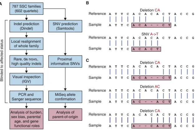
To assess the burden of de novo indels in ASD, we analyzed WES data derived from whole-blood DNA from 787 families (602 quartets and 185 trios) in the SSC (Iossifov et al., 2012; O’Roak et al., 2012; Sanders et al., 2012; Willsey et al., 2013; Table S1). Accurate prediction of indels is complicated by difficulties with alignment (Figure 1B) and multiple possible representations of the same indel in variant call format (VCF; Figure 1C). To overcome these difficulties, we developed an analysis pipeline optimized for de novo indel detection (Fig-ure 1A) using Dindel local realignment (Albers et al., 2011) to cor-rect alignment errors, and the LeftAlignIndels tool from GATK (McKenna et al., 2010) to resolve problems with multiple repre-sentations of the same variant.
Using this approach, we identified a total of 307 putative de novo indels (258 coding and 49 intronic) in cases and controls. All 307 were submitted for confirmation by PCR amplification and Sanger sequencing, blinded to affected status. High-quality confirmation data were generated for 284 indels (93%), 146 of which were confirmed as being de novo (119 in coding regions and 27 in intronic regions), reflecting an overall confirmation rate of 51% (Table S2). Although a 78% confirmation rate was achieved with more stringent detection thresholds, there was a corresponding 18% reduction in indel detection, so we elected to use the less stringent thresholds to maximize sensitivity.
To further assess the pipeline, we first evaluated our ability to detect 54 previously confirmed de novo indels within our current data set (Iossifov et al., 2012; O’Roak et al., 2012). We correctly identified 52 (96%) of these, and the remaining two indels were not detected by Dindel in the first step of our pipeline. In addition, we detected and confirmed six de novo indels in these samples. Furthermore, use of the latest iteration of GATK re-sulted in an 8% reduction in indel detection, with no new de novo indels detected (Table S3). Although the absence of a gold standard precludes an accurate estimation of sensitivity, these results suggest that the method outlined in this work compares favorably with other widely used tools.
In addition to the 59 previously confirmed de novo coding in-dels in the SSC (Table S2), we confirmed an additional 16 previ-ously predicted de novo coding indels and identified and confirmed 44 de novo coding indels.
Increased Burden of De Novo Frameshift Indels in ASD Probands
In total, we observed and confirmed 119 de novo coding indels (79 in 787 probands and 40 in 602 unaffected siblings). To assess the burden of de novo indels in cases versus controls, we relied solely on the 100 indels detected in 602 quartet families that included both a proband and an unaffected sibling. We found 47 confirmed de novo indels that altered the reading frame (frameshift) in probands (0.078 per sample), compared with 30 (0.050 per sample) in siblings (OR = 1.6; 95% confidence interval [CI]: 1.0–2.7; p = 0.03, one-sided Wilcoxon paired test;Figure 2A; Table S2). Considering only brain-expressed genes resulted in a higher OR of 1.7 (95% CI = 1.0–3.0; p = 0.02;Figure 2A;Table S2). For de novo indels that did not alter the reading frame (in-frame), no such excess was observed, with 13 (0.022 per sam-ple) in probands and 10 (0.017 per samsam-ple) in siblings (OR = 1.3; 95% CI = 0.5–3.2; p = 0.28, one-sided Wilcoxon paired test;Figure 2A). Similarly, no excess of intronic de novo indels was observed in ASD probands versus unaffected sibling con-trols (Figure S1). We observed a similar burden of frameshift de novo indels when we applied increasingly stringent quality met-rics to the 258 putative de novo coding indels instead of visual-ization and confirmation (Figure S2).
As expected, these results mirrored the previously reported burden of de novo LoF (nonsense or canonical splice-site) SNVs (OR = 2.4; 95% CI = 1.3–4.3; p = 0.0002, one-sided Wil-coxon paired test; Figure 2A), whereas de novo missense SNVs showed a trend toward overrepresentation in cases (OR = 1.1; 95% CI = 0.9–1.4; p = 0.07) (Iossifov et al., 2012; O’Roak et al., 2012; Sanders et al., 2012; Willsey et al., 2013).
Two Genes Show Multiple Independent De Novo LoF Mutations
Given the similar functional impact of frameshift indels and LoF SNVs, as well as the similarity between the observed OR and frequency in ASD cases (Figure 2A), we concluded that we could treat these mutations as a single class of LoF mutations when considering the implications of observing multiple de novo disruptive mutations in the same gene. Using a permuta-tion test (Sanders et al., 2012) that simulated de novo LoF mu-tations based on gene size and guanine-cytosine (GC) content at the rate observed in siblings (0.083 per sample), we found that a gene with a single disruptive de novo mutation had a 50.4% probability of being associated with ASD (q = 0.496), whereas a gene with at least two disruptive de novo mutations had a 97.6% (q = 0.024) probability of being associated with ASD.
Using this approach, we identified two ASD-associated genes (Table 1): KMT2E (also called MLL5) Figure 2B) and RIMS1 (Figure 2C).
De Novo Frameshift Indels Support a Role forFMRP Targets in the Pathophysiology of ASD
The identification of genes that overtly reflected chromatin modi-fication and synaptic function in ASD led us to evaluate the puta-tive functions of all 62 unique genes that carried de novo
frame-shift indels in the 787 probands (Table S2). We first assessed enrichment in Gene Ontology (GO) categories and KEGG path-ways, as well as for connectivity of protein-protein interaction networks (DAPPLE). We found no significant results after correc-tion for multiple comparisons.
We then turned to an assessment of mRNA targets of fragile X mental retardation protein (FMRP) in light of a recent analysis that showed enrichment of de novo SNVs in this set of genes among affected individuals in the SSC (Iossifov et al., 2012). We assessed the intersection of genes in this study with 842 FMRP targets identified in mouse brain (Darnell et al., 2011) and 939 FMRP targets identified in human embryonic kidney 293 (HEK293) cells (Ascano et al., 2012); 178 of these targets are present in both tissue types.
To ensure that factors known to influence de novo mutation rates did not confound the analysis, we used a generalized linear model of exome coverage, gene size and GC content, brain expression, and identification as an FMRP target as predictors of genes carrying a de novo frameshift indel. We observed a strong signal for FMRP targets identified in mouse brain, but not in HEK293 cells (p = 6 3 10 9, mouse brain; p = 0.13, HEK293 cells; p = 1 3 10 6, combined list). No enrichment was observed for the 29 unique genes with frameshift de novo indels in siblings (p = 0.55 and p = 0.43 for mouse brain and HEK293 cells, respectively).
787 SSC families (602 quartets) Indel prediction (Dindel) Local realignment of whole family Rare, de novo, high quality indels
Visual inspection (IGV) PCR and Sanger sequence Proximal informative SNVs MiSeq allele confirmation Blinded to af fected status Analysis of burden, sex bias, parental
age, and gene functional roles Analysis of parent-of-origin SNV prediction (Samtools) A Reference Sample A T T C A C A C A C T A C C G A T T C A C A - - C T A Deletion CA A T T C A C A C A C T A C C G A T T C A C A C T SNV A- T A T T C A C A C A C T A C C G A T T C A C A - - C T A C C G Deletion CA A T T C A C A C A C T A C C G A T T C A C - - A C T A C C G Deletion AC A T T C A C A C A C T A C C G A T T C A - - C A C T A C C G Deletion CA Reference Sample Reference Sample Reference Sample Reference Sample B C
Figure 1. Experimental Overview
(A) Indels were predicted in 787 families from the SSC using Dindel. Throughout the analytical pipeline, probands and siblings are treated equally to allow accurate assessment of de novo indel burden. Informative SNPs were used to establish the parent of origin of de novo indels.
(B) Alignment errors at the end of reads lead to indels being miscalled as SNVs. (C) An indel can be represented in multiple ways in VCF files.
We then considered our findings in light of the ASD-associated spatiotemporal coexpression networks recently reported by our group (Willsey et al., 2013). Since our prior work relied on over-lapping sequencing data, including previously reported de novo indels, here we focused only on the intersection of 18 newly identified frameshift indels detected in probands. The gene RIMS1 was found to be present in an ASD-associated network in the cerebellum and mediodorsal nucleus of the thalamus in early postnatal life.
Female Probands Have a Greater Burden of De Novo Frameshift Indels
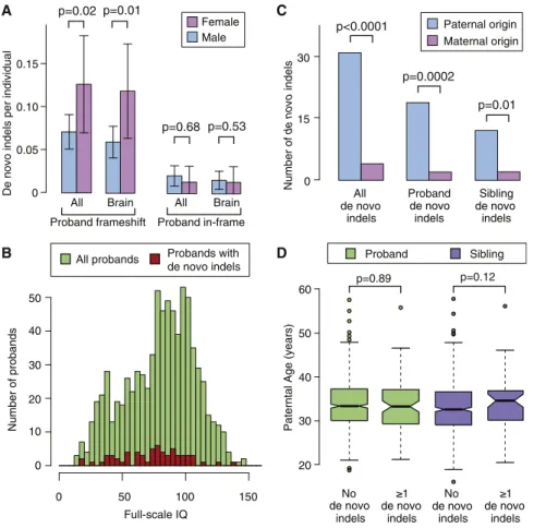
Female probands have previously been noted to have a higher burden of de novo CNVs than their male counterparts (Levy et al., 2011; Sanders et al., 2011); therefore, we assessed the de novo indel burden by sex. A similar pattern was observed for de novo frameshift indels in probands, with 0.126 per sample in the 151 female cases compared with 0.071 per sample in the 636 male cases (OR = 1.9; 95% CI = 1.0–3.4; p = 0.02, one-sided Wilcoxon unpaired test; Figure 3A). This sex-related burden was not observed for the de novo in-frame indels (OR = 0.6; 95% CI = 0.1–3.0; p = 0.68, one-sided Wilcoxon un-paired test;Figure 3A).
De Novo Frameshift Indels Are Associated with Lower IQ
Given the significant clinical overlap between intellectual disability (ID) and ASD, and long-standing interest in the relative contribution of genetic risk to social disability versus ID (Skuse, 2007), we evaluated the relationship between IQ and mutation status. The presence of a de novo frameshift indel was associ-ated with a 6.3 point decrement in proband full-scale IQ (FSIQ) (p < 0.0001, Mann-Whitney U test) compared with probands with no known de novo LoF indel or SNV. However, de novo frameshift indels only explained a small fraction of variance in FSIQ (R2= 0.004), and 43% of probands with de novo frameshift indels had FSIQ measures greater than the proband mean of 80.2 (Figure 3B). The current absence of FSIQ data for the par-ents prevpar-ents an analysis of the genetic deviation in FSIQ due to de novo mutations, as was recently performed for IQ in
individ-uals with 16p11.2 CNVs (Zufferey et al., 2012) and for head circumference in the SSC (Chaste et al., 2013).
De Novo Indels Arise Predominantly from the Paternal Chromosome
Given the observation that the majority of de novo SNVs arise on the paternal chromosome (Kong et al., 2012; O’Roak et al., 2012), we assessed the parent of origin for the de novo indels. Informative SNPs (i.e., those unique to one parent and trans-mitted to the child) within 1,000 bp of de novo indels were iden-tified in WES data. The regions were amplified with PCR and sequenced on an Illumina MiSeq. Visual inspection of the data al-lowed us to determine the parent of origin.
We observed a significant excess of de novo indels arising from the paternal chromosome (31 paternal versus 4 maternal; p < 0.001; binomial exact test;Figure 3C), as was observed for de novo SNVs.
Correlation between Parental Age and De Novo Indels
Multiple prior studies, including our own (Kong et al., 2012; O’Roak et al., 2012; Sanders et al., 2012), have demonstrated a robust correlation between paternal age and the rate of de novo SNVs. Consequently, we tested for this relationship with re-gard to de novo indels by fitting a linear model with paternal age (years) at the child’s birth as a predictor for the presence of a de novo indel. Surprisingly, we found no association with paternal age (slope b = 0.01, SE± 0.01, p = 0.33, regression). This result was not altered by considering maternal age (b = 0.01, p = 0.41), probands only (b = 0.00, p = 0.89;Figure 3D), or siblings only (b = 0.03, p = 0.12;Figure 3D), or by excluding frameshift indels (b = 0.02, p = 0.34). In comparison, applying the same model to the de novo SNVs continued to show a robust association for paternal age (b = 0.02, SE± 0.01, p = 0.0002) equivalent to an ex-tra 0.2 de novo coding mutations per decade of the father’s age.
Contribution of De Novo Indels and SNVs to ASD Population Risk
Based on the observed difference in de novo mutation burden between cases and controls (Figure 2A), we predict that 3% of
p=0.43 p=0.28 p=0.02 p=0.03 Proband Sibling 0 0.05 0.10 0.15 De novo
mutations per individual
Nonsense/ splice-site All Brain All Brain All Brain
Frameshift In-frame p=0.0002 p=0.0001 A B 14299.p1 KMT2E
Lysine (K)-specific methyltransferase 2E
104.65Mb 104.70Mb
Regulating synaptic membrane exocytosis 1 C RIMS1 72.6Mb 72.8Mb 73.0Mb 13162.p1 +A +A -C -C 7q22.3 6q13
Figure 2. De Novo Indel Burden and Genes with Multiple Hits
(A) The rate of de novo indels and SNVs is shown for 602 probands (red) and matched unaffected sib-lings (blue). ‘‘All’’ refers to all RefSeq genes in hg19. ‘‘Brain’’ refers to the subset of genes that are brain expressed. ‘‘Nonsense’’ refers to single-nucleotide substitutions that result in a premature stop codon. ‘‘Splice site’’ refers to single-nucleotide sub-stitutions that disrupt the canonical splice site. Error bars represent the 95% CIs and p values were calculated with a one-sided paired Wilcoxon test. (B) Two de novo frameshift indels in independent samples are shown in the gene KMT2E. Both indels are likely to induce nonsense-mediated decay
(Nagy and Maquat, 1998).
(C) Two de novo frameshift indels in independent samples are shown in the gene RIMS1. Both indels are likely to induce nonsense-mediated decay
(Nagy and Maquat, 1998).
affected individuals carry de novo risk frameshift indels and an additional 4% carry de novo risk LoF SNVs. Should an ASD as-sociation be demonstrated for de novo missense and de novo in-frame mutations (as is likely with increased power), such muta-tions would potentially account for a further 7% of ASD individuals.
DISCUSSION
Our analysis of 787 ASD families from the SSC, including 602 unaffected sibling controls, demonstrates the association of de novo frameshift indels with ASD. Furthermore, the similar-ity between the OR and mutation rate observed for de novo frameshift indels and those observed for de novo LoF SNVs, as well as the overlap in the functional consequences, fits with the assumption that de novo frameshift indels and de novo LoF SNVs can be considered as a single group of high-ly disruptive mutations. Overall, these disruptive mutations are predicted to contribute to risk in 7% of the ASD population.
By reanalyzing WES data from the SSC cohort using this more sensitive and reliable approach for discovering de novo indels, we identified two ASD genes: KMT2E (MLL5) and RIMS1. KMT2E is a chromatin regulator that is recruited to methylated histones found at the promoters of actively expressed genes, specifically H3K4me3. It was initially identified as a tumor-sup-pressor gene and its role in hematopoietic stem cell homeosta-sis and self-renewal has been well documented. However, the gene is highly pleiotropic, with roles in cytokinesis, response to DNA damage, and genome maintenance (Ali et al., 2013). Although KMT2E has not previously been associated with neurological disorders, chromatin regulation in fetal develop-ment has been identified as a key risk factor for ASD (O’Roak et al., 2012; Willsey et al., 2013) and the gene is highly expressed throughout the brain, especially during fetal development (Kang et al., 2011).
RIMS1 is a RAS signaling gene that is essential for multiple aspects of neurotransmitter release. It plays a role in presynap-tic plaspresynap-ticity (Kaeser et al., 2012), with mouse knockouts showing deficits in learning and memory (Powell et al., 2004) and increased seizure frequency following induced status epi-lepticus (Pitsch et al., 2012). RIMS1 is expressed throughout the human brain, with levels increasing throughout
develop-ment and reaching a plateau in the third trimester that persists throughout adulthood (Kang et al., 2011). The gene is present in an ASD-associated postnatal coexpression network in the cer-ebellum and mediodorsal nucleus of the thalamus (8-10 MD-CBC) due to its coexpression with the ASD gene SCN2A (Will-sey et al., 2013).
The FSIQ was below the proband average of 81 in the SSC (range 46–74) in all four individuals with mutations in KMT2E and RIMS1. The anxious/depressed, withdrawn/depressed, somatic complaints, and thought problems scales of the Child Behavior Checklist were elevated for both individuals with RIMS1 muta-tions only. Inconsistent results were observed for other pheno-typic measures, including seizures and head circumference.
Although the ASD-associated de novo indels do not form a highly connected protein-protein interaction network or show enrichment for GO terms, we do confirm the previously docu-mented enrichment of FMRP target genes carrying de novo LoF mutations (Iossifov et al., 2012). In light of the strength and reproducibility of this relationship, the identification of mRNAs targeted by FMRP in the developing human brain is likely to be a valuable resource for ASD gene discovery.
Given the observed similarities between de novo frameshift indels and de novo LoF SNVs, a marked overrepresentation of mutations on the paternal allele might have been anticipated. However, we did not observe the expected correlation between these paternally enriched de novo mutations and paternal age. Given the relatively small number of indels, it is likely that this negative result reflects inadequate statistical power. We will test this hypothesis as substantially larger WES data sets from ASD families become available in the near future (Bux-baum et al., 2012).
Finally, we investigated the relationship between de novo frameshift indels and IQ. Given the association of many estab-lished ASD mutations with decrements in cognitive functioning and the frequent phenotypic overlap seen in clinical samples, there has been speculation that de novo disruptive mutations may only carry a risk for ID and not for the core social deficits that define ASD. Our data do not support this hypothesis. Although we observe lower IQ among probands that carry de novo frameshift indels compared with probands without any de novo LoF mutations, the difference is small (6.3 IQ points) and accounts for only a fraction of the variance in IQ (R2= 0.004), and the distribution of IQ is similar to that in other probands
Table 1. Identified De Novo Indels in Genes with Previously Reported De Novo Nonsynonymous Mutations
Gene Sample hg19 Location Variant Effect Source
CHD2 10C100480 chr15:93518170 C->T missense Neale et al., 2012
13618.p1 chr15:93524060 AAAG frameshift Identified herein
KMT2E 14299.p1 chr7:104702706 C frameshift Identified herein
12952.p1 chr7:104748101 C frameshift Iossifov et al., 2012
PHF3 14133.p1 chr6:64413433 CG frameshift Identified herein
14110.p1 chr6:64423242 C->T missense Sanders et al., 2012
RIMS1 13162.p1 chr6:72889392 +A frameshift Iossifov et al., 2012
13497.p1 chr6:73102488 +A frameshift Identified herein
(Figure 3B). Moreover, given the emerging picture of shared risks for de novo SNVs among a wide range of neurodevelopmental syndromes (Allen et al., 2013; Fromer et al., 2014; Moreno-De-Luca et al., 2014), the most parsimonious explanation is that a subset of highly disruptive risk mutations are associated with a range of phenotypic outcomes, including ID, ASD, schizo-phrenia, and epilepsy.
Based on current estimates, the detection of de novo frame-shift indels and LoF SNVs identifies a genetic risk factor in approximately 7% of affected individuals, rivaling the contribu-tion of de novo CNVs (Sanders et al., 2011). Moreover, in addicontribu-tion to confirming important recent observations regarding the genomic architecture of ASD, including the paternal origin of the majority of small de novo mutations, the approach is yielding a growing list of ASD risk genes, pointing to chromatin modifica-tion, synaptic functioning, and binding to FMRP as key patho-physiological mechanisms.
EXPERIMENTAL PROCEDURES
Sample Collection and Initial Data Processing
Whole-exome data for 2,963 samples from 787 families (602 quartets and 185 trios) in the SSC were obtained (Table S1). Exome capture was per-formed using a NimbleGen custom array (N5210) or NimbleGen EZExo-meV2.0 (N5718) followed by sequencing on Illumina GAIIx or HiSeq2000 instruments. Reads were aligned to hg19 with BWA. This research was re-viewed by the Yale institutional review board under HIC protocol number 0301024156.
20 30 40 50
Paterntal Age (years)
No indels indels No indels 1 1 indels Proband Sibling C 60 p=0.89 p=0.12 Number of de novo indels 0 15 30 Paternal origin Maternal origin All indels p<0.0001 Proband indels Sibling indels p=0.01 B Female Male 0 0.05 0.10 0.15
De novo indels per individual
A p=0.02
All Brain All Brain Proband frameshift Proband in-frame
p=0.01
p=0.68 p=0.53
p=0.0002
Number of probands
0 50 100 150
All probands Probands with de novo indels 20 30 40 50 10 0 Full-scale IQ D
de novo de novo de novo
de novo de novo de novo de novo
Figure 3. Sex Difference, Parent of Origin, and Parental Age
(A) A consistently higher rate of de novo frameshift indels was observed in female probands (pink) compared with male probands (blue), but this dif-ference was not observed in unaffected siblings. ‘‘All’’ describes all de novo frameshift indels and ‘‘Brain’’ indicates only those expressed in the brain. Error bars represent the 95% CIs and p values were calculated with a one-sided paired Wilcoxon test.
(B) Histogram of full-scale IQ in all probands (green) and probands with a de novo frameshift indel (red).
(C) The majority of de novo indels for which the parent of origin could be resolved were found to be on the paternal (blue) rather than the maternal (pink) chromosome (p < 0.001; binomial). This result was observed in both probands and siblings separately.
(D) No clear relationship between the presence of a de novo indel and increased paternal age was observed for probands (green) or siblings (purple); p values were estimated with a Poisson regression.
Family-Based De Novo Indel Detection Indels were predicted in children using Dindel (
Alb-ers et al., 2011) followed by Dindel local
realign-ment for all family members. The LeftAlignIndels tool from GATK (McKenna et al., 2010) was applied to all of the resulting BAM files, and indels were as-sessed in the realigned files. Rare inherited hetero-zygous indels were used to set appropriate quality filters to identify rare de novo indels, includingR10 unique reads in all family members, indel not observed in other SSC families, and <5% of reads with an indel in either parent.
Realigned BAM files for the resulting 522 putative de novo coding indels (0.39 per sample in probands and 0.37 per sample in siblings) were visualized using Integrative Genome Viewer (IGV) (Thorvaldsdo´ttir et al., 2013) by two in-dependent researchers who were blinded to affected status. High concor-dance between the two researchers was observed (kappa coefficient = 0.94) and any indel that was potentially de novo according to either researcher was submitted for confirmation. In total, 258 indels (50%, 0.27 per sample in probands and 0.16 per sample in siblings) were selected. In addition, the 49 intronic de novo indels with the best indel quality scores were submitted for confirmation as an additional control, yielding a total of 307 confirmations.
Indel Confirmations
Indels were confirmed using PCR amplification of whole-blood DNA and Sanger sequencing. Of 307 putative de novo indels, high-quality confirmation data were generated for 284 (96%). Of these, no indel was observed in the child for 44 (15%), while an inherited indel was observed in 93 (33%). One confirmed indel was observed in both the proband and sibling, but not in either parent, suggesting germline mosaicism. This left 146 confirmed de novo indels and a confirmation rate of 51%.
Identifying the Parent of Origin
Informative SNPs within 1,000 bp of a confirmed de novo indel were identified in WES data. The regions were amplified from whole-blood DNA of the index child and both parents using PCR. Amplified DNA was normalized using PicoGreen quantitation and pooled separately for children, fathers, and mothers. Each pool underwent indexed library preparation and was run on an Illumina MiSeq with 250 bp paired-end reads. The aligned sequence data were assessed in IGV.
SUPPLEMENTAL INFORMATION
Supplemental Information includes Supplemental Experimental Procedures, two figures, and three tables and can be found with this article online at
http://dx.doi.org/10.1016/j.celrep.2014.08.068.
AUTHOR CONTRIBUTIONS
S.D., M.W.S., L.W., and S.J.S. designed the study. S.D., N.J.C., A.J.W., A.Y.Y., V.H.B., J.F.K., N.A.T., J.G., and S.J.S. developed analysis methods and analyzed the data. S.D., M.F.W., M.D., Z.W., L.E.G., J.D.D., S.F., J.D., J.D.M., C.A.S., N.M.D., R.O.K., Z.Y., S.M.S., A.G.E., A.R.G., S.M.M., M.S., and A.I.B. confirmed the indels. S.D., K.R., B.D., M.W.S., L.W., and S.J.S. wrote the manuscript.
ACKNOWLEDGMENTS
We are grateful to the families participating in the Simons Foundation Autism Research Initiative (SFARI) Simplex Collection (SSC). We thank the SSC prin-cipal investigators A.L. Beaudet, R. Bernier, J. Constantino, E.H. Cook, Jr., E. Fombonne, D. Geschwind, D.E. Grice, A. Klin, D.H. Ledbetter, C. Lord, C.L. Martin, D.M. Martin, R. Maxim, J. Miles, O. Ousley, B. Peterson, J. Piggot, C. Saulnier, M.W. State, W. Stone, J.S. Sutcliffe, C.A. Walsh, and E. Wijsman. We also thank the coordinators and staff of the SSC clinical sites, the SFARI staff, the Rutgers University Cell and DNA Repository for accessing biomate-rials, N. Buenaventura and L. Chow for their help in administering the project at UCSF, and T. Brooks-Boone, N. Wright-Davis, and M. Wojciechowski for their help in administering the project at Yale. This work was supported by grants from the Simons Foundation (to M.W.S.), the CIHR (DRA to A.J.W.), the HHMI (International Student Research Fellowship to S.J.S.), the NIMH (R37 MH057881 to K.R. and B.D.), and the National Center for Research Resources (UL1 TR000142 and KL2 TR000140 to A.G.E.). L.W. was supported by the Na-tional Natural Science Foundation of China (no. 31025014) and the Ministry of Science and Technology of China (no. 2012CB837600).
Received: April 9, 2014 Revised: June 4, 2014 Accepted: August 27, 2014 Published: October 2, 2014 REFERENCES
Albers, C.A., Lunter, G., MacArthur, D.G., McVean, G., Ouwehand, W.H., and Durbin, R. (2011). Dindel: accurate indel calls from short-read data. Genome Res. 21, 961–973.
Ali, M., Rinco´n-Arano, H., Zhao, W., Rothbart, S.B., Tong, Q., Parkhurst, S.M., Strahl, B.D., Deng, L.W., Groudine, M., and Kutateladze, T.G. (2013). Molecu-lar basis for chromatin binding and regulation of MLL5. Proc. Natl. Acad. Sci. USA 110, 11296–11301.
Allen, A.S., Berkovic, S.F., Cossette, P., Delanty, N., Dlugos, D., Eichler, E.E., Epstein, M.P., Glauser, T., Goldstein, D.B., Han, Y., et al.; Epi4K Consortium; Epilepsy Phenome/Genome Project (2013). De novo mutations in epileptic en-cephalopathies. Nature 501, 217–221.
Ascano, M., Jr., Mukherjee, N., Bandaru, P., Miller, J.B., Nusbaum, J.D., Cor-coran, D.L., Langlois, C., Munschauer, M., Dewell, S., Hafner, M., et al. (2012). FMRP targets distinct mRNA sequence elements to regulate protein expres-sion. Nature 492, 382–386.
Buxbaum, J.D., Daly, M.J., Devlin, B., Lehner, T., Roeder, K., and State, M.W.; Autism Sequencing Consortium (2012). The autism sequencing consortium: large-scale, high-throughput sequencing in autism spectrum disorders. Neuron 76, 1052–1056.
Chaste, P., Klei, L., Sanders, S.J., Murtha, M.T., Hus, V., Lowe, J.K., Willsey, A.J., Moreno-De-Luca, D., Yu, T.W., Fombonne, E., et al. (2013). Adjusting head circumference for covariates in autism: clinical correlates of a highly her-itable continuous trait. Biol. Psychiatry 74, 576–584.
Darnell, J.C., Van Driesche, S.J., Zhang, C., Hung, K.Y., Mele, A., Fraser, C.E., Stone, E.F., Chen, C., Fak, J.J., Chi, S.W., et al. (2011). FMRP stalls ribosomal translocation on mRNAs linked to synaptic function and autism. Cell 146, 247–261.
Fromer, M., Pocklington, A.J., Kavanagh, D.H., Williams, H.J., Dwyer, S., Gormley, P., Georgieva, L., Rees, E., Palta, P., Ruderfer, D.M., et al. (2014). De novo mutations in schizophrenia implicate synaptic networks. Nature
506, 179–184.
Iossifov, I., Ronemus, M., Levy, D., Wang, Z., Hakker, I., Rosenbaum, J., Yam-rom, B., Lee, Y.H., Narzisi, G., Leotta, A., et al. (2012). De novo gene disrup-tions in children on the autistic spectrum. Neuron 74, 285–299.
Kaeser, P.S., Deng, L., Fan, M., and Su¨dhof, T.C. (2012). RIM genes differen-tially contribute to organizing presynaptic release sites. Proc. Natl. Acad. Sci. USA 109, 11830–11835.
Kang, H.J., Kawasawa, Y.I., Cheng, F., Zhu, Y., Xu, X., Li, M., Sousa, A.M., Ple-tikos, M., Meyer, K.A., Sedmak, G., et al. (2011). Spatio-temporal transcrip-tome of the human brain. Nature 478, 483–489.
Kong, A., Frigge, M.L., Masson, G., Besenbacher, S., Sulem, P., Magnusson, G., Gudjonsson, S.A., Sigurdsson, A., Jonasdottir, A., Jonasdottir, A., et al. (2012). Rate of de novo mutations and the importance of father’s age to dis-ease risk. Nature 488, 471–475.
Levy, D., Ronemus, M., Yamrom, B., Lee, Y.-H., Leotta, A., Kendall, J., Marks, S., Lakshmi, B., Pai, D., Ye, K., et al. (2011). Rare de novo and transmitted copy-number variation in autistic spectrum disorders. Neuron
70, 886–897.
McKenna, A., Hanna, M., Banks, E., Sivachenko, A., Cibulskis, K., Kernytsky, A., Garimella, K., Altshuler, D., Gabriel, S., Daly, M., and DePristo, M.A. (2010). The Genome Analysis Toolkit: a MapReduce framework for analyzing next-generation DNA sequencing data. Genome Res. 20, 1297–1303.
Moreno-De-Luca, D., Moreno-De-Luca, A., Cubells, J.F., and Sanders, S.J. (2014). Cross-disorder comparison of four neuropsychiatric CNV loci. Curr. Genet. Med. Rep. 2, 1–11.
Nagy, E., and Maquat, L.E. (1998). A rule for termination-codon position within intron-containing genes: when nonsense affects RNA abundance. Trends Bio-chem. Sci. 23, 198–199.
Neale, B.M., Kou, Y., Liu, L., Ma’ayan, A., Samocha, K.E., Sabo, A., Lin, C.F., Stevens, C., Wang, L.S., Makarov, V., et al. (2012). Patterns and rates of exonic de novo mutations in autism spectrum disorders. Nature 485, 242–245. O’Roak, B.J., Vives, L., Girirajan, S., Karakoc, E., Krumm, N., Coe, B.P., Levy, R., Ko, A., Lee, C., Smith, J.D., et al. (2012). Sporadic autism exomes reveal a highly interconnected protein network of de novo mutations. Nature 485, 246–250.
Pitsch, J., Opitz, T., Borm, V., Woitecki, A., Staniek, M., Beck, H., Becker, A.J., and Schoch, S. (2012). The presynaptic active zone protein RIM1a controls epileptogenesis following status epilepticus. J. Neurosci. 32, 12384–12395.
Powell, C.M., Schoch, S., Monteggia, L., Barrot, M., Matos, M.F., Feld-mann, N., Su¨dhof, T.C., and Nestler, E.J. (2004). The presynaptic active zone protein RIM1alpha is critical for normal learning and memory. Neuron
42, 143–153.
Sanders, S.J., Ercan-Sencicek, A.G., Hus, V., Luo, R., Murtha, M.T., Moreno-De-Luca, D., Chu, S.H., Moreau, M.P., Gupta, A.R., Thomson, S.A., et al. (2011). Multiple recurrent de novo CNVs, including duplications of the 7q11.23 Williams syndrome region, are strongly associated with autism. Neuron 70, 863–885.
Sanders, S.J., Murtha, M.T., Gupta, A.R., Murdoch, J.D., Raubeson, M.J., Will-sey, A.J., Ercan-Sencicek, A.G., DiLullo, N.M., Parikshak, N.N., Stein, J.L., et al. (2012). De novo mutations revealed by whole-exome sequencing are strongly associated with autism. Nature 485, 237–241.
Sebat, J., Lakshmi, B., Malhotra, D., Troge, J., Lese-Martin, C., Walsh, T., Yamrom, B., Yoon, S., Krasnitz, A., Kendall, J., et al. (2007). Strong association of de novo copy number mutations with autism. Science 316, 445–449.
Skuse, D.H. (2007). Rethinking the nature of genetic vulnerability to autistic spectrum disorders. Trends Genet. 23, 387–395.
Thorvaldsdo´ttir, H., Robinson, J.T., and Mesirov, J.P. (2013). Integrative Geno-mics Viewer (IGV): high-performance genoGeno-mics data visualization and explora-tion. Brief. Bioinform. 14, 178–192.
Willsey, A.J., Sanders, S.J., Li, M., Dong, S., Tebbenkamp, A.T., Muhle, R.A., Reilly, S.K., Lin, L., Fertuzinhos, S., Miller, J.A., et al. (2013). Coexpression
net-works implicate human midfetal deep cortical projection neurons in the path-ogenesis of autism. Cell 155, 997–1007.
Zufferey, F., Sherr, E.H., Beckmann, N.D., Hanson, E., Maillard, A.M., Hippo-lyte, L., Mace´, A., Ferrari, C., Kutalik, Z., Andrieux, J., et al.; Simons VIP Con-sortium; 16p11.2 European Consortium (2012). A 600 kb deletion syndrome at 16p11.2 leads to energy imbalance and neuropsychiatric disorders. J. Med. Genet. 49, 660–668.