Purposes
V. Hasirci,1 A. Tezcaner,1 N. Hasirci,2 S¸. Su¨zer3
1Middle East Technical University, Department of Biological Sciences, Biotechnology Research Unit, 06531 Ankara, Turkey
2Middle East Technical University, Chemistry Department, 06531 Ankara, Turkey 3Bilkent University, Chemistry Department, 06533 Ankara, Turkey
Received 6 November 2000; accepted 2 April 2002
ABSTRACT: Plasma glow-discharge application is known as a technique to coat or modify the surfaces of various materials. In this study, the influence of oxygen rf-plasma treatment on surface and bulk properties of a biological polyester, poly(3-hydroxybutyrate-co-3-hydroxyvalerate), were studied by determining water content and water con-tact angle, and by using X-ray photoelectron spectroscopy (XPS) and scanning electron microscopy (SEM). The plasma-treated films absorbed more water than the unplasma-treated film, and the absorbance increased with the total power applied. The water contact angles decreased and O/C atomic ratio increased on treatment, indicating that the material became more hydrophilic due to increases in the oxygen-containing
functional groups on the surface of the polymer. A direct relation could be observed when the O/C ratio was plotted against the total power applied (treatment duration⫻ treat-ment power). SEM revealed a visual record of surface mod-ification, the extent of which increased with increased total power. It was thus possible to alter the surface chemistry and relevant properties of the polymer film using oxygen plasma as a tool.© 2002 Wiley Periodicals, Inc. J Appl Polym Sci 87: 1285–1289, 2003
Key words:plasma polymerization; hydrophilic polymers; biodegadable biomaterials
INTRODUCTION
Polyhydroxyalkanoates (PHAs) are polyesters of mi-crobial origin. The chemical structure of PHAs is very similar to those of polylactides (PLAs), the highly popular synthetic biodegradable polymers used in biomedical applications, and copolymers of PLAs with glycolides, [i.e., poly(lactide-co-glycolide) (PLGA)]. PHAs, however, generally degrade at a much slower rate than PLAs and PLGA.1Poly(3-hydroxybutyrate)
(P3HB) is generally found as a copolymer containing hydroxyvalerate (HV) as the comonomer, which is called poly(hydroxybutyrate-co-hydroxyvalerate) (PHBV). The copolymers have varying HV ratios, and their mechanical (tensile and compressive strengths) and chemical (surface energy, hydrolysis rate) prop-erties depend on the HV content. They are biodegrad-able and biocompatible, and are reported to induce new bone formation due to their piezoelectricity. The interest in PHA is increasing rapidly, and exciting new applications in the fields of drug delivery and tissue engineering are being reported.2–5 The biomedical
field has been using PLGAs for sometime to fulfill its
need for biodegradable materials. The biological ori-gin of PHBV, however, makes it environmentally more acceptable, which is a major advantage over PLGA. In addition, it is possible to obtain very differ-ent copolymers of PHAs with exceptional properties by just changing the carbon source of the growth medium of the PHA-producing microorganisms.
PHAs are produced by a large number of microor-ganisms, including Alcaligenes latus, Alcaligenes eutro-phus, and Pseudomonas oleovorans.6 – 8Genes for PHBV growth were introduced to other biological systems that are normally nonproducers (like E. coli)9and even
into plants.10 As a result, this polyester family has great potential in the biomedical field, especially as biodegradable implant materials, drug release sys-tems, and tissue engineering scaffolds. In all these applications, the surface properties of the polymer are of utmost importance because the surface is the region of contact with the biological system. For example, bovine aortic endothelial cell growth was linearly cor-related with the oxygen content obtained by the oxy-gen plasma treatment of the polymeric matrix.11As a
result of this interaction, cell loading and attachment to a biomaterial in tissue engineering applications, and the biocompatibility and biodegradability of the poly-meric matrices are influenced.12In this study, PHBV8
(PHBV with 8% HV) was treated at varying levels
Correspondence to: V. Hasirci ([email protected]).
Journal of Applied Polymer Science, Vol. 87, 1285–1289 (2003) © 2002 Wiley Periodicals, Inc.
with oxygen plasma, the modifications created on the surface of this material were studied by X-ray photo-electron spectroscopy (XPS), and the water content and water contact angle values were measured.
MATERIALS AND METHODS Plasma treatment of polymer films
Poly(3-hydroxybutyrate-co-3-hydroxyvalerate) (PHBV) containing 8% hydroxyvalerate (molar) in its structure was purchased from Aldrich Chemical Company (Germany) and films (8 –10 m thickness) were pre-pared by solvent casting of 0.6% solutions in chloro-form in Petri plates (9 cm in diameter). The films, in their Petri plates, were placed in a plasma chamber (Advanced Plasma Systems, Inc., (St. Petersburg, FL), with a SEREN IPS R 300 13.56 MHz power supply) and subjected to oxygen plasma (pressure: 40 mTorr initial and 300 mTorr final; see Table I for conditions). The membranes were then removed and used in fur-ther tests, taking care to identify the treated sides.
Water content determination
Water content of the polymeric films was determined by gravimetry. A sample (5–10 mg) was weighed to the nearest 0.1 mg, immersed in physiological phos-phate buffer (PBS; pH 7.4, 0.1 M), and stored at 37° for 24 and 72 h. At 24 h, the sample was removed, super-ficial water was removed by gently blotting with a filter paper, and the sample was weighed and then re-introduced into the swelling medium. At 72 h, this weighing procedure was repeated and then the film was dried in a vacuum oven at 50°C overnight and weighed again. The water content was calculated as: (Wweight of wet sample⫺ Wweight of dry sample)
⫼ Wweight of dry sample⫻ 100
Water contact angle determination
Advancing contact angle measurements were made with a sessile drop of deionized water on a Cam-Micro
(Tantec Inc., (Schaumberg, IL)) contact angle meter. The values reported are an average of at least five measurements.
X-ray photoelectron spectroscopy (XPS)
XPS measurements were performed on films prepared on glass slides using a Kratos ES300 spectrometer with MgK␣ X-rays at 1253.6 eV. The films on the slides were directly inserted into the spectrometer, which had a base pressure ⬎10⫺8 Torr. Power was kept at ⬍100 Watt/cm2to prevent any radiation damage.
Scanning electron microscopy (SEM)
Scanning electron micrographs of PHBV8 films, before and after plasma treatment, were obtained after coat-ing with a thin layer of gold with a Leitz (Model AMR 1000) SEM microscope.
RESULTS AND DISCUSSION Water content
Water contents of polymer films treated with different plasma powers were determined (Table I). The un-treated polymer was very hydrophobic, as are all PHBV, and did not absorb any water in the first 24 h and only 8.2% after 72 h of immersion. All the treated films absorbed more water than the untreated film, and the absorbance increased with the total power applied. The film with the highest water absorbing capability (60.2%) was the one treated with the most oxygen plasma (100 W⫻ 20 min). These results con-firm the expectation that the film treated with the highest level of oxygen plasma would be the most hydrophilic. The cause for high water absorption with this specific treatment mode could be (a) very high surface hydrophilicity and (b) degradation of the sam-ple during treatment. We believe both these phenom-ena are valid because the contact angle of this sample could not be determined due to rapid absorption of water by the material.
Water contact angle
It is known that PHBs and their copolymers are quite hydrophobic polyesters, with the only polar function-ality residing in the ester groups. The water content results reported in the previous section confirm this hydrophobicity. PHBV20 (HV content of 20%) was reported to have a water contact angle of⬃68°, which decreased to 66° on hydrolytic degradation.13 Ammo-nia gas plasma treatment of polyhydroxyoctanoate) led to a water contact angle decrease of 20 –30°from a very high value of 95°as a result of stable incorpora-tion of nitrogen in the structure.14Following treatment
TABLE I
Water Content (%) of PHBV8 Polymer Films in Pristine and Oxygen Plasma-Treated State after Incubation in
PBS at 37 °C for Various Durations
Power applied (Watt⫻ min) Water content (%) 24 h Incubation 72 h Incubation 0 0 8.2⫾ 1.1 50⫻ 10 4.6⫾ 0.3 11.4⫾ 0.0 50⫻ 20 4.1⫾ 0.6 19.2⫾ 4.9 100⫻ 10 11.2⫾ 1.8 29.2⫾ 5.9 100⫻ 20 13.3⫾ 5.6 60.2⫾ 11.0
with plasma (oxygen, ammonia,15 etc.), other poly-mers are also known to have moved towards higher hydrophilicities as indicated by lower contact angles and surface energies. In the present study, the un-treated polymer had a significantly high contact angle (68.3 ⫾ 7.8°) that was substantially reduced (43.3 ⫾ 3.3°) following oxygen plasma treatment (100 W for
10 min), as shown in Table II and Figure 1(a). It was also observed that the decrease in the contact angle was directly related with the extent of treatment
(power⫻ duration; i.e., no significant difference was observed between 50 W⫻ 20 min and 100 W ⫻ 10 min treatments), indicating that it is possible to modify surface hydrophilicity on demand. This observation is very important because it has been reported that cell adhesion on polymers is controlled by the surface energy. For example, a study of adherence of fibro-blasts, endothelial cells, and L cells on polymer sur-faces indicates that 70°is optimum for adherence and that at higher or lower contact angles, adherence de-creases sharply.16Also, it was reported that fibroblast spreading on polymer surfaces showed a sudden in-crease at a surface energy of 40 erg/cm2and further increases in the surface energy did not result in higher spreading. Thus, this technique becomes an important tool for tissue engineering applications, where surface chemistry is of utmost importance for cell loading, viability, and functionality. The porosity of the poly-mer films, however, increased on oxygen plasma treat-ment, and it was not possible to measure the contact angle of the 100 W ⫻ 20 min sample because of the rapid transference of the water across the membrane. This result indicates that an excess of treatment not only leads to surface modification but could actually result in damage to the surface. Thus, plasma treat-ment needs to be applied carefully so as not to exten-sively modify and damage the surface.
X-ray photoelectron spectroscopy
The wide XPS spectra of the various films are shown in Figure 2. In addition to the strong O1s and C1s
peaks, N1speaks are observed in plasma-treated films.
The N1speaks are probably due to a small leak,
resid-ual N2in the plasma chamber, or interaction with N2
in the air on opening of the chamber. C1speaks exhibit
multiplet structure and are curve-fitted to (i) carbon bonded to hydrogen, (ii) etheric, and (ii) esteric com-ponents, as shown in Figure 2 and Table II.17The O1s
peaks are broad and become broader after plasma
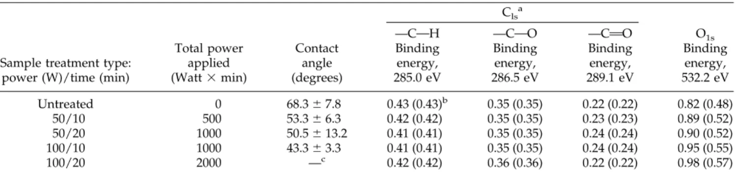
TABLE II
Properties of the PHBV8 Samples after Treatment with Oxygen Plasma
Sample treatment type: power (W)/time (min)
Total power applied (Watt⫻ min) Contact angle (degrees) Clsa O1s
—COH —COO —CAO
Binding energy, 285.0 eV Binding energy, 286.5 eV Binding energy, 289.1 eV Binding energy, 532.2 eV Untreated 0 68.3⫾ 7.8 0.43 (0.43)b 0.35 (0.35) 0.22 (0.22) 0.82 (0.48) 50/10 500 53.3⫾ 6.3 0.42 (0.42) 0.35 (0.35) 0.23 (0.23) 0.89 (0.52) 50/20 1000 50.5⫾ 13.2 0.41 (0.41) 0.35 (0.35) 0.24 (0.24) 0.90 (0.52) 100/10 1000 43.3⫾ 3.3 0.41 (0.41) 0.35 (0.35) 0.24 (0.24) 0.95 (0.55) 100/20 2000 —c 0.42 (0.42) 0.36 (0.36) 0.22 (0.22) 0.98 (0.57) a Determined by XPS. b
Corrected atomic composition in parentheses.
c
Could not be measured because of the high porosity created by the plasma treatment.
Figure 1 Variation of (a) water contact angle and (b) O/C atomic ratio (by XPS) of the oxygen plasma-treated PHBV8 films with the applied plasma power and duration.
treatment. The O/C atomic ratio derived from XPS measurements (0.48) matches exactly the expected ra-tio from the chemical formula (0.49) but increases up to 0.57 as a function of the plasma power and dura-tion, as shown in Figure 1(b). To this end it is neces-sary to note that the changes in the O/C atomic ratio are indistinguishable (within the experimental uncer-tainty) for the 50 W ⫻ 20 min and 100 W ⫻ 10 min treatments, which is similar to our water contact angle results.
The XPS sampling depth was ⬃5 nm. The atomic compositions involve both surface and subsurface lay-ers and, in general, a direct comparison with water contact angle measurements is not expected.18,19 How-ever, the decrease of the water contact angle as a function of plasma treatment surprisingly correlates directly with the O/C atomic ratio increase, as de-picted in Figure 1.
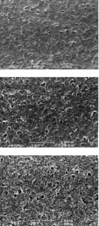
Scanning electron microscopy
Scanning electron microscopy of the PHBV8 films re-vealed a gradually increased surface roughness with increase in the total power applied (Figure 3). A
sim-ilar observation was reported following argon treat-ment of poly(phenylene vinylene), where the uni-formly smooth-surfaced pristine films became granu-lar and rougher.20
CONCLUSIONS
In this study we showed, by changes in water absorb-ing capacity and water contact angle and by XPS and SEM investigations, that the surface properties of PHBV films containing 8% HV could be controllably modified by varying the power and the duration of the oxygen rf-plasma application. Our results showed that there was direct correlation between the gradual in-crease in the concentration of oxygen-containing groups on the membrane surface and increases in total power applied. This effect was manifest in the prop-erties of the material as an increase in hydrophilicity. It is known that plasma treatment modifies only the surface of treated samples. In this study, all the sam-ples showed some wettability even before treatment, and after treatment they revealed low but consistent water absorptions. Only the samples treated with 100 W ⫻ 20 min plasma application seemed to absorb a
Figure 2 XPS spectra (wide) and the detailed C1s region (inset) following oxygen plasma treatment of PHBV8 films using different power and durations.
significant amount of water. This result was possibly due to very high surface hydrophilicity and degrada-tion of sample during this treatment.
Hence, plasma modification of PHBV surfaces could help modify surface topography and tailor-make sur-face chemistry for tissue engineering applications in which surface properties are critical for cell adhesion and growth.
We acknowledge the METU AFP and TU¨ BI˙TAK NATO grants for A. Tezcaner, and the State Planning Organization of Turkey for the chemicals.
References
1. Hasirci, V. In: Wise, D.L., Ed. Biomaterials and Bioengineering Handbook; Marcel Dekker: New York, 2000; pp. 141–155. 2. Rivard, C.H.; Chaput, C.J.; DesRosiers, E.A.; Yahia, L.H.;
Sel-mani, A. J Appl Biomater 1995, 6, 65– 68.
3. Sendil, D.; Gursel, I.; Wise, D.L.; Hasirci, V. J Controlled Release 1999, 59, 207–217.
4. Yagmurlu, M.F.; Korkusuz, F.; Gursel, I.; Korkusuz, P.; Ors, U.; Hasirci, V. J Biomed Mater Res 1999, 46, 494 –503
5. van der Giessen, W.J.; Lincoff, A.M.; Schwartz, R.S.; van Beusekom, H.M.; Serruys, D.R.; Holmes, S.G.; Ellis, E.J. Topol. Circulation 1996, 94(7), 1690 –1697.
6. Lee, M.Y.; Park, W.H.; Lenz, R.W. Polymer 2000, 41(5),1703– 1709.
7. Pouton, C.W.; Akhtar, S. Adv Drug Delivery Rev 1996, 18(2), 133–162.
8. Nakamura, S.; Doi, Y.; Scandola, M. Macromolecules 1992, 25(17), 4235– 4240.
9. Pool, R.G. Science. 1989, 245, 1187–1189.
10. Poirier, Y.; Somerville, C.; Schechtman, L.A.; Satkowski, M.M.; Noda, I. Int J Biol Macromol 1995, 17, 7–12.
11. Ertel, S.I.; Ratner, B.D.; Horbett, T.A. J Biomed Mater Res 1990, 24,1637–1659.
12. Hasirci, N. J Appl Polym Sci 1987, 34, 2457–2468.
13. Yasin, M.; Holland, S.J.; Jolly, A.M.; Tighe, B.J. Biomaterials 1989, 10, 400 – 412.
14. Williams, S.F.; Martin, D.P.; Horowitz, D.M.; Peoples, O.P. Int J Biol Macromol 1999, 25, 111–121.
15. Mason, M.; Vercruysse, K.P.; Kirker, K.R.; Frisch, R.; Marecak, D.M.; Prestwich, G.D.; Pitt, W.G. Biomaterials 2000, 21, 31–36. 16. Saltzman, W.M. In: Principles of Tissue Engineering; Lanza,
R.P.; Langer, R.; Chick, W.L., Eds.; R.G. Landes Company: Aus-tin, TX, 1996; pp. 225–246.
17. Beamson, G.; Briggs, D. High Resolution XPS of Organic Poly-mers; Wiley: New York, 1992.
18. Su¨zer, S.; Argun, A.; Vatansever, O.; Aral, O. J Appl Polym Sci 1999, 74, 1846 –1850.
19. Occhiello, E.; Mora, M.; Morini, G.; Garbassi, F.; Humphrey, P. J Appl Polym Sci 1991, 42, 551–559.
20. Nguyen, T.P.; Lahman, A.; Jonnard, P. J Adhesion 1998, 66, 303–317.
Figure 3 A composite SEM of untreated and oxygen plas-ma-treated PHBV8 films: (a) untreated; (b) treated with 50 W for 10 min; (c) treated with 100 W for 20 min.