Comparing biometry in normal eyes of children with unilateral
cataract/corneal disease to age-matched controls
Adem Gul1 ,Adnan Cinal2, Cagatay Caglar3, Tekin Yasar4,Adil Kilic5 1Ondokuz Mayis University, Ophthalmology Department, Samsun, Turkey
2Beyoglu Eye Research and Training Hospital, Istanbul,34000, Turkey 3Hitit University, Ophthalmology Department, Corum, 19000, Turkey 4Yuzuncu Yil University, Ophthalmology Department, Van, 65000, Turkey 5Balikesir University, Ophthalmology Department, Balikesir, 10000, Turkey
Abstract
Objective: To compare ocular biometry and central corneal thickness of unaffected
healthy eyes of pediatric patients with monocular cataracts/corneal opacities and age-matched controls. Materials and methods: We studied 329 eyes of 329 children who were between 1 and 12 years old. The study group (n: 164) consisted of healthy fellow eyes of children operated for unilateral congenital/traumatic cataract and corneal laceration. Axial length, anterior chamber depth, lens thickness, vitreous chamber depth, and central corneal thickness were measured by ultrasound biometry/ pachymetry. Results: Axial length was 22.16 mm in the study group and 21.99 mm in the control group. Anterior chamber depth, lens thickness, and vitreous chamber depth results were 3.35; 3.64 and 15.20 in the treatment group and 3.20; 3.63, and 15.15 mm in the control group, respectively. The axial length and all the components, i.e. anterior chamber depth, lens thickness, and vitreous chamber depth are higher in the unaffected healthy eyes of the pediatric patients than that of the control group but only the difference in the anterior chamber depth was statistically significant. The central corneal thickness was 548 microns and 559 microns in the study and the control groups, respectively, and the difference was found to be significant. Conclusion: Greater anterior chamber depth was chiefly responsible for the overall increase in the axial length in the study group. The central corneal thickness was significantly thinner in the study group than that of the control group.
Keywords: Ocular biometry, central corneal thickness, anterior chamber depth, lens
thickness, vitreous chamber depth
Original article
Received on: 11/09/14 Accepted on: 30/6/15
Address for correspondence
Adem Gul , MD,
Assist. Prof. , Ondokuz Mayis University, Ophthalmology Department, Samsun, Turkey.
Postal address: Korfez Mah. Mehmet Akif Ersoy Bulvari. No:84/15, Atakum, Samsun, 55000, Turkey.
Phone: +90505 768 36 44 Email: [email protected]
Introduction
Axial length (AL) measurements are mostly
performed to calculate intraocular lens power and to observe refractive changes for better understanding of both ocular structures and growth, especially in children (Augusteyn et al, 2012).
There have been numerous studies on axial length measurement according to the age groups in pediatric population. The axial length reaches from 16-17 millimeters at birth
to 20 millimeters in the first two years of life and then becomes slow until the seventh year and reaches adult size at about the age of ten (Isenberg, 1994; Gordon and Donzis, 1985; Zadnik et al 2003; Larsen, 1975; Trivedi and Wilson, 2007).
Visual disturbances or abnormal visual input in early life period may lead to impaired modulation of axial growth. Furthermore, studies report that the visual deprivation results with increased eye elongation (Wiesel and Raviola, 1979; Wiesel and Raviola, 1977; Rasooly and Ben Ezra, 1988; Gee and Tabbara, 1988; Rabin et al 1981; Smith et al 1987; Raviola and Wiesel, 1985; Calossi A, 1994; Leiba et al 2006).
There are several studies, which compare the affected, and the fellow healthy eyes in same patients with congenital/traumatic cataract. Most of the previous studies were performed by accepting the fellow healthy eyes of the same patients as the control group. This is a controversial issue for which some authors claimed that it might not give exact results. The studies reported that bilateral structural changes were detected in the amblyopic patients (Kim et al 2013; Pineles and Demer, 2009; Bruce et al 2013). It was thought that the fellow non-amblyopic eye of anisometropic non-amblyopic children might not be accepted as ‘normal’ due to the presence of relative scotoma in scanning laser ophthalmoscopy and a decrease in contrast sensitivity (Leguire et al 1990; Johnson, 2006). There is a paucity of the comparative data between the unaffected fellow healthy eyes and the age-matched normal population. Very rare relevant studies reported that there is no difference between the healthy eyes of unilateral amblyopic patients and the age-matched control group. These studies have relatively small sample sizes (Nishi et al 2014; Marjanovic et al 2008). Therefore, primary goal of this study is to investigate if there is any
eyes of the unilaterally operated patients due to congenital/traumatic cataract and corneal laceration and the eyes of the age- and sex-matched normal population.
There is also limited comparative data for central corneal thickness between the healthy eyes of the unilaterally operated patients and the normal controls. Therefore, in this study, we also aimed to investigate if there is any central corneal thickness difference between the unaffected fellow eyes of the unilaterally operated patients and the healthy control group.
Materials and methods
The study was carried out prospectively in a tertiary referral hospital. The study was approved by the local ethics committee and adhered to the tenets of the Declaration of Helsinki.
The study included 329 children with ages ranging from 1 to 12 years. A total of 164 fellow unoperated eyes and 165 right eyes of normal control group were studied. Central corneal thickness (CCT), axial length (AL), anterior chamber depth (ACD), lens thickness (LT), and vitreous chamber depth (VCD) were measured. The study did not include measurements of axial length of operated eyes and refractive error of both eyes. Subjects were divided into four age groups namely: 1 and <4 years, 4 and<7 years, 7 and <10 years, and 10-12 years. All the participants were of Caucasian origin. Inclusion criteria for treatment group were unilateral congenital/infantile cataracts, unilateral traumatic cataracts, unilateral corneal lacerations (corneal penetration and corneal perforation) with subsequent corneal scarring. Exclusion criteria were; bilateral cataract, uveitis, microphthalmia, glaucoma, bilateral corneal scarring after trauma.
The distribution of 164 eyes of the unilaterally operated patients was as follows. Ninety-seven eyes had sutured corneal lacerations, 46 eyes
unilateral congenital/infantile cataract. The control group was composed of the right eyes of 165 healthy subjects. Measurements were taken in the clinic setting or under general anesthesia. All measurements were carried out by the same investigator.
Axial length measurements were performed with the US-1800 Echoscan (Nidek, Japan) using A-scan ultrasound biometry handheld probe via the contact method when the children were in the supine position. The device was calibrated and switched to automatic mode before measurements. Probe was handled by the right hand and the head of the subjects with the left hand. Readings were taken after instilling anesthetic drops (proparacaine) on the cornea. Children were told to look at the light on the probe. Artificial tears were instilled on the cornea when it was dry. Measurements were taken to be on-axis without indentation. Readings with weak retinal peaks were deleted. An average of ten readings was used. When the standard deviation was ≤ 0.10, the averaged value was accepted as axial length. All measurements were taken between 08:30 and 11:30 a.m.
CCT measurements were taken by contact method with the same device with the ultrasound pachymetry probe just before AL measurement. By this way, any wrong measurement due to the probe effect on cornea was prevented. Measurements were taken from central cornea and the mean value of five measurements was selected as CCT of the eye. More than ten percent deviation was accepted as wrong and new measurement was undertaken.
One-way Anova and independent samples t-tests were used for statistical analysis.
Results
The mean age of the participants were 6.42 (range: 1-12) in the study/treatment group and 6.52 (range: 1-12) years in the control group.
There was no significant age difference between the groups. The average time after trauma was 36.48 months (range: 8-134 months).
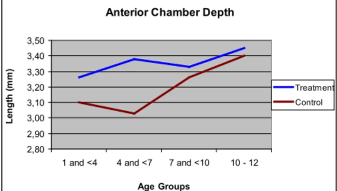
The percentages of the boys which were higher both in the study and the control groups were 59.7% (98/66) and 61.2% (101/64), respectively. The ratio of boys to girls was 1.48 in the study group and 1.58 in the control group. The axial length was 22.16 (range:20.02-25.14) mm in the treatment group and 21.99 (range:18.51-25.03) mm in the control group (Figure-1). Although AL was longer in the study group, the difference was not found to be significant (p>0.05). ACD, LT, and VCD results were 3.35 (range:2.30-4.37), 3.64 (range:2.82-5.98), and 15.20 (range:12.87-19.27) in the treatment group and 3.20 (range:2.44-4.23), 3.63 (range:3.00-5.98), and 15.15 (range:11.80-17.99) mm in the control group (p<0.05, p>0.05, p>0.05, respectively). Although all components of AL in the study group were higher than that of the control group, only the difference in ACD result was statistically significant (p<0.05) (Figure-2). The anterior chambers of the unoperated eyes were deeper when compared to the age-matched normal controls.
In both groups, there was no association between CCT and AL, ACD, LT, or VCD (p>0.05), but a positive relationship between AL, ACD, and VCD but a negative one with LT. The negative relationship between AL and VCD with LT suggests emmetropization. As a matter of fact the increase in ACD already accompanies the increases in AL and VCD. Central corneal thickness was 548 microns (range: 487-632) and 559 microns (range: 455-668) in the study and the control groups, respectively. CCT was thinner in the study group than that of the control group and the difference was found to be statistically significant (p<0.05) (Figure-3:).
In 1 and <4 year age group the results of study and control groups were: CCT: 555 vs 551, AL: 21.22 vs 20.96, ACD: 3.26 vs 3.10, LT: 3.63 vs 3.68, VCD: 14.42 vs 14.18. Only the differences in ACD and LT were statistically significant in this sub-group.
In 4 and <7 year age group: CCT: 549 vs 564, AL: 21.96 vs 21.78, ACD: 3.83 vs 3.03, LT: 3.73 vs 3.76, VCD: 14.84 vs 14.99. In 7 and <10 year age group: CCT: 546 vs 563, AL: 22.48 vs 22.29, ACD: 3.33 vs 3.26, LT: 3.69 vs 3.55, VCD: 15.46 vs 15.47. In 10-12 year age group: CCT: 544 vs 560, AL: 23.19 vs 22.93, ACD: 3.45 vs 3.40, LT: 3.58 vs 3.56, VCD: 16.16 vs 15.96. In all three sub-groups (4 and <7 years, 7 and <10 years, 10-12 years) a significant difference between the unoperated eyes and that of the control group was found only in CCT and ACD results (Table-1).
The mean +/- standard deviations (SD) were shown for each biometric measure for the control group and the treatment group in Table-1.
Discussion
Axial length
In this study, the results have shown that the total axial length in the unoperated eyes was longer than that of the age-matched control group, but the difference was not statistically significant. Regarding the components of AL, all individual parameters (ACD, LT, and VCD) were also longer in the treatment group than that of the control group but only the difference in ACD result was statistically significant (p<0.05). Hence the greater ACD in the treatment group was chiefly responsible for the overall increase in axial length.
The literature is extremely limited in terms of the comparison of biometry data in fellow healthy eyes of the unilaterally operated patients with that of normal control eyes. Nishi et al (2014) reported axial lengths in three groups
as 21.55 mm in healthy eyes of unilateral amblyopia, 22.14 mm in eyes with amblyopia and 21.72 mm in age-matched normal subjects and found no significant difference between the three groups. In that study, the axial lengths ofthe healthy eyes of the unilateral amblyopic subjects were shorter than those of the eyes of the age-matched normal subjects (21.55 vs 21.72). This finding is inconsistent with our findings in which the axial length was founded as longer in the healthy eyes of the unilaterally affected subjects. There are limitations in their study since they had studied hyperopic patients whose axial length was already shorter. Secondly, the quite small sample size in the study might have given the opposite results. Marjanovic et al (2008) studied axial elongation in the first three years of life in 48 pediatric cataractous eyes and also found that there was no significant difference between the unaffected healthy eyes and the normal control eyes.
Although several studies carried out in last decades (1993-2012) reported no significant difference in axial length within-subject inter-ocular comparisons, there are conflicting results as some studies report elongation in fellow unoperated eyes, while the others report shortening (Zhou et al 2000; Huang and Xie, 2005; Urban et al 2007; Filipek et al 2006; Hussin and Markham, 2009; Inatomi et al 2004; Vasavada et al 2004; Flitrcroft et al 1999; Sminia et al 2010). Lal et al (2005) reported both results. They examined 171 pediatric cataractous eyes and reported that while 24% of the operated eyes were longer, 24% of the operated eyes were shorter than the fellow eye.
Trivedi and Wilson (2007) reported that the fellow unoperate d eyes (control group) were significantly longer than the operated eyes. In another study, Cass and Tromans (2008) examined axial length and components in four
and anisometropic amblyopia-control group. They reported that the eyes of the control group were longer than the anisometropic amblyopic eyes. They also reported that anterior chambers were longer in the eyes of the control group but there was no difference between lens thicknesses. Our study accepts the fellow unaffected eye as the study group and reports a slight increase in axial length in these eyes. This result is also consistent with the findings of the studies of Trivedi-Wilson and Cass Tromans. In our study the most significant difference was concerning with ACD in that ACD in the untreated eyes was 0.15 mm longer on average in comparison to the eyes of the normal children. This finding is in agreement with the results of Kora et al (1993) who reported that ACD in fellow untreated eyes were larger than the cataractous ACD. In another study Debert et al (2011) reported that there was no significant difference in ACD but a significant difference in LT and VCD.
Central corneal thickness
There are several studies regarding corneal thickness in children but comparative studies are very rare. In the reports, the range of corneal thickness changes from 529 to 564 µm and the average is likely 550 µm (Faramarzi et al 2010; Bradfield et al 2011; Fern et al 2012; Resende et al 2012; Sahin et al 2008; Coste et al 2008; Hussein et al 2004; Mendes et al 2011). In our study, central corneal thickness is 548 µm in the study group and 559 µm in the control group. CCT was significantly thinner in the study group than that in the control group. In a study, Mendes et al (2011) investigated
CCT in patients with congenital glaucoma and control group. They found the results as 539 µm in the congenital glaucoma without Haab striae group and 559 µm in the control group. In another study, Faramarzi et al (2010) investigated 47 eyes of 30 patients with congenital cataract for pre- and post-operative results and those of the patients in the age-matched control groups. In that study, only 10 eyes had been evaluated as unoperated eyes. Despite we could not exactly compare our results with those of their studies due to the lower sample size of number of unoperated eyes, the mean CCT in the unoperated eyes (540 µm) was thinner than that in the control group (558 µm).
The presumption to have thinner corneas in our study group can be explained possibly by collagen bundle rearrangement and remodelization, which was mentioned in a study by Parentin and Pensiero (2010).
One of the disadvantages of our study is the not using the immersion method, which gives more accurate results than the contact method
Conclusions
Axial length and all components of biometry, i.e. anterior chamber depth, lens thickness, and vitreous chamber depth are higher, but not statistically significant, in the unaffected healthy eyes of the pediatric patients than those in the control group. Greater anterior chamber depth in this group was chiefly responsible for the overall increase in axial length. Central corneal thickness was significantly thinner in the study group than that in the control group.
Table 1: Mean results in age groups. CCT: central corneal thickness, AL: axial length, ACD: anterior chamber depth, LT: lens thickness, VCD: vitreous chamber depth, n: number, mm: millimeter, µm: micron, SD: Standard deviation
Mean Results in Age Groups in Both Group
CCT AL ACD LT VCD Ages n: µm ± SD mm ± SD mm ± SD mm ± SD mm ± SD 1 and <4 Treatment 42 555 ± 21 21.22 ± 0.76 3.26 ± 0.32 3.53 ± 0.27 14.42 ± 0.83 Control 37 551 ± 37 20.96 ± 1.27 3.10 ± 0.33 3.68 ± 0.25 14.18 ± 1.21 4 and <7 Treatment 38 549 ± 28 21.95 ± 0.59 3.38 ± 0.25 3.73 ± 0.42 14.84 ± 0.72 Control 47 564 ± 34 21.78 ± 0.83 3.03 ± 0.31 3.76 ± 0.42 14.99 ± 0.93 7 and <10 Treatment 44 546 ± 17 22.48 ± 0.79 3.33 ± 0.38 3.69 ± 0.40 15.46 ± 0.88 Control 43 563 ± 40 22.29 ± 0.93 3.26 ± 0.30 3.55 ± 0.27 15.47 ± 0.89 10-12 Treatment 41 544 ± 20 23.18 ± 0.97 3.45 ± 0.29 3.58 ± 0.28 16.16 ± 1.04 Control 37 560 ± 39 22.92 ± 0.87 3.40 ± 0.33 3.56 ± 0.21 15.96 ± 0.78
Axial Lengths in Both Groups
19,50 20,00 20,50 21,00 21,50 22,00 22,50 23,00 23,50
1 and <4 4 and <7 7 and <10 10-12
Age Groups m illim et er s Treatment Control
Figure 1: Axial lengths in both groups
Individiual Components of Axial Length ACD - LT - VCD 0,00 2,00 4,00 6,00 8,00 10,00 12,00 14,00 16,00 18,00
Treatment Control Treatment Control Treatment Control Treatment Control
(1 and <4 Ages) (4 and <7 Ages) (7 and <10 Ages) (10-12 Ages)
Age Groups Le ngt h ( m m )
Anterior Chamber Depth
2,80 2,90 3,00 3,10 3,20 3,30 3,40 3,50
1 and <4 4 and <7 7 and <10 10 - 12 Age Groups Le ng th (m m ) Treatment Control
Figure 2: Axial length and components in
age groups. ACD: Anterior Chamber Depth, LT: Lens Thickness, VCD: Vitreous Chamber Depth
Central Corneal Thickness
530 535 540 545 550 555 560 565 570
1 and <4 4 and <7 7 and <10 10 - 12 Age Groups m ic ro ns Treatment Control
Figure 3: Anterior chamber depth in age groups.
Figure 4: Central corneal thickness in both
References
Augusteyn RC, Nankivil D, Mohamed A, Maceo B, Pierre F, Parel JM (2012). Human ocular biometry. Exp Eye Res; 102:70-5.
Bradfield YS, Melia BM, Repka MX, Kaminski BM, Davitt BV, Johnson DA, et al (2011). Pediatric Eye Disease Investigator Group. Central corneal thickness in children. Arch Ophthalmol;129 (9):1132-8.
Bruce A, Pacey IE, Bradbury JA, Scally AJ, Barrett BT (2013). Bilateral changes in foveal structure in individuals with amblyopia. Ophthalmology; 120(2):395–403
Calossi A (1994). Increase of ocular axial length in infantile traumatic cataract. Optom Vis Sci; 71(6):386-91.
Cass K, Tromans C (2008). A biometric investigation of ocular components in amblyopia. Ophthalmic Physiol; Opt 28(5):429-40.
Coste R, Cornand E, Denis D (2008). Central corneal thickness in a pediatric population using a noncontact specular microscope: a study of 405 cases. J Fr Ophtalmol; 31(3):273-8.
Debert I, de Alencar LM, Polati M, Souza MB, Alves MR (2011). Oculometric parameters of hyperopia in children with esotropic amblyopia. Ophthalmic Physiol Opt; 31:389-97.
Faramarzi A, Javadi MA, Jabbarpoor Bonyadi MH, Yaseri M (2010). Changes in central corneal thickness after congenital cataract surgery. J Cataract Refract Surg; 36(12): 2041-7.
Fern KD, Manny RE, Gwiazda J, Hyman L, Weise K, Marsh-Tootle W; COMET Study Group (2012). Intraocular pressure and central corneal thickness in the COMET cohort. Optom Vis Sci; 89(8):1225-34.
Filipek E, Koraszewska B, Samochowiec
E, Nawrocka L, Pieczara E (2006). [Variability of the eyeballs axial length in children with pseudophakia]. Klin Oczna; 108(7-9): 301-5.
Flitrcroft DI, Knight-Nanan D, Bowell R, et al (1999). Intraocular lenses in children changes in axial length, corneal curvature and refraction. Br J Ophthalmol; 83:265–269.
Gee SS, Tabbara KF (1988). Increase in ocular axial length in patients with corneal opacification. Ophthalmology; 95(9): 1276-8.
Gordon RA, Donzis PB (1985). Refractive development of the human eye. Arch Ophthalmol; 103:785-789.
Huang YS, Xie LX (2005). [Refractive change and axial growth after bilateral intraocular lens implantation in children with congenital cataract]. Zhonghua Yan Ke Za Zhi; 41(4):335-9.
Hussein MA, Paysse EA, Bell NP, et al (2004). Corneal thickness in children. Am J Ophthalmol; 138(5):744-8.
Hussin HM, Markham R (2009). Changes in axial length growth after congenital cataract surgery and intraocular lens implantation in children younger than 5 years. J Cataract Refract Surg; 35(7):1223-8.
Inatomi M, Kora Y, Kinohira Y, et al (2004). Long-term follow-up of eye growth in pediatric patients after unilateral cataract surgery with intraocular lens implantation. J AAPOS; 8(1):50-5.
Isenberg SJ. Physical and refractive characteristics of the eye at birth and during infancy. In : Isenberg SJ, editor. The eye in infancy. 2nd edition. St. Louis: Mosby; 1994; p.36-51.
Johnson DA (2006). The use of the scanning laser ophthalmoscope in the evaluation of amblyopia (an American Ophthalmological Society thesis). Trans Am Ophthalmol Soc; 104:414–36
Kim YW, Kim SJ, Yu YS (2013). Spectral-domain optical coherence tomography analysis in deprivational amblyopia: a pilot study with unilateral pediatric cataract patients. Graefes Arch Clin Exp Ophthalmol; 251(12):2811-9.
Kora Y, Shimizu K, Inatomi M, et al (1993). Eye growth after cataract extraction and intraocular lens implantation in children. Ophthal Surg; 24:467–475.
Lal G, Trivedi RH, Wilson ME Jr, Scarlett LC, Peterseim MM (2005). Interocular axial length difference in eyes with pediatric cataracts. J AAPOS; 9(4):358-62.
Larsen JS (1971). The sagittal growth of the eye. IV. Ultrasonic measurement of the axial length of the eye from birth to puberty. Acta Ophthalmol; 49(6):873– 886.
Leguire LE, Rogers GL, Bremer DL (1990). Amblyopia: the normal eye is not normal. J Pediatr Ophthalmol Strabismus; 27(1):32-8
Leiba H, Springer A, Pollack A (2006). Ocular axial length changes in pseudophakic children after traumatic and congenital cataract surgery. J AAPOS; 10(5):460-3.
Marjanović I, Stefanović I, Vlajković G (2008). [Axial length of the eye in the first three years of life in children with congenital cataract in one or both eyes]. Srp Arh Celok Lek; 136(1-2):7-11.
Mendes MH, Sakata L, Betinjane AJ (2011). [Central corneal thickness and its correlations with other ocular biometric data in patients with congenital glaucoma]. Arq Bras Oftalmol; 74(2):85-7.
Nishi T, Ueda T, Hasegawa T, Miyata K, Ogata N (2014). Choroidal thickness in children with hyperopic anisometropic amblyopia. Br J Ophthalmol; 98(2):228-32.
Parentin F, Pensiero S (2010). Central corneal thickness in children with growth
hormone deficiency. Acta Ophthalmol; 88(6):692-4
Pineles SL, Demer JL (2009). Bilateral abnormalities of optic nerve size and eye shape in unilateral amblyopia. Am J Ophthalmol; 148(4):551–57
Rabin J, Van Sluyters RC, Malach R (1981). Emmetropization: a vision-dependent phenomenon. Invest Ophthalmol Vis Sci; 20(4):561-4.
Rasooly R, BenEzra D (1988). Congenital and traumatic cataract. Arch Ophthalmol; 106:1066–1068.
Raviola E, Wiesel TN (1985). An animal model of myopia. N Engl J Med; 312:1609
Resende GM, Lupinacci AP, Árieta CE, Costa VP (2012). Central corneal thickness and intraocular pressure in children undergoing congenital cataract surgery: a prospective, longitudinal study. Br J Ophthalmol; 96(9):1190-4.
Sahin A, Basmak H, Yildirim N (2008). The influence of central corneal thickness and corneal curvature on intraocular pressure measured by tono-pen and rebound tonometer in children. J Glaucoma; 17(1):57-61.
Sminia ML, de Faber JT, Doelwijt DJ, Wubbels RJ, Tjon-Fo-Sang M (2010). Axial eye length growth and final refractive outcome after unilateral paediatric cataract surgery. Br J Ophthalmol; 94(5):547-50.
Smith EL, Harwerth RS, Crawford ML, von Noorden GK (1987). Observations on the effects of form deprivation on the refractive status of the monkey. Invest Ophthalmol Vis Sci; 28(8):1236-45.
Trivedi RH, Wilson ME (2007). Biometry data from caucasian and african-american cataractous pediatric eyes. Invest Ophthalmol Vis Sci; 48(10):4671-8.
Urban B, Bakunowicz-Lazarczyk A, Sredzińska-Kita D, Mrugacz M (2007). [Retrospective evaluation of ocular axial length after unilateral cataract surgery with intraocular lens implantation in children and adolescents]. Klin Oczna; 109(10-12):425-7.
Vasavada AR, Raj SM, Nihalani B (2004). Rate of axial growth after congenital cataract surgery. Am J Ophthalmol; 138(6):915-24.
Wiesel TN, Raviola E 1979 (). Increase in axial length of the macaque monkey eye after corneal opacification. Invest Ophthalmol Vis Sci; 18(12):1232-6.
Wiesel TN, Raviola E (1977). Myopia and eye enlargement after neonatal lid fusion in monkeys. Nature; 266(5597):66-8.
Zadnik K, Manny RE, Yu JA, et al (2003). Collaborative Longitudinal Evaluation of Ethnicity and Refractive Error (CLEERE) Study Group. Ocular component data in schoolchildren as a function of age and gender. Optom Vis Sci; 80(3):226-36.
Zhou J, Zhou L, Wu Y, Hu D, Hui Y (2000). [Ocular axial length and refractive changes in pediatric pseudophakia]. Yan Ke Xue Bao; 16(4):270-5.