Corresponding author: Ali SAMANCIOGLU E-mail: [email protected]
Original Investigation
Published Online: 05.03.2016Ali SAMANCIOGLU
1, Erhan AKINCI
2, Arif OSUN
3, Ozan GANIUSMEN
4, Umit OZKAN
3, Cuneyt TEMIZ
51Buca Seyfi Demirsoy State Hospital, Neurosurgery Clinic, İzmir, Turkey 2Buca Seyfi Demirsoy State Hospital, Psychiatry Clinic, İzmir, Turkey
3Dumlupınar University, School of Medicine, Department of Neurosurgery, Kütahya, Turkey 4Şifa University, School of Medicine, Department of Neurosurgery, İzmir, Turkey
5Celal Bayar University, School of Medicine, Department of Neurosurgery, Manisa, Turkey
Actigraphic Analysis of Patients with Cervical Disc Herniation
ABSTRACT
consulting a physician in these patients. Sleep disorders are in the second place after brachialgia with a ratio of 64% (5, 12,24,27).
Sleep disorders are frequently seen in patients with chronic low back pain (17). In another study, pain was shown to be the most important factor impairing quality of sleep (23). Other studies demonstrated a significant relationship between the severity of pain and the impairment in sleep quality. It has been also shown that quality of sleep improves after the treat-ments focusing on the cause of pain (5,6).
In addition to chronic pain syndromes, there are a lot of studies on sleep disorders in patients who underwent open heart surgery, major abdominal surgery and thoracic surgery which were targeted to the disease underlying the pain (29). In the █
INTRODUCTION
C
ervical disc herniation (CDH) is characterized by compression of the motor and sensory tracts of cervical nerve roots (radiculopathy) and/or compression of the motor and sensory tracts of the long pathways in the spinal cord (myelopathy) as a result of degenerative events in the cervical vertebrae. An intractable and severe pain radiating from the neck down to the arm (brachialgia) and progressive neurological deficits associated with radiculopathy and/or myelopathy findings in cervical magnetic resonance imaging (MRI) are the major surgical indications in patients with CDH. The surgical procedure in CDH is at the same time a surgical procedure for pain relief. Brachialgia associated with radiculopathy and myelopathy is the main reason forAIm: To analyze the relationship between the severity of pain and sleep disorder using wrist actigraphy in patients with cervical disc herniation (CDH).
mATERIAl and mEThODS: Fifty patients with the diagnosis of CDH underwent subjective tests and actigraphic analysis in preoperative period, and at the end of postoperative first week and postoperative first month. The data of the subjective tests and actigraphic analysis were compared.
RESUlTS: There was a strong and statistically significant negative correlation between the subjective tests of Visual Analog Scale (VAS) for Pain 0-1-2 and the objective parameters of Sleep Onset Latency (SOL) 0-1-2 (rs= -0.798, p=0.009 – rs= - 0.832, p=0.006 – rs=- 0.710, p=0.004). There was a positive correlation between the subjective tests of VAS for Pain 0-1-2 and the objective parameters of Sleep Efficiency (SEF) 0-1-2 (rs=0.721, p=0.006 – rs= 0.768, p=0.001 – rs= 0.748, p=0.001).
CONClUSION: Actigraphy may be used for the evaluation of cervical disc surgery, as an alternative and objective test for sleep disorders.
studies, the assessments of the patients have generally been relied on patient’s statements and tests based on subjective criteria (1-Visual Analog Scale for Pain (VAS Pain), 2-Oswestry Disability Index (ODI), 3-Beck Depression Index (BDI), 4-Pittsburgh Sleep Quality Index (PSQI), 5-Insomnia Severity Index (ISI), and SF-36 Quality of Life Index (SF-36 QLI)) (10, 25,27,28). Polysomnography (PSG) and actigraphy have been used for objective sleep measurements. Although PSG is still the gold standard in objective measurement of sleep, it has some disadvantages such as need for being hospitalized, poor patient compliance and need for a special setting. Actigraphy is now used as an alternative method for objective sleep measurement (4,8,11). Actigraphy, which was validated by the American Sleep Academy through scientific research to be an objective sleep measurement method alternative to PSG, does not require hospitalization and it records the measurements with a wrist device that is easier for patients to comply with. These records are stored in a computer and the data are obtained through analysis programs (7,15,16,18). Although the second most frequent complaint is sleep disorder in CDH patients, who were hospitalized in neurosurgery clinics for a surgical intervention, we realized that the relationship between CDH and sleep disorders has not been adequately investigated in the literature. Sleep disorders have only been assessed in some CDH patients who did not need any surgery and for whom medication and physical exercise were planned (14). In 2013, Madsen et al. reported that there was no study in the literature evaluating the sleep disorders using actigraphy in patients who underwent surgery for CDH. So, they were pointed out a deficiency in the literature (15).
In our study, we aimed to compare the effects of pre- and postoperative pain and sleep disorders on the quality of life in patients who underwent surgery for CDH. We also investigated the relationship between severity of pain and sleep disorder in pre- and postoperative periods using wrist actigraphy test. █
mATERIAl and mEThODS
Our study is a multi-center study involving Buca Seyfi Demirsoy State Hospital Neurosurgery and Psychiatry Clinics, Şifa University Medical School Neurosurgery Clinic, Dumlupınar University Medical School Neurosurgery Clinic, and Celal Bayar University Medical School Neurosurgery Clinic. Ethical approval was obtained from the Clinical Trials Medical Ethics Committee of Şifa University (Date: 12.02.2014, number: 144-44).
Our study was carried out in 50 patients, who were informed about the study and whose consents were obtained. The patients were admitted to our clinics between February and December 2013. They were between 40 and 60 years of age, and they were advised to undergo surgical treatment with the diagnosis of CDH after radiological and neurological examinations.
Exclusion criteria were; being treated for another disease causing sleep disorder, having a metabolic disease such as diabetes mellitus, hypertension, heart disease or a psychiatric condition, and using narcotic or psychotic drugs.
We did not use a healthy control group for this study because we aimed to investigate the correlation between pain severity levels, quality of life and sleep quality before and after the surgery for CDH.
Following the information of the patients about sleep hygiene procedures and recording their demographic data, we carried out the following procedures in preoperative period and at the postoperative first week and first month:
1. Demographic data: The patients were inquired about their names-surnames, ages, genders, economic statuses, known diseases, drug and substance abuse, operations and treatments.
2. Visual Analog Scale for Pain (VAS Pain): This scale was used in assessing the pain of the patients with CDH. The patients were explained about the meanings of the figures from 0 to 10 placed on a 10 cm scale. They were asked to show on the scale the numeric value that best described their pain (21,27, 28,30).
3. Oswestry Disability Index (ODI): This index consists of 10 items measuring the patients’ pain intensity, personal care, lifting, walking, sitting, standing, sex life, social life, sleeping, and traveling. Each item is scored between 0 and 5. Higher total scores indicate a higher level of disability. The maximum score is 50 points; 31-50 points was considered as severe, 11-31 as moderate and 1-10 as mild disability (1,22,27,28, 30). 4. Pittsburgh Sleep Quality Index (PSQI): This index was developed by Buysse et al. to assess an individual’s sleep quality, sleep duration and sleep disorders. Consisting of nineteen questions, the scale is scored between 0 and 3. PSQI contains seven self-rated sub-components; sleep quality, sleep latency, sleep duration, habitual sleep efficiency, sleep disturbance, use of sleeping medication and daytime dysfunction. Higher scores indicate lower sleep quality. The scale was tested for validity and reliability in Turkish by Agargun et al. (2,20,23).
5. Beck Depression Index (BDI): This scale was developed by Beck et al. to measure the severity of depression symptoms. The scale has 21 items, which are scored between 0 and 3. The scale was adapted to Turkish and shown to be acceptable by Hisli (1,3,11,26,30).
6. Insomnia Severity Index (ISI): Developed to assess insomnia severity, this is a measurement instrument having high validity and reliability. Its items consist of seven questions and are scored between 0 and 4. The score obtainable from the scale range between 0 and 28. Based on the ratings of the individuals and their partners, ISI is used to measure difficulty in falling asleep, difficulty in staying asleep, problems waking up too early, satisfaction with sleeping pattern, interference with daily functioning, noticeability of sleep-related impairments, and level of stress caused by the problem (18,20,28,30).
7. The 36-Item Short Form Health Survey (SF-36): Developed to measure quality of life particularly in the patients who have a physical illness, SF-36 is the most widely used survey in measuring quality of life. It can assess the positive aspects of health conditions as well as their negative aspects. The
survey’s most prominent superiority is its ability to measure physical functioning and skills associated with it. The survey consists of 36 items, which enable measurement of 8 domains; physical functioning (10 items), social functioning (2 items), role limitations due to physical health (4 items), role limitations due to emotional problems (3 items), mental health (5 items), energy/vitality (4 items), pain (2 items) and perceived general health (5 items). SF-36 is a patient-reported survey (20,22,26, 30).
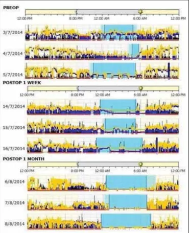
8. Use of actigraphic wristband: Actigraph (Actiwatch 2 Philips Respironics Co. Inc. 1001 Murry Ridge Lane Murrysville, Pennsylvania, USA) is a lightweight, portable and watch-like device that can precisely sense and record motor activities with its accelerometer (Figure 1). After being read to a computer, the recorded data are analyzed on an actigraphy program (Figure 2). It provides objective information on measuring and assessing continuity of sleep and sleep-related disorders. In addition to the subjective tests (consisting of reported information by the patient) being used today in sleep studies, actigraphy devices have been accepted and approved as an alternative to the PSG device. Actigraphy is accepted as being the gold standard among the objective sleep tests, has the advantage of practical usage without the need of hospitalization (4,7,8,11,13,16,18). The standards have been established for its usage (14,15).
After briefing our patients about sleeping, we gave them information about the device to eliminate their worries. We asked our patients to wear the actigraphic wristband on their non-dominant wrists at least 3 consecutive days before the surgery and at least 3 days after the postoperative 7th day (the
medical support process after anesthesia and surgery) and at least another 3 days after postoperative first month.
We used the actigraph during the main sleep at night to measure the objective values for sleep quality and insomnia such as total bed time (TBT), total sleep time (TST), sleep efficiency (%)(SEF), Wake time after sleep onset (minutes) (WASO), sleep-onset latency (SOL) and numbers of awakening (Awake).
Actigraphs are accelerometers that can record the main motor movements of a person for a long time and can measure motions of at least 0.01 G in amplitude. Our wrist actigraphy devices had the capacity of detecting a motion up to 32 micro-deflexions (32 Hz) a second. The data is stored in the device’s memory in 30-second epochs. The data was uploaded to a computer to be analyzed by an actigraphic program and daily actograms were collected. The analysis data were obtained electronically in preoperative period and postoperative first week and first month separately for each patient. From our study group of 50 patients, 2 were excluded from the study at the end of postoperative first week because they needed additional medical treatment due to incision site complications. They were replaced by new patients.
CDH was located in the right side in 30 patients (60%) and in the left side in 20 patients (40%) in our study. The level of CDH was C4-C5 in 9 (18%) patients, C5-C6 in 21 (42%) patients and C6-C7 in 20 (40%) patients. The disc was located laterally in 32 (64%) patients and mediolaterally in 18 (36%) patients.
All of our patients had single-level CDH. Same surgical method was performed to all patients, which involved a microsurgical cervical discectomy with anterior approach and placing a titanium cage in the size of the removed disc.
Statistical Analysis
Statistical analyses were performed using the Rstudio software version 0.98.501 via R language. The variables were investigated using visual (histograms, probability plots) and analytical methods (Kolmogorov-Simirnov/Shapiro-Wilk test)
Figure 1: Device of wrist actigraph.
█
RESUlTS
Fifty patients were included in this study. Twenty nine (58%) patients were male and 21 (42%) were female. The mean age of male patients was 52.62±5.86 years and that of female patients was 53.24±4.57 years. There was no statistically significant difference between male and female patients according to their ages. All patients stated that they had sleep disorders together with pain complaints.
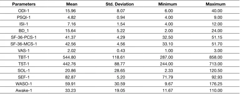
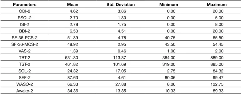
The descriptive statistics of the subjective surveys which were administered to each patient before the operation (preop-0) and at the first week after the operation (postop-1) and at the first month after the operation (postop-2) and those of the objective actigraphic parameters are presented in Tables I - III, respectively. The comparison of the parameters is presented in Tables IV - VI.
to determine whether or not they are normally distributed. Descriptive analyses were presented using means, standard deviations and minimum-maximum for variables-parameters (ODI; PSQI; ISI; BDI; SF-36-PCS; SF-36-MCS; VAS; TBT; TST; SOL; %SEF; WASO; Awake Time ).
Since the variables were not normally distributed; non-parametric tests were conducted to compare these parameters. The Wilcoxon test was used to compare the change in these parameters between PreOp, PostOp -1 Week Later and PostOp-1 Month Later. The Mann-Whitney U test was used to compare these parameters between genders. While investigating the associations between non-normally distributed variables, the correlation coefficients and their significance were calculated using the Spearman test. A p-value of less than 0.05 was considered as statistically significant.
Table I: Descriptive Statistics of Parameters for Preoperative Period
Parameters mean Std. Deviation minimum maximum
ODI-0 45.58 4.85 41.00 50.00 PSQI-0 11.06 1.68 5.00 14.00 ISI-0 21.46 3.08 11.00 28.00 BDI-0 45.86 9.66 13.00 56.00 SF-36-PCS-0 23.28 4.72 18.19 41.55 SF-36-MCS-0 28.31 5.96 18.60 39.25 VAS-0 8.98 0.39 8.00 10.00 TBT-0 608.74 147.32 330.00 970.00 TST-0 404.60 115.86 187.00 745.00 SOL-0 79.93 56.25 18.50 294.50 SEF-0 65.45 9.71 33.49 82.37 WASO-0 53.12 36.85 5.92 138.33 Awake-0 53.34 28.00 8.33 153.25
Table II: Descriptive Statistics of Parameters for 1 Week After Surgey
Parameters mean Std. Deviation minimum maximum
ODI-1 15.96 8.07 6.00 40.00 PSQI-1 4.82 0.94 4.00 9.00 ISI-1 7.16 1.54 4.00 12.00 BD_1 15.64 5.22 2.00 24.00 SF-36-PCS-1 41.37 4.29 32.50 51.15 SF-36-MCS-1 42.56 4.56 33.10 51.70 VAS-1 2.02 0.43 1.00 3.00 TBT-1 544.80 118.61 287.00 858.00 TST-1 442.76 88.77 244.00 713.00 SOL-1 20.86 28.65 2.33 120.50 SEF-1 82.87 5.20 71.79 92.93 WASO-1 59.91 30.59 9.67 176.25 Awake-1 33.23 19.05 11.67 110.00
and very strong (rs= 0.90 – 1.00) relationships. We gave the
results concerning the correlation between the subjective and objective test parameters at preoperative and postoperative first week and postoperative first month in Tables VII, VIII and IX, respectively. Color scales in Tables VII, VIII and IX show statistically significant p values and correlation coefficients between the objective actigraphic analysis parameters and the subjective parameters as well as between the objective parameters’ own analyses.
It may be an inconsistency between our clinical observations and the test results based on patients’ subjective criteria about quality of life and sleep quality at preoperative period and postoperative first week and first month. The causes of this inconsistency may be too self-conscious patients, or patients might be influenced by the environment. On the other In order to increase the statistical reliability and accuracy of
our study, we used a patient population aged between 40 and 60 years whose sleep duration and sleep quality were rather homogenous. Statistical analyses did not show any statistical difference between the variables of male/female patients and patient age, and the study parameters in any of the three periods. A statistically significant conformity that supported each other appeared in the tables in the mean results of subjective parameters and objective actigraphic analysis parameters at preoperative period and postoperative first week and postoperative first month.
Additionally, accepting that p value<0.05 indicates a statistically significant result, a four-step color scale from light green to dark green was designed according to correlation coefficients to show unaccountably low (rs= 0.000 – 0.19), weak (rs= 0.20
– 0.39), moderate (rs= 0.40 – 0.69), strong (rs= 0.70 – 0.89)
Table III: Descriptive Statistics of Parameters for Postoperative (One Month After Surgery)
Parameters mean Std. Deviation minimum maximum
ODI-2 4.62 3.86 0.00 20.00 PSQI-2 2.70 1.30 0.00 5.00 ISI-2 2.78 1.75 0.00 8.00 BDI-2 6.50 4.51 0.00 20.00 SF-36-PCS-2 51.39 4.78 40.75 65.50 SF-36-MCS-2 48.92 2.95 43.50 54.45 VAS-2 1.39 0.46 1.00 2.00 TBT-2 531.30 113.37 384.00 889.00 TST-2 461.82 101.69 319.00 885.00 SOL-2 24.32 17.05 2.75 84.32 SEF-2 87.63 4.61 80.06 99.47 WASO-2 66.33 27.88 8.06 122.75 Awake-2 34.36 13.85 10.33 89.33
Table IV: p-values of Parameters between PreOp and PostOp-1 Week Later Parameters p-Value ODI 0.000 PSQI 0.000 ISI 0.000 BDI 0.000 SF-36-PCS 0.000 SF-36-MCS 0.000 VAS 0.000 TBT 0.032 TST 0.066 SOL 0.000 SEF 0.000 WASO 0.037 Awake 0.001
Table V: p-values of Parameters between PreOp and PostOp-1 Month Later Parameters p-Value ODI 0.0001 PSQI 0.0001 ISI 0.0001 BDI 0.0001 SF-36-PCS 0.0001 SF-36-MCS 0.0001 VAS 0.0001 TBT 0.007 TST 0.013 SOL 0.0001 SEF 0.0001 WASO 0.015 Awake 0.0001
hand, the objective actigraphic analysis parameters gave us the impression of being more reliable improvement criteria. █
DISCUSSION
The studies on the actigraphic analyses showed that this technique is still the gold standard for sleep disorders. Actigraphic analyses were accepted and approved by the American Sleep Academy and used in many centers as an alternative to PSG with a 90% rate of consistency (4,8,13,16, 18). Its administration is easy and it has a cost advantage over PSG. Recently, researchers have begun to use actigraphic analyses not only in studies related to sleep disorders and quality, but also in pain studies. Hartwell et al. reported that pain is an important factor affecting sleep and demonstrated the relationship between pain and sleep disorders using actigraphic analysis parameters. They also found statistically consistent results on the use of actigraphic TBT, TST, SEF, SOL, WASO and Awake analysis parameters through drug treatments against pain (9).
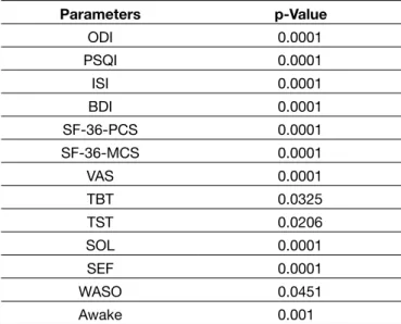
Table VI: p-values of Parameters between PostOp-1 Week and PostOp-1 Month Later
Parameters p-Value ODI 0.0001 PSQI 0.0001 ISI 0.0001 BDI 0.0001 SF-36-PCS 0.0001 SF-36-MCS 0.0001 VAS 0.0001 TBT 0.0325 TST 0.0206 SOL 0.0001 SEF 0.0001 WASO 0.0451 Awake 0.001
Table VII: Correlation between Objective and Subjective Measurement Techniques (PreOp)
Variables Spearman’s rho
ODI-0 PSQI-0 ISI-0 BDI-0 SF-36- PCS-0 SF-36- mCS-0 VAS-0 TBT
-0 TST -0 SO l-0 SEF-0 wASO-0
PSQI-0 Correlation Coefficient 0.434
p-Value 0.002
ISI-0 Correlation Coefficientp-Value 0.0001 0.2320.515 0.472
BDI-0 Correlation Coefficient 0.135 0.188 0.409
p-Value 0.349 0.191 0.003 SF-36-PCS-0 Correlation Coefficient -0.072 -0.104 -0.145 -0.012 p-Value 0.617 0.473 0.316 0.932
SF-36-mCS-0 Correlation Coefficient -0.067 0.171 -0.070 0.170 0.440p-Value 0.642 0.234 0.629 0.237 0.016 VAS-0 Correlation Coefficientp-Value 0.464 0.026 0.225 -0.083 0.094 0.1640.001 0.857 0.115 0.566 0.517 0.257
TBT-0 Correlation Coefficient -0.124 -0.333 0.454 -0.384 0.097 0.309 -0.234
p-Value 0.041 0.018 0.009 0.023 0.044 0.049 0.048
TST-0 Correlation Coefficient -0.234 -0.326 0.102 -0.121 0.472 0.425 -0.226 0.714p-Value 0.032 0.015 0.049 0.034 0.032 0.085 0.042 0.000
SOl-0 Correlation Coefficient 0.293 0.287 0.279 0.192 0.165 -0.177 -0.798 0.286 -0.017
p-Value 0.019 0.043 0.040 0.041 0.046 0.018 0.009 0.044 0.006
SEF-0 Correlation Coefficientp-Value 0.167 -0.170 0.156 0.129 0.207 0.137 0.721 -0.154 0.345 -0.2280.044 0.027 0.001 0.027 0.018 0.043 0.006 0.035 0.014 0.111
wASO-0 Correlation Coefficient -0.287 0.257 -0.249 0.124 -0.130 0.131 -0.218 0.556 0.589 0.370 0.457
p-Value 0.044 0.002 0.082 0.047 0.034 0.044 0.029 0.000 0.000 0.029 0.047
was used for 7 consecutive days and a healthy control group was also included in this study studies because they investigated the changes in sleep patterns associated with the blood flow changes in the sleep regulating centers of the central nervous system (29).
Campbell et al. showed that endogenous opioid peptides were strongly bound to the Mu opioid receptors (MOR), which are located especially in the prefrontal cortex, primary and secondary somatosensorial cortex, insula, thalamus and anterior cingulate lobes, in the brain of healthy subjects in whom supraspinal dysfunctional pain was created. Using positron emission tomography (PET) and cranial MRI, they showed same changes in metabolic activity and blood oxygen level signals in the same cerebral regions depending on the level of pain. They also showed the hypoactivation in the dorsolateral prefrontal cortex in cases where sleep disorders and shortening of sleep duration accompanied pain. They stressed that in assessing the progress and outcomes of treatments of pain and pain-causing diseases (fibromyalgia, peripheral neuropathic pain, rheumatoid arthritis, etc.), sleep Similarly, actigraphic analyses were used to compare the
subjective tests on the medical treatment and outcomes of fibromyalgia, osteoporosis, rheumatoid arthritis, chronic lower back pain, shoulder pain and neck pain (7,17,21,22,24,25). In addition, actigraphic analyses have been used to evaluate surgical cases. Madsen et al. examined 32 studies with appropriate criteria out of 143 studies where sleep disorders were assessed using actigraphic analyses in surgically managed cases and they concluded that there was no study from the field of neurosurgery about the use of actigraphic analyses (15).
In our study, the method and duration of actigraphy, as well as the analysis parameters, were similar to those of the recent 32 studies which were evaluated by Madsen et al. These parameters were generally studied for 3 consecutive days before the surgery and postoperative first week and first month in patients who underwent a surgery in the studies assessed by Madsen et al (15).
Yılmaz et al. reported the use of actigraphy in patients who underwent a cardiovascular surgery. In this study, actigraphy
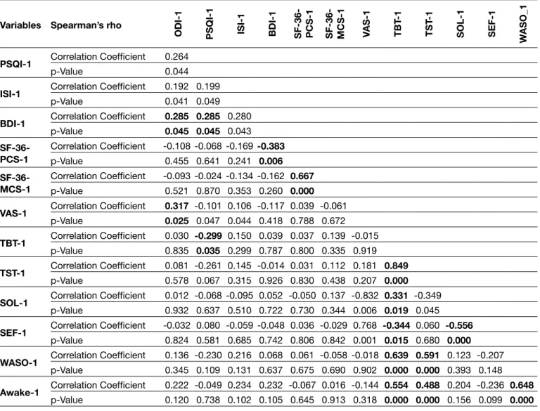
Table VIII: Correlation between Objective and Subjective Measurement Techniques (PostOp-1 Week Later)
Variables Spearman’s rho
ODI-1 PSQI-1 ISI-1 BDI-1 SF-36- PCS-1 SF-36- mCS-1 VAS-1 TBT
-1 TST -1 SO l-1 SEF-1 wASO_1
PSQI-1 Correlation Coefficientp-Value 0.2640.044
ISI-1 Correlation Coefficient 0.192 0.199
p-Value 0.041 0.049
BDI-1 Correlation Coefficientp-Value 0.285 0.285 0.2800.045 0.045 0.043
SF-36-PCS-1 Correlation Coefficient -0.108 -0.068 -0.169 -0.383
p-Value 0.455 0.641 0.241 0.006
SF-36-mCS-1 Correlation Coefficientp-Value -0.093 -0.024 -0.134 -0.162 0.6670.521 0.870 0.353 0.260 0.000
VAS-1 Correlation Coefficient 0.317 -0.101 0.106 -0.117 0.039 -0.061
p-Value 0.025 0.047 0.044 0.418 0.788 0.672
TBT-1 Correlation Coefficientp-Value 0.030 -0.299 0.150 0.039 0.037 0.139 -0.0150.835 0.035 0.299 0.787 0.800 0.335 0.919
TST-1 Correlation Coefficient 0.081 -0.261 0.145 -0.014 0.031 0.112 0.181 0.849
p-Value 0.578 0.067 0.315 0.926 0.830 0.438 0.207 0.000
SOl-1 Correlation Coefficientp-Value 0.012 -0.068 -0.095 0.052 -0.050 0.137 -0.832 0.331 -0.3490.932 0.637 0.510 0.722 0.730 0.344 0.006 0.019 0.045
SEF-1 Correlation Coefficient -0.032 0.080 -0.059 -0.048 0.036 -0.029 0.768 -0.344 0.060 -0.556
p-Value 0.824 0.581 0.685 0.742 0.806 0.842 0.001 0.015 0.680 0.000
wASO-1 Correlation Coefficientp-Value 0.136 -0.230 0.216 0.068 0.061 -0.058 -0.018 0.639 0.591 0.123 -0.2070.345 0.109 0.131 0.637 0.675 0.690 0.902 0.000 0.000 0.393 0.148
Awake-1 Correlation Coefficient 0.222 -0.049 0.234 0.232 -0.067 0.016 -0.144 0.554 0.488 0.204 -0.236 0.648
Academy (15). We planned the recovery process on the basis of the results of subjective tests and objective actigraphic analyses. As we aimed to compare the preoperative and postoperative patient data, we did not include a control group in our study.
The homogeneity of 50 patients in our study is statistically reliable and valid based on gender and age of the patients. Patients who had another disease affecting the patient’s life, quality of life and sleep quality, such as metabolic disease and heart disease or a psychiatric disease, were excluded from the study. We also excluded the patients who had surgery-related complications in the postoperative period.
We know that age affects sleep parameters. Sleep quality and TST are less, and SL and WASO times are longer in older patients (particularly 60 years and over). In addition, TST, SEF, SOL, WASO and Awake parameter values change significantly in older patients than the younger’s (40 years and less) (14,25, 27). By the inclusion criteria, we tried to create a homogenous group of patients aged between 40 and 60 years in our study. disorders should also be taken into consideration (3). We
suggest that, besides being a mechanical pain, CDH also causes the occurrence of a sensorial and emotional response involving sleep disorders as a result of the chain reaction, which is started by the proinflammatory cytokines that are secreted secondary to the herniation and which is perceived by our patients as pain. Consequently, these patients have severe pain and sleep disorder complaints that enforce them to undergo surgical treatment. The inflammatory chain reaction gradually loses its effect during the course of recovery from the operation. It falls below the limits that would not trigger the reaction as the surgical wound recovers.
Patients, who undergo a CDH surgery, usually do not need a postoperative intensive care and are mobilized within 8-12 hours. We removed the surgical wound sutures at the postoperative 5th or 6th days and let the patients have a
bath. We suggested that they may go back to work after the postoperative 20th day. In our study, the assessment processes
was determined with this schedule. Moreover, our study was performed according to the criteria of the American Sleep
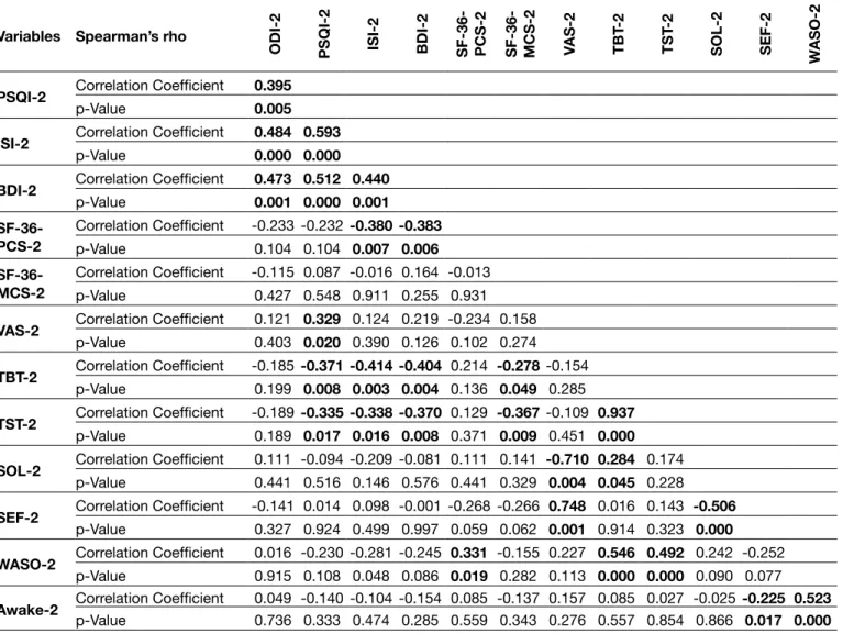
Table IX: Correlation between Objective and Subjective Measurement Techniques (PostOp-1 Months Later)
Variables Spearman’s rho
ODI-2 PSQI-2 ISI-2 BDI-2 SF-36- PCS-2 SF-36- mCS-2 VAS-2 TBT
-2 TST -2 SO l-2 SEF-2 wASO-2
PSQI-2 Correlation Coefficientp-Value 0.3950.005
ISI-2 Correlation Coefficient 0.484 0.593
p-Value 0.000 0.000
BDI-2 Correlation Coefficientp-Value 0.473 0.512 0.4400.001 0.000 0.001
SF-36-PCS-2 Correlation Coefficientp-Value -0.233 -0.232 -0.380 -0.3830.104 0.104 0.007 0.006
SF-36-mCS-2 Correlation Coefficientp-Value -0.115 0.087 -0.016 0.164 -0.0130.427 0.548 0.911 0.255 0.931
VAS-2 Correlation Coefficient 0.121 0.329 0.124 0.219 -0.234 0.158
p-Value 0.403 0.020 0.390 0.126 0.102 0.274
TBT-2 Correlation Coefficientp-Value -0.185 -0.371 -0.414 -0.404 0.214 -0.278 -0.1540.199 0.008 0.003 0.004 0.136 0.049 0.285
TST-2 Correlation Coefficient -0.189 -0.335 -0.338 -0.370 0.129 -0.367 -0.109 0.937
p-Value 0.189 0.017 0.016 0.008 0.371 0.009 0.451 0.000
SOl-2 Correlation Coefficientp-Value 0.111 -0.094 -0.209 -0.081 0.111 0.141 -0.710 0.284 0.1740.441 0.516 0.146 0.576 0.441 0.329 0.004 0.045 0.228
SEF-2 Correlation Coefficient -0.141 0.014 0.098 -0.001 -0.268 -0.266 0.748 0.016 0.143 -0.506
p-Value 0.327 0.924 0.499 0.997 0.059 0.062 0.001 0.914 0.323 0.000
wASO-2 Correlation Coefficientp-Value 0.016 -0.230 -0.281 -0.245 0.331 -0.155 0.227 0.546 0.492 0.242 -0.2520.915 0.108 0.048 0.086 0.019 0.282 0.113 0.000 0.000 0.090 0.077 Awake-2 Correlation Coefficientp-Value 0.049 -0.140 -0.104 -0.154 0.085 -0.137 0.157 0.085 0.027 -0.025 -0.225 0.5230.736 0.333 0.474 0.285 0.559 0.343 0.276 0.557 0.854 0.866 0.017 0.000
objective parameters in the concentrated regions of the color scales in these tables.
The subjective tests are in conformity with the objective actigraphic analysis parameters, but the previous studies mostly focused on demonstrating the relationship between pain-related diseases and sleep disorders (9,15,17,20-22). In our study, as shown in Tables VIII, IX and X, subjective tests were in conformity with objective actigraphic analysis parameters and we suggest that objective actigraphic analysis parameters can be used as an alternative to subjective tests in pain studies.
█
CONClUSION
The pain in CDH is mechanical and causes health problems, including sleep disorders, enforcing the patient to undergo surgical treatment. This pain is the result of chain reaction, which is started by the proinflammatory cytokines after disc herniation. We should be aware of these problems that negatively affect the patient’s quality of life before and after the surgery. We should also consider sleep disorder and sleep quality when assessing our patients for their quality of life. Actigraphic analyses provide objective data about the sleep disorders of patients with CDH.
We suggested SOL and SEF as actigraphic analysis parameters instead of VAS test for the objective pain measurement. Further studies are needed on this issue with larger patient population.
█
REFERENCES
1. Abdülkadiroğlu Z, Bayramoğlu F, İlhan N: Sleep and sleep disorders. Genel Tıp Dergisi 7(3):161-166,1997
2. Boysan M, Güleç M, Beşiroğlu L, Kalafat T: Psychometric properties of the insomnia severity index in Turkish sample. Anatolian Journal of Psychiatry 11: 248-252, 2010
3. Campbell CM, Bounds SC, Kuwabara H, Edwards RR, Campbell JN, Haythornthwaite JA, Smith MT: Individual variation in sleep quality and duration is related to cerebral Mu Opioid Receptor Binding potential during tonic laboratory pain in healthy subjects. Pain Med 14(12):1882-1892, 2013 4. Clete KA, Ghang A, Gadkary C, Guilleminault C, Carrillo O,
Dement WC: Comparison of actigraphic, polysomnographic and subjective assessment of sleep parameters in sleep-disordered patients. Sleep Med 2:389-396, 2001
5. Cunningham JM, Blake C, Power CK, O’keeffe D, Kelly V, Horan S, Spencer O, Fullen BM: The impact on sleep of a multidisciplinary cognitive behavioural pain management programme: A pilot study. BMC Musculoskelet Disord 12:15-21, 2011
6. Evren B, Evren C, Yapıcı A, Güler HM: Severity of pain and relationship with psychiatric symptoms in patients with fibromyalgia. Anatolian Journal of Psychiatry 6: 69-74, 2005 7. Fung MM, Peters K, Ancoli IS, Redline S, Stone KL,
Barrett-Connor E: Total sleep time and other sleep characteristics measured by actigraphy do not predict incident hypertension in a cohort of community-dwelling older men. J Clin Sleep Med 9(6):585-591, 2013
The improvement of sleep disorders in patients who undergo large abdominal surgery or cardiovascular surgery is shown by actigraphic analyses between postoperative 2nd month
and 6th month. Yılmaz et al. reported that the sleep quality
levels of a healthy control group could be achieved at the end of postoperative 2nd month. They also reported that
sleep quality was achieved earlier in young patients than in older ones, and women had longer TST and higher SEF, and shorter SOL and WASO times than men (29). In our study, we found that the subjective criteria and actigraphic analysis objective parameters had similar statistical patterns. We obtained a positive increase in the correlation coefficient and statistically significant recovery and quality of life outcomes in both the subjective and objective parameters particularly at the end of postoperative first month when compared with the preoperative values. This suggested that many factors, such as patient anxiety, the type of surgery, the method of anesthesia, the intraoperative pharmacological drugs, the duration of the surgery, and the care services provided to the patient, contribute to the improvement in the postoperative sleep quality (9,15,19,30).
There is no significant evidence in the literature related to the effect of gender on the sleep quality. Madsen et al. and O’Donoghue et al. emphasized the need for further studies in this issue (2,18,19,23,24). We also did not find any statistical difference in terms of gender in our study. Moreover, patients with sleep disorders and fibromyalgia, rheumatoid arthritis, osteoporosis, chronic low back pain, and chronic neck pain have been frequently assessed subjectively in the literature using VAS, ODI, BDI, ISI, PSQI, SF-36 QSI tests in different combinations depending on the study. In recent years, the use of actigraphic analysis, as an objective test, is increased along with the subjective tests. In these studies, TST, WASO, SOL, SEF and Awake have been used as objective testing criteria in actigraphic analysis of the patients. The statistical consistency between subjective tests and objective actigraphic analysis data parameters has been found significant in all the studies. Unlike the other studies, we also investigated the correlation coefficients between the subjective tests and objective actigraphic analysis parameters in preoperative and postoperative periods. The results are presented in Tables VII, VIII and IX. The relationship between the VAS values and the SEF, WASO and Awake parameters is obvious in these tables. Moreover, there was a strong (in dark green color) and negative correlation between the subjective tests of VAS 0-1-2 and the objective parameters of SOL 0-1-0-1-2 (rs= -0.798, p=
0.009 – rs= - 0.832, p= 0.006 – rs= - 0.710, p= 0.004), whereas
there was a strong (in dark green color) and positive correlation between the subjective tests of VAS 0-1-2 and the objective parameters of SEF 0-1-2 (rs= 0.721, p= 0.006 – rs= 0.768,
p= 0.001 – rs= 0.748, p= 0.001). They also show a moderate
and statistically significant positive correlation between the objective actigraphic analysis data of WASO and Awake, WASO and TBT, and WASO and TST results and a very strong and statistically significant correlation between TBT and TST. Positive correlation, ranging from weak to moderate and statistically significant, can be seen between subjective and
19. O’Donoghue GM, Fox N, Heneghan C, Hurley DA: Objective and subjective assessment of sleep in chronic low back pain patients compared with healthy age and gender matched controls: A pilot study. BMC Musculoskelet Disord 10:122-131, 2009
20. Onat ŞŞ, Deliağaoğlu SÜ, Biçer S, Özel S: Effect of sleep quality on quality of life in patients with osteoporosis. Turk J Osteoporos 19:32-37, 2013
21. Patel A, Culbertson MD, Patel A, Hashem J, Jacob J, Edelstein D, Choueka J: The negative effect of carpal tunnel syndrome on sleep quality. Sleep Disord 1:1-7, 2014
22. Sayar K, Arikan M, Yontem T: Sleep quality in chronic pain patients. Can J Psychiatry 47(9):844-848, 2002
23. Şendir M, Acaroğlu R, Kaya H, Erol S, Akkaya Y: Evaluation of quality of sleep and effecting factors in hospitalized neurosurgical patients. Neurosciences 112(3):226-231, 2007 24. Tekeoğlu I, Ediz L, Hız O, Toprak M, Yazmalar L, Karaaslan
G: The relationship between shoulder impingement syndrome and sleep quality. Eur Rev Med Pharmacol Sci 17:370-374, 2013
25. Uluğ N, Yılmaz OT: Comparison of patients with cervical and lumbar pain problems for pain, emotional status, and quality of life. Fizyoterapi Rehabilitasyon 23(2):90-99, 2012
26. Utku S, Baysal H, Zileli M: Spine surgery database: A Turkish Registry for spinal disorders. Turk Neurosurg 20(2):223-230, 2010
27. Yıldız M, Tuna H, Tokuç B, Kokino S: Investigation of factors associated chronic mechanic neck pain. Romatizma 20(2): 15-21, 2005
28. Yılmaz A, Altuğ F, Coşkun E: Pain, disability status and psychological factors in patients with chronic low back pain. Türkiye Klinikleri J Med Sci 32(5):1278-1283, 2012
29. Yılmaz H, Iskesen I: Follow-up with objective and subjective tests of the sleep characteristics of patients after cardiac surgery. Circ J 71: 1506-1510, 2007
30. Yin W, Pauza K, Olan WJ, Doerzbacher JF, Thorne KJ: Intradiscal injection of fibrin sealant for the treatment of symptomatic lumbar internal disc disruption: Results of a prospective multicenter pilot study with 24 -month follow-up. Pain Med 15: 16-31, 2014
8. Gironda RJ, Lloyd J, Clark ME, Walker RL: Preliminary evaluation of the reliability and criterion validity of the actiwatch-score. J Rehabil Res Dev 44(2):223-230, 2007 9. Hartwell EE, Pfeifer JG, McCauley JL, Moran-Santa Maria M,
Back SE: Sleep disturbances and pain among individuals with prescription opioid dependence. Addict Behav 39(10):1537-1542, 2014
10. Karkela J, Vakkuri O, Kaukinen S, Huang WQ, Pasanen M: The influence of anaesthesia and surgery on the circadian rhythm of melatonin. Acta Anaesthesiol Scand 46(1):30-36, 2002
11. King MA, Jaffre MO, Morrish E, Shneerson JM, Smith IE: The validation of a new actigraphy system for the measurement of periodic leg movements in sleep. Sleep Med 6(4):1-7, 2005 12. Korfalı E, Zileli M, Ziyal İ, Ünlü A: Temel Nöroşirürji. Vol:2.
Ankara: Turkish Neurosurgical Society, 2010: 1461-1480 13. Lichstein KL, Stone KC, Donaldson J, Nau SD, Soeffing JP,
Murray D, Lester K, Aguillard N: Actigraphy validation with insomnia. Sleep 29(2):232-239, 2006
14. Littner M, Kushida CA, Anderson WM, Bailey D, Berry RB, Davila DG, Hirshkowitz M, Kapen S, Kramer M, Loube D, Wise M, Johnson SF: Standards of practice Committee of American Academy of Sleep Medicine. 2003 Practice parameters for the role of actigraphy in the study of sleep and circadian rhythms: An update for 2002. Sleep 26(3):337-341, 2003
15. Madsen MT, Rosenberg D, Gögenur I: Actigraphy for measurement of sleep and sleep-wake rhythms in relation to surgery. J Clin Sleep Med 9(4):387-394, 2013
16. Martin JL, Hakim AD: Wrist actigraphy. Chest 139(6):1514-1527, 2011
17. Marty M, Rozenberg S, Duplan B, Thomas P, Duquesnoy B, Allaert F: Quality of sleep in patients with chronic low back pain: a case -control study. Eur Spine J 17: 839-844, 2008 18. Morgenthaler T, Allesi C, Friedman L, Owens J, Kapur V,
Boehlecke B, Brown T, Chesson A, Coleman J, Leo-Chiong T, Pancer J, Swick TJ; Standards of practice Committee, American Academy of Sleep Medicine: Practice parameters for the use of actigraphy in the assessment of sleep and sleep disorders: An Update for 2007. Sleep 30(4):519-529, 2007