Normative values of thymus in healthy children; stiffness by shear
wave elastography
Zuhal Bayramoğlu
Mehmet Öztürk
Emine Çalışkan
Hakan Ayyıldız
İbrahim Adaletli
Diagn Interv Radiol 2020; 26:147–152 © Turkish Society of Radiology 2020
PEDIATRIC R ADIOLOGY
ORIGINAL AR TICLEYou may cite this article as: Bayramoğlu Z, Öztürk M, Çalışkan E, Ayyıldız H, Adaletli İ. Normative values of thymus in healthy children; stiffness by shear wave elastography. Diagn Interv Radiol 2020; 26:147–152.
From the Department of Pediatric Radiology (Z.B [email protected], H.A., İ.A.), İstanbul University, İstanbul School of Medicine, İstanbul, Turkey; Departmant of Radiology, Division of Pediatric Radiology (M.Ö.), Selçuk University School of Medicine, Konya, Turkey; Clinic of Pediatric Radiology (E.Ç.), Dr. Lütfi Kırdar Education and Research Hospital, İstanbul, Turkey.
Received 04 July 2019; revision requested 23 July 2019; last revision received 07 November 2019; accepted 10 December 2019.
DOI 10.5152/dir.2019.19344
T
hymus is a soft, roughly triangular shaped lymphoreticular organ presenting an age dependent appearance and content. Embryologically, the thymus develops bilater-ally at approximately the 5th week of gestation from the third and fourth pharyngeal pouches and migrates caudally and medially along with thymopharyngeal duct (1). Ventral wing of the third pharyngeal pouch forms the thymic tissue. Along the descent pathway, thymic tissue can be observed anywhere from the angle of the mandible to the manubrium sterni. An abnormally positioned thymus could be determined either as aberrant if thymus is located along the normal descent pathway, or as ectopic if it is located at different loca-tions such as the pharynx, trachea, posterior neck, or esophagus (2).Ultrasonographic (US) features of mediastinal thymus have been published in a recent study (3). Thymus is a homogeneous soft tissue hypoechogenic to thyroid gland and includes punctuate echogenicities. The echogenicity is equal to or lower than strap muscles especial-ly in newborns and increases with age. On unenhanced computed tomography (CT), thymus density is lower than thyroid gland due to the internal iodine content of thyroid gland and similar with mediastinal lymph nodes. Therefore, it would be easy to differentiate plunging
PURPOSE
Thymus grows after birth, reaches maximal size after the first few years and involutes by puber-ty. Because of the postnatal developmental and involutional duration, we aimed to investigate normal stiffness values of mediastinal thymus by shear wave elastography (SWE) in different age groups of children and discuss imaging findings of thymus.
METHODS
We prospectively examined 146 children (90 girls, 56 boys) who underwent a thyroid or neck ultrasound examination. All subjects underwent ultrasound and SWE evaluation of mediastinal thymus by parasternal and suprasternal approach. We grouped the subjects based on age as 0 to 2 months, >2 to 6 months, >6 months to 2 years, >2 to 5 years, >5 to 8 years, and greater than 8 years old. We investigated differences of mean shear wave elasticity (kPa) and shear wave veloc-ity (m/s) values among age groups and the association of SWE values with age, body mass index (BMI), height, and weight of the patients.
RESULTS
Median and range of age, height, weight, and BMI were 24 months (2–84 months), 85 cm (55– 120 cm), 12 kg (4.55–22 kg), 15.37 kg/m2 (13.92–17.51 kg/m2), 11 cc (2.64–23.15 cc), respectively. Mean shear wave elasticity of thymus of all participants was 6.76±1.04 kPa. Differences of mean elasticity values among gender and consecutive age groups were not statistically significant. There were significant decreases of mean elasticity values after 2 years and 6 years in several age group classifications. Differences of mean elasticity values among gender and consecutive age groups were not statistically significant, except 0-6 and 6-18 years. Thymus elasticity and veloc-ity values showed highly significant negative correlations with age (r= –0.3), height (r= –0.26), weight (r=–0.3) (P < 0.001).
CONCLUSION
Quantitative evaluation of the thymus by SWE provides normative stiffness values based on age and gender groups. The thymus elasticity decreases with increased age, height, and weight.
goiter or ectopic thyroid from thymus on CT due to radiodensity differences. However, it will be difficult to differentiate rebound thymic hyperplasia from mediastinal lym-phoreticular malignancy recurrence only by density-based CT evaluation. Thymic tissue is hyperintense than salivary glands and isointense to lymph nodes on T2-weighted magnetic resonance imaging (MRI). Along the descent pathway, ectopic or aberrant thy-mic tissue should be kept in mind among the differential diagnosis of anterior cervical and superior mediastinal lymph nodes depicted on T2-weighted MRI. Despite well-described imaging features, many cases regarding ab-errant thymic tissue either located as cervical or intrathyroidal have been published as a diagnostic challenge (4, 5). In such cases an ultrasound-based evaluation that is noninva-sive and quantitative, such as SWE, is required as an additional imaging modality.
US is a noninvasive, radiation-free, repro-ducible, first-choice imaging modality in pediatric patients with anterior mediastinal enlargement, especially in the first few years of life. US is indispensable for newborns with mediastinal widening, because of partial os-sification of the sternal segments and costal cartilages and also homogeneous echo tex-ture of an enlarged thymus gland providing an acoustic window. As a descriptive quali-tative evaluation, contour lobulation and heterogeneity in the thymus would lead to consideration of neoplasia or parenchymal diseases. In terms of quantitative evaluation, the strain ratio of intrathyroidal ectopic thy-mic tissue compared to thyroid gland has been investigated by strain elastography re-vealing semiquantitative data (6). However, there is lack of quantitative criteria for diag-nosing thymic tissue by US-based applica-tions such as shear wave elastography (SWE).
Thymus presents a postnatal growth peri-od followed by involution. In this study, we
aimed to investigate SWE values as diagnos-tic quantitative data according to age and gender groups during developmental and involutional processes. Being aware of nor-mative data would provide diagnostic con-tribution in distinguishing thymic tissue by a radiation-free diagnostic modality.
Methods
Subjects and study design
This prospective study included 146 chil-dren, 56 boys and 90 girls, between March and June 2019. The local ethics committee of the institutional review board approved this study (File number: 2019/99). Before parasternal and suprasternal US and SWE examinations, informed consent was ob-tained from the parents of participants. Due to lack of sufficient number of cases to con-stitute nomograms for each age, the sub-jects were divided into several age groups in order to reveal changes in thymus stiffness. Subjects were analyzed in classes A, B and C, consisting of six, four, and two age groups, respectively. The classifications include neo-nate, infant, preschool age children, school age group, and adolescent age group (Table 1). Demographic data were noted as gender, height, weight, and body mass index (BMI). We included patients who are proposed to be healthy during SWE examination based on physical examinations. Exclusion criteria were rejecting participation in the study, hematological malignancy or chemoradio-therapy history, prematurity for newborns, fever during assessment, weight loss, previ-ous mediastinal surgery, primary immuno-deficiency, immunosuppressant medication use, or focal intraglandular lesion.
Ultrasonographic evaluation
All gray-scale and SWE evaluations were performed by using an AplioTM 500Platinum
US device (Canon Medical Systems Co. Ltd.)
and a high-frequency linear probe that was set to small parts preset (frequency range, 5–14 MHz). SWE evaluations were per-formed by a pediatric radiologist with more than seven years of pediatric radiology and three years of SWE experience and a radiol-ogist with a year of pediatric radiology and SWE experience, in consensus. The patients were in the supine position. Once the whole thymus gland was evaluated carefully in terms of absence of a local heterogeneity or contour lobulation (Fig. 1), the best acqui-sitions demonstrating parallel propagation lines were obtained by consensus of the two radiologists. Propagation map along with SWE window were evaluated when the lines were parallel and smooth on the prop-agation mode (arrival time contour). For SWE measurements, the entire linear probe surface was covered with ultrasonic gel pad of 5 mm in thickness. Any external pressure was not applied to the probe, and the op-erator was stationary. The long axis of the probe was positioned such that it was per-pendicular to the tissue surface. When the tissue with parallel propagation lines was obtained, five circular regions of interest (ROI) of 3 mm in diameter were selected on the acquisition by consensus of the two ra-diologists (Fig. 2). The medians of five shear wave elasticity and shear wave velocity val-ues were selected to reveal closest value for each thymus gland. The SWE parameters were measured in kilopascal (kPa) for shear wave elasticity (SWe) and meters/second (m/s) for shear wave velocity (SWv). The elas-tographic scale was set to 0 to 40 kPa and 0 to 8 m/s with a real-time propagation map. Overall examination regarding the scan of the gland with gray-scale US, obtaining optimal propagation lines, selection of the ROIs by consensus took 5–6 minutes in gen-eral, but the examination took 2–3 minutes longer in newborns and infants.
Main points
•
Several thymic pathologies such as intrathy-roidal ectopic thymus, undescended cervical thymus, or thymic hyperplasia would be con-founding entities.•
Being aware of the normal shear wave elastic-ity and velocelastic-ity values of thymus according to age groups would limit the differential diagno-ses and decrease unnecessary interventions.•
SWE evaluation of the thymus is anoninva-sive, quantitative diagnostic tool for distin-guishing thymic tissue.
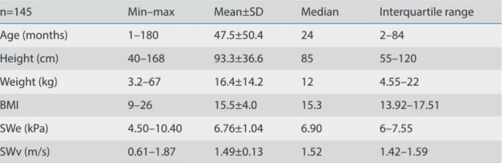
Table 1. Characteristics of the patients and elasticity and velocity values of the thymus
n=145 Min–max Mean±SD Median Interquartile range
Age (months) 1–180 47.5±50.4 24 2–84 Height (cm) 40–168 93.3±36.6 85 55–120 Weight (kg) 3.2–67 16.4±14.2 12 4.55–22 BMI 9–26 15.5±4.0 15.3 13.92–17.51 SWe (kPa) 4.50–10.40 6.76±1.04 6.90 6–7.55 SWv (m/s) 0.61–1.87 1.49±0.13 1.52 1.42–1.59
Statistical analysis
All data were processed in Microsoft Of-fice Excel and transferred to SPSS (version 21.0, IBM Corp.) for statistical analysis. The distribution of the data was assessed with the Kolmogorov-Smirnov test. Differenc-es of SWE parameters among the age and gender groups were compared using the Student t test and ANOVA. The groups demonstrating differences among mean values have been tested with Tukey’s HSD (honestly significant difference) test.
Correlation analysis of the SWE param-eters with BMI, height, weight, and age was performed using the Spearman ρ (r) correlation coefficient. Quantitative
vari-ables were shown as mean with standard deviation, or median with range. Variables were studied at the 95% confidence inter-val with P < 0.05 accepted as statistically significant.
Results
Descriptive statistics of the demograph-ic data including age, height, weight, and BMI of all the participants are given in Ta-ble 1. Mean values of thymus SWe and SWv for all participants were 6.76±1.04 kPa and 1.49±0.13m/s, respectively.
Table 2 represents the comparison of the mean SWe and SWv values among the age
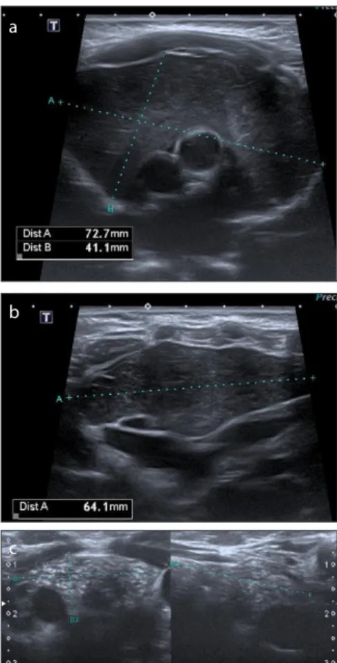
Figure 1. a–c. Gray-scale images (a, b) of the diffusely hypoechogenic mediastinal thymus in a male newborn. Gray-scale image (c) reveals thymus of a 13-year-old female patient.
a
b
c
Figure 2. a, b. Shear wave elasticity (SWe) measurements and propagation map on shear wave elastography (SWE) are seen. On SWE image (a) of a newborn, mean SWe value was 8.74 kPa for the thymus. B. On SWE image (b) of an adolescent girl, mean SWe value was 4.82 kPa for the thymus.
a b
Table 2. Comparison of gender and age groups according to different group classifications (A, B, C)
Age SWe SWv
Age range Mean±SD n Mean±SD P Mean±SD P
Age groups A 1 0–2 m 1±0.5 25 7.14±1.21 0.005a 1.53±0.13 0.005a 2 2–6 m 3.3±1.3 30 7±0.96 1.52±0.12 3 >6 m to 2 y 17.4±5.3 19 6.9±1.04 1.47±0.23 4 >2 to 5 y 43.5±8.8 24 6.59±0.95 1.49±0.11 5 >5–8 y 81±7.9 25 6.43±0.89 1.46±0.09 6 >8 y 136±25.7 22 6.31±0.95 1.45±0.11 B 1 0–3 m 1.5±0.77 41 7.1±1.14 0.001a 1.53±0.13 0.001a 2 >3 m to 2 y 17.1±11.5 42 6.96±0.94 1.49±0.17 3 >2 to 10 y 66.1±16.3 37 6.35±0.83 1.46±0.01 4 >10 y 132.2±27.3 25 6.4±1 1.46±0.11 C 1 0–6 y 9.6±11.4 83 7.05±1.04 0.001b 1.51±0.15 0.003b 2 6–18 y 94.3±39.3 62 6.37±0.90 1.46±0.11
Gender groups Male 46.2±50.7 86 6.65±0.97 0.11b 1.5±0.17 0.70b
Female 49.5±50.2 59 6.93±1.12 1.49±0.11
Tukey's HSD test results. Group A, SWe; group 1 vs. 5 (P = 0.022), group 1 vs. 6 (P = 0.013) and SWv; group 1 vs. 5 (P = 0.037), group 1 vs. 6 (P = 0.033). Group B, SWe; group 1 vs. 3 (P = 0.003), group 1 vs. 4 (P = 0.018), group 2 vs. 3 (P = 0.034) and SWv; group 1 vs. 3 (P = 0.007), group 1 vs. 4 (P = 0.02), group 2 vs. 3 (P = 0.04).
SWe, shear wave elasticity; SWv, shear wave velocity; SD, standard deviation, m, months; y, years. aP values obtained by ANOVA test.
groups. Among the six consecutive age groups under classification A (0–2 months, n=25; 2 to 6 months, n=30; 6 months to 2 years, n=19; 2 to 5 years, n=24; 5 to 8 years, n=25; >8 years, n= 22), differences of mean SWe and SWv values were not statistically
significant. However, we found significant difference within the age groups in classifi-cation A (P = 0.005). Mean values of SWe of group 1 vs. 5 (P = 0.022) and group 1 vs. 6 (P = 0.013) were significantly different. Simi-larly, mean values of SWv of group 1 vs. 5 (P
= 0.037) and group 1 vs. 6 (P = 0.033) were significantly different.
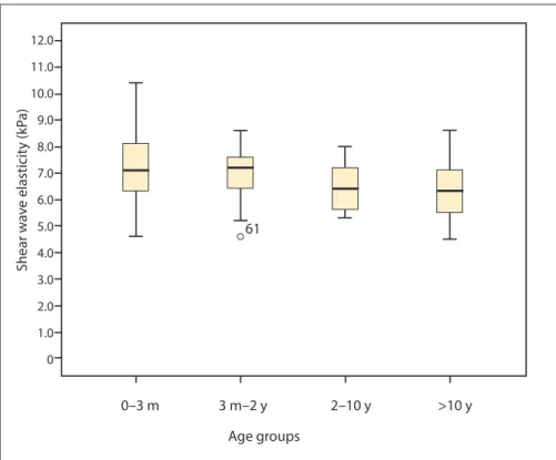
Among the four consecutive age groups under classification B (0–3 months, n=41; 3 months to 2 years, n=42; 2 to 10 years, n=37; > 10 years, n=25), no significant dif-ference was found in terms of mean values of SWe and SWv. However, we found sig-nificant difference within the age groups in classification B (P = 0.001). Mean values of SWe of group 1 vs. 3 (P = 0.003), group 1 vs. 4 (P = 0.018), group 2 vs. 3 (P = 0.034) were significantly different. Similarly mean values of SWv of group 1 vs. 3 (P = 0.007), group 1 vs. 4 (P = 0.02), group 2 vs. 3 (P = 0.04) were significantly different.
Under classification C, with two age groups of children 0–6 years (n=83) and 6–18 years (n=62), both SWe (P < 0.001), and SWv values (P < 0.003) decreased after six years and dif-ferences of SWe (group 1 vs. 2; P = 0.001) and SWv (group 1 vs. 2; P = 0.003) values were statistically significant. The mean SWe (P = 0.11) and SWv (P = 0.7) values did not differ significantly among gender groups.
The SWe values presented statistically significant mild negative correlation with age (P = 0.001, r=-0.31) (Fig. 4), height (P = 0.003, 0.26) and weight (P = 0.001, r=-0.30) (Table 3). The SWv values were also negatively correlated with age (P = 0.001, r=-0.26) (Fig. 4), height (P = 0.027, r=-0.19) and weight (P = 0.008, r=0.22).
Discussion
The thymus components involving true thymic epithelial space and the perivascu-lar space are fully developed before birth, and thymus upregulates functions during the early neonatal period (7, 8). Mean thy-mus volume has been found to be signifi-cantly greater in term infants compared to preterm neonates among children younger than two years old (9). Thymus is a common initial consideration in children with a wide superior mediastinum at early childhood. On chest roentgenogram, the wavy contour of an enlarged mediastinum in an asymp-tomatic newborn reflects the softness of the thymic tissue (10). In addition, such as a typical geographic shape in case of ectopic intrathyroidal location, and pliable proper-ties such as elongation through the carotid space and positioning between the major vessels in either ectopic or eutopic location in absence of mass effect help to differen-tiate a normal thymus from its mimickers. Beyond these qualitative and descriptive
Figure 3. Box plot presents SWe values of thymus in all four age groups (class B).
Age groups Shear w av e elasticit y (kP a) 0–3 m 3 m–2 y 61 12.0 11.0 10.0 9.0 8.0 7.0 6.0 5.0 4.0 3.0 2.0 1.0 0 2–10 y >10 y
Figure 4. Scatter plot diagram presents the negative correlation of age with SWe of the thymus.
Age (months) (kP a) Shear w av e elasticit y .0 10.0 8.0 6.0 4.0 50.0 100.0 150.0 200.0 Scale 14 12 10 8 6 4 2 0
radiological features, there has been only one study investigating relative stiffness of intrathyroidal ectopic thymus with strain elastography compared with thyroid gland, revealing a mean strain ratio value as 1.02 (range, 0.95–1.09) (6). However, no study investigating the normal elasticity range for thymic stiffness via US-based applications has been conducted.
SWe values of several superficial neck organs in children have been published in recent studies (11–15). In these studies, mean±standard deviation / median (inter-quartile range) for SWe values of thyroid gland, submandibular gland, parotid gland, cervical lymph nodes and palatine tonsils in healthy pediatric population have been reported as 6.38±1.97 kPa (11), 11.8±2.2 kPa (12), 8.37±2.09 kPa (13), 11.19 kPa (5.9–16.6 kPa) (14), 9.38±1.27 kPa (15), respectively. Mean SWe of the thyroid gland has been found as 14.6±3.3 kPa in a recent study (12). The preliminary results of the present study suggest the softness of the thymic tissue by revealing the mean stiffness of the thymus as 6.76±1.04 kPa, which is lower than previ-ously determined stiffness values for parotid gland, submandibular gland, thyroid gland, lymph nodes and palatin tonsils in the pedi-atric population. We reported SWE results as SWe (kPa) and also as SWv (m/s). We propose that the preliminary results would help to identify thymic tissue, especially in case of aberrant location. An ectopic thymic tissue around the angle of the mandible would be considered in differential diagnosis of palatin tonsils or lymph nodes due to the their hypoechogenic appearance because of having a lymphoreticular origin. Lower SWe values would suggest thymic tissue rather than a lymph node or tonsil. Pathological conditions mimicking an ectopic thymus in children include lymph node metastasis of papillary thyroid carcinoma and neuroblas-toma. As the elasticity of the reactive lymph nodes (14) is already higher than the thymus,
the lymph node elasticity is expected to in-crease in the presence of metastasis. In ad-dition, our results would be used in the daily practice for eliminating suspected diffusely infiltrative lymphoreticular disorders of the thymus such as leukemia and lymphoma.
The primordial thymopharyngeal tract remnants can appear anywhere from the pir-iform sinus to the manubrium along the thy-rohyoid membrane and carotid artery (16). In case of a mass detected along the descent pathway of the thymus, presenting with hyperintensity on T2-weighted MRI and ho-mogeneous contrast enhancement, further evaluation with high-resolution US as well as SWE can support the diagnosis of ectopic thymus if lower stiffness is found compared with superficial neck organs; thus, a diagno-sis can be reached using a noninvasive, ra-diation-free diagnostic method without the need for sedation or contrast administration.
Dyshormonogenetic glands in children with congenital hypothyroidism tend to be enlarged, hypoechogenic relative to normal thyroid gland and cricoid cartilage on US, and also associated with increased vascu-larity relative to healthy thyroid glands on Doppler US examination (17). Patients with congenital hypothyroidism may be misdiag-nosed as thyroid agenesia with ectopic cer-vical thymic tissue, due to the significantly decreased echogenicity. The tiny echogenic reticulations in dyshormonogenetic gland and punctate echogenic foci within the thy-mic tissue would help to differentiate the origin of the tissue (17). Because there has not been a significant difference in elastici-ty among dyshormonogenetic and normal thyroid glands in children (median values of SWe, 9.75 kPa vs. 9.5 kPa) (17), and we have depicted the mean SWe of thymus gland as 6.76±1.04 kPa; an ectopic thymus could be distinguished by SWE even when it is locat-ed in a dyshormonogenetic thyroid gland. During routine cervical and thyroid exam-inations, SWE together with gray-scale US
can be very helpful for diagnosing ectopic thymic tissue, which has a considerable re-ported incidence of 0.99% for intrathyroidal ectopy (3), by obtaining SWe values equal to or smaller than 7 kPa. Thus, SWE would help to achieve final diagnosis noninvasively.
Rebound thymic hyperplasia and also ectopic cervical thymus extending into the highest mediastinal lymphatic stations and supraclavicular region would be confusing in patients with lymphoreticular malignan-cy (18). In addition, positive interim FDG-PET/CT examinations may have poor pre-dictive values for prediction of recurrence of primary mediastinal B-cell lymphoma (19). The Deauville criteria (20) interpret uptake values of the lesions in comparison with mediastinum and liver. In cases with Deauville score 3 (mediastinum< FDG up-take < liver) or more, US and elastography evaluation could be valuable in decision making of residual or recurrent lymphop-roliferative disorders by excluding eutopic and ectopic normal thymic tissue along with thymus hyperplasia.
There have been several US based elas-tography methods. Qualitative and semi-quantitative results could be obtained with quasi-static strain imaging and acoustic radiation force impulse (ARFI) imaging by measuring the shape deformation. Transient elastography (TE) and ARFI quantification, as well as point shear wave elastography (p-SWE) could quantitatively calculate tissue stiffness in a target region. However, two-di-mensional SWE evaluates viscoelastic tissue properties reflected by shear or Young’s modulus quantitatively as a more popular and also more operator-independent meth-od (21, 22). We used two-dimensional SWE to provide easily measurable, quantitative and objective data in the pediatric age group.
There are several limitations to our study. First, we had a limited number of cases per age groups. Nevertheless, our study popu-lation for the SWE of the thymus is the larg-est one to date in a single study. Second, the measurements were made by two op-erators in consensus but we did not obtain separate SWE values for each participant. Repeated acquisitions and many regions of interest would be performed to reduce in-ter and intraobserver variability. Third, it is too difficult to stabilize patients especially infants and younger children resulting in suboptimal SWE images and propagation lines. We excluded the data that could not be assessed and asked the mothers to
Table 3. Association of the demographic data with SWE parameters
Shear wave elasticity (kPa) Shear wave velocity (m/s)
P r P r
Age 0.001 -0.31 0.001 -0.26
Height 0.003 -0.26 0.027 -0.19
Weight 0.001 -0.30 0.008 -0.22
Body mass index 0.18 -0.11 0.31 -0.08
Data represent Spearman correlation analysis results. SWE, shear wave elastography; r, correlation coefficient.
breastfeed or put the infants to sleep in or-der to stabilize them during acquisitions. We kept the gel warm to avoid waking the infants. Moreover, especially infants and patients with small thymic tissue around vessels are prone to cardiac or vascular pul-sation artifacts, which was a limiting factor on calculations. Suprasternal approach and selection of the regions of the interest from the periphery of the gland were used to avoid pulsation artifacts. In addition breath holding in patients who can comply would facilitate achieving optimal acquisitions rapidly.
In conclusion, mean thymus stiffness rep-resents a narrow range in terms of elasticity. Thymus elasticity decreases with age, inde-pendently from gray-scale and Doppler imag-ing characteristics. SWE evaluation of the thy-mus is a noninvasive, quantitative diagnostic tool for distinguishing the thymic tissue.
Conflict of interest disclosure
The authors declared no conflicts of interest.
References
1. Spigland N, Bensoussan AL, Blanchard H, Russo P. Aberrant cervical thymus in children: three case reports and review of the literature. J Pe-diatr Surg 1990; 25:1196–1199. [CrossRef]
2. Song I, Yoo SY, Kim JH, Hong E, Yoon HK. Aber-rant cervical thymus: imaging and clinical find-ings in 13 children. Clin Radiol 2011; 66:38–42.
[CrossRef]
3. Erol OB, Sahin D, Bayramoglu Z, et al. Ectopic intrathyroidal thymus in children: Prevalence, imaging findings and evolution. Turk J Pediatr 2017; 59:387–394. [CrossRef]
4. Frates MC, Benson CB, Dorfman DM, Cibas ES, Huang SA. Ectopic intrathyroidal thymic tissue mimicking thyroid nodules in children. J Ultra-sound Med 2018; 37:783–791. [CrossRef]
5. Tanrivermis Sayit A, Elmali M, Hashimov J, Cey-han Bilgici M, Dagdemir A. Bilateral ectopic cervical thymus presenting as a neck mass: Ultrasound and magnetic resonance imaging. Pediatr Int 2016; 58:943–945. [CrossRef]
6. Stasiak M, Adamczewski Z, Stawerska R, Kraw-czyk T, Tomaszewska M, Lewinski A. Sono-graphic and elastoSono-graphic features of extra- and ıntrathyroidal ectopic thymus mimicking malignancy: differential diagnosis in children. Front Endocrinol (Lausanne) 2019; 10:223.
[CrossRef]
7. Steinmann GG, Klaus B, Muller-Hermelink HK. The involution of the ageing human thy-mic epithelium is independent of puberty. A morphometric study. Scand J Immunol 1985; 22:563–575. [CrossRef]
8. Shanley DP, Aw D, Manley NR, Palmer DB. An evolutionary perspective on the mechanisms of immunosenescence. Trends Immunol 2009; 30:374–381. [CrossRef]
9. Yekeler E, Tambag A, Tunaci A, et al. Analysis of the thymus in 151 healthy infants from 0 to 2 years of age. J Ultrasound Med 2004; 23:1321– 1326. [CrossRef]
10. Alves ND, Sousa M. Images in pediatrics: the thymic sail sign and thymic wave sign. Eur J Pediatr 2013; 172:133. [CrossRef]
11. Uysal E, Ozturk M. Quantitative assessment of thyroid glands in healthy children with shear wave elastography. Ultrasound Q 2019; 35:297–300. [CrossRef]
12. Arioz Habibi H, Memis Durmaz ES, Qarayeva V, et al. Quantitative assessment of thyroid, submandibular, and parotid glands elasticity with shear-wave elastography in children. Ul-trasound Q 2018; 34:58–61. [CrossRef]
13. Caliskan E, Ozturk M, Bayramoglu Z, Comert RG, Adaletli I. Evaluation of parotid glands in healthy children and adolescents using shear wave elastography and superb microvascu-lar imaging. Radiol Med 2018; 123:710–718.
[CrossRef]
14. Bayramoglu Z, Caliskan E, Karakas Z, et al. Di-agnostic performances of superb microvas-cular imaging, shear wave elastography and shape index in pediatric lymph nodes catego-rization: a comparative study. Br J Radiol 2018; 91:20180129. [CrossRef]
15. Ozturk M, Caliskan E, Bayramoglu Z, Adaletli I. Quantitative assessment of palatine ton-sils in healthy children and adolescents with shear-wave elastography. Ultrasound Q 2018; 34:213–218. [CrossRef]
16. Zarbo RJ, McClatchey KD, Areen RG, Baker SB. Thymopharyngeal duct cyst: a form of cervi-cal thymus. Ann Otol Rhinol Laryngol 1983; 92:284–289. [CrossRef]
17. Adaletli I, Bayramoglu Z, Caliskan E, et al. Multi-parametric ultrasound evaluation of pe-diatric thyroid dyshormonogenesis. Ultrasound Med Biol 2019; 45:1644–1653. [CrossRef]
18. Smith CS, Schoder H, Yeung HW. Thymic exten-sion in the superior mediastinum in patients with thymic hyperplasia: potential cause of false-pos-itive findings on 18F-FDG PET/CT. AJR Am J Roentgenol 2007; 188:1716–1721. [CrossRef]
19. Lazarovici J, Terroir M, Arfi-Rouche J, et al. Poor predictive value of positive interim FDG-PET/ CT in primary mediastinal large B-cell lym-phoma. Eur J Nucl Med Mol Imaging 2017; 44:2018–2024. [CrossRef]
20. Barrington SF, Kluge R. FDG PET for therapy monitoring in Hodgkin and non-Hodgkin lym-phomas. Eur J Nucl Med Mol Imaging 2017; 44:97–110. [CrossRef]
21. Bamber J, Cosgrove D, Dietrich CF, et al. EF-SUMB guidelines and recommendations on the clinical use of ultrasound elastography. Part 1: Basic principles and technology. Ultra-schall Med 2013; 34:169–184. [CrossRef]
22. Cosgrove D, Piscaglia F, Bamber J, et al. EFSUMB guidelines and recommendations on the clin-ical use of ultrasound elastography. Part 2: Clinical applications. Ultraschall Med 2013; 34:238–253. [CrossRef]