LIPOSOME ENCAPSULATION OVERCOMES D-TYPE AND K-TYPE CpG ODN DICHOTOMY AND INDUCES SYNERGISTIC IMMUNE
ACTIVATION
A THESIS
SUBMITTED TO THE DEPARTMENT OF MOLECULAR BIOLOGY AND GENETICS
AND THE GRADUATE SCHOOL OF ENGINEERING AND SCIENCE OF BILKENT UNIVERSITY
IN PARTIAL FULFILLMENT OF THE REQUIREMENTS FOR THE DEGREE OF
MASTER OF SCIENCE
By
Begüm Han Horuluoğlu August, 2014
I certify that I have read this thesis and that in my opinion it is fully adequate, in scope and in quality, as a thesis for the degree of Master of Science.
Prof. Dr. İhsan Gürsel
(Advisor)
I certify that I have read this thesis and that in my opinion it is fully adequate, in scope and in quality, as a thesis for the degree of Master of Science.
Prof. Dr. Kamil Can Akçalı
I certify that I have read this thesis and that in my opinion it is fully adequate, in scope and in quality, as a thesis for the degree of Master of Science.
Assist. Prof. Dr. Ali Osmay Güre
Approved for the Graduate School of Engineering and Science
Prof. Dr. Levent Onural Director of the Graduate School
ABSTRACT
LIPOSOME ENCAPSULATION OVERCOMES D-TYPE AND K-TYPE CpG ODN DICHOTOMY AND INDUCES SYNERGISTIC IMMUNE
ACTIVATION
Begüm Han Horuluoğlu
M.S. in Molecular Biology and Genetics Advisor: Prof. Dr. İhsan Gürsel
August, 2014
Liposomes are one of the best candidates for the encapsulation of labile bioactive agents due to their safety and high entrapment efficiency. In human, two structurally distinct classes of CpG ODN are capable of activating different signaling pathways, leading to differential immune activation. While K-type ODN triggers plasmacytoid dendritic cells (pDCs) to mature and produce TNFα, D-type ODN leads to IRF-7 dependent IFNα secretion. Strikingly, when K-and D-type ODN are co-incubated in their free forms, K-ODN masks the D-ODN specific immune activation. Identifying proper delivery vehicles that provide both ODN types to display their superior features upon stimulation is of great clinical importance. In this study, first we investigated the synergistic effects of K- and D-ODN upon encapsulating them within five different liposome types. Then with the selected potential liposome combinations, we identified synergistic activation capacities both on human PBMCs and on mice splenocytes. In PBMC cytokine results revealed that D-ODN loaded in all five liposome types stimulated more IFNα than free D-ODN. Similarly, liposomal K-ODN triggered more TNFα than free K-ODN type. While incubation of free K and D- type ODN as expected, abrogated D-specific IFNα production from PBMC, simultaneous incubation with neutral or anionic D-ODN loaded liposomes plus cationic liposomes loaded with K-ODN significantly increased K-specific as well as D-specific effect rather than masking it (i.e. more production of TNFα and IFNα specific for K and D, respectively). This improved synergistic immune activity for both D and K ODN observed with ND+CK combination in 100% of individuals (TNFa) and 90% of individuals for IFNa. Additionally, intracellular cytokine staining findings supported improved TNFα and IFNα, from pDC population of PBMCs. Co-stimulatory molecule expressions and APC activation also significantly upregulated compared with free treatment. In mice contrary to ND+CK combination, sterically stabilized cationic liposome encapsulated K-ODN combined with i) neutral, ii) anionic, iii) cationic or iv) stealth encapsulated D-ODN increased IL6, IL12 and IFNγ levels, when stimulated simultaneously. Moreover, ex vivo experiments showed that cellular uptake and pro-inflammatory cytokine gene expressions significantly increased with combined liposomal formulations. This study established that by selecting proper liposome type(s) we reverse antagonistic action of K-ODN on D-ODN and induce a synergistic effect leading to a more robust immunostimulatory activity in both human and mice. This approach could broaden the immunotherapeutic application of these two important CpG ODN classes in clinic.
ÖZET
LİPOZOMA YÜKLENMİŞ D-TİPİ VE K-TİPİ ODN’LERİN SİNERJİK İMMUN AKTİVASYONU
Begüm Han Horuluoğlu
Moleküler Biyoloji ve Genetik Yüksek Lisans Danışman: Prof. Dr. İhsan Gürsel
Ağustos, 2014
Yüksek yükleme verimi ve güvenliği sayesinde, lipozomlar ; kararsız biyoaktif ajanların taşınmasında en iyi adaylardan biridir. İnsanda, yapı olarak farklı gruplandırılan CpG ODN’ler farklı sinyal yolaklarını aktive ederek, değişik immün cevaplara yol açarlar. K-tipi ODN plazmasitoid dendiritik hücrelerin (pDC) olgunlaşması ve aktifleşmesi sonucu TNFα salgılanmasına sebep olurken, D-tipi ODN IRF’ye bağımlı sinyal yolaklarını aktive ederek IFNα üretilmesine neden olur. Çarpıcı bir şekilde, K-tipi ve D-tipi ODN’ler serbest hallerinde hücrelere verildiklerinde, K-tipi D-tipi etkisini bloke eder ve sadece K-tipine özel cevap gözlemlenir. Her iki tip ODN için de en uygun ve en önemli özelliklerini kaybetmeyecekleri bir taşıyıcı sistemin geliştirilmesi klinik uygulamalar için çok önemlidir. Bu çalışmada, öncelikle K- ve D- tipi ODN’leri 5 farklı lipozoma yükleyerek sinerjik etkilerini araştırdık. Bu lipozomlara yüklü ODN’lerin insan periferik kan mononükleer hücreleri ve fare dalak hücreleri üzerinde sinerjik bir etki gösterebilme kabiliyeti en yüksek olan potansiyel gruplarını seçtik. İnsan periferik kan hücreleri üzerinde yapılan sitokin ELİSA deneyleri sonucunda, kullanılan 5 lipozomal kombinasyonun da serbest ODN’lere göre daha fazla IFNα ve TNFα ürettiğini gözlemledik. Serbest haldeki K-ve D-tipi ODN’ler beraber inkübe edildiklerinde beklendiği gibi D-spesifik olan IFNα salımında azalma gözlemlenmiştir. Nötral ve anyonik lipozoma yüklü D-tipi ODN (ND ve AD) ile katyonik lipozoma yüklü K-tipi ODN (CK) beraber hücreye verildiklerinde ise, K-tipi D-tipini engellemek yerine, hem K-tipine özgü etkiyi arttırmakta hem de D-tipine spesifik etkiler göstermektedir. Bu gelişmiş sinerjik etki, ND+CK grubunda TNFα salımı için kullanılan donörlerin %100’ünde gözlemlenirken, IFNα için %90’ında gözlemlenmektedir. Bunlara ek olarak, hücre içi sitokin boyama deney sonuçlarına göre TNFα ve IFNα sekresyonu özel olarak insan pDC’lerinden artmaktadır. Yardımcı uyaran molekül ifadeleri ve antijen sunucu hücrelerin de lipozomal kombinasyonlarda, serbest ODN’lere göre belirgin bir şekilde arttığı gözlemlenmiştir. Fare deneylerinde, insan deneylerinden farklı olarak ND+CK grubu yerine, K-tipi ODN yüklü sterik olarak stabilize edilmiş katyonik lipozom (SSCL) ile D-tipi ODN yüklü i) nötral, ii) anyonik, iii) katyonik ve iv) stealth kombinasyonlarında sinerjik etkiler görülmüştür (IL6, IL12 ve IFNγ üretiminde artış). Ex-vivo deneyleri sonucunda, lipozomal kombinasyonların CpG ODN’lerin hücre içine alımını ve proinflamatuar sitokinlerin gen ifadelerini arttırdığını ortaya çıkarmıştır. Bu çalışmada hem insan, hem de farede doğru lipozom tipi seçimiyle K-tipi ODN’in D-tipi ODN’e karşı gösterdiği antagonist etkiyi, sinerjik bir etkiye çevirebileceğimizi kanıtladık. Yaklaşımımız bu önemli iki tip ODN’in immunoterapötik uygulamalarda kullanımını arttırabilecek niteliktedir.
Acknowledgement
First, I would like to thank my advisor Prof. Dr. Ihsan Gursel, for giving me a chance to work with him. His continued support, guidance and patience with me throughout the completion of this thesis is invaluable. I am grateful for his trust in me.
My heartfelt thanks go to Assoc. Prof. Dr. Mayda Gursel for her scientific guidance throughout my studies. I would also like to thank to MG Group members.
I would like to thank to past and present IG members, Gizem, Tamer, Fuat, Arda, Gözde, Defne, Merve Deniz, Kübra, Begum, Banu, Ihsan and Mehmet for their help, support and friendship during my MSc studies.
Especially, I would like to thank to my dearest friends, for giving me support and love at all times, Ayse, Cagatay Yigit, Merve Mutlu, Defne, Merve Deniz, Dilan, and Deniz Cansen as well as all the MBG family. Most importantly, I am thankful to my roommate and best friend Gozde Gucluler throughout this whole journey.
Without the support and the endless love from my mother and brother, I would not have found the courage to chase my dreams. I would like to express my deepest love gratitude and dedicate my thesis to my mother, Esin and my brother Cahit.
DEDICATED
Content
1. Introduction ... 1
1.1The Immune System ... 1
1.2 Innate Immunity ... 2
1.2.1.Pathogen Recognition Receptors ... 2
1.2.2 Toll-like Receptor Family ... 3
1.3 Synthetic CpG ODNs ... 9
1.3.1 D or A Type ODN ... 10
1.3.2 K or B Type ODN ... 10
1.3.3 C-type CpG ODN ... 11
1.3.4 Other Types of CpG ODNs ... 11
1.3.5 The Dichotomy of D and K Type ODN ... 12
1.4 Drug Delivery with Nanoparticles ... 14
1.4.1 Delivery of CpG ODN in liposomes ... 16
2. Aim of this study ... 18
3. Materials and Methods ... 19
3.1. Materials ... 19 3.1.1.ELISA Reagents ... 19 3.1.2.TLR Ligands ... 19 3.1.3 Lipids ... 20 3.2. Methods ... 20 3.2.1. Cell Culture ... 20 3.2.2 Cell Lines ... 21
3.2.3 Time Lapse detection of Labeled Liposome Uptake within RAW 264.7 cells ... 22
3.2.4 Cell Counting by Flow Cytometer ... 22
3.2.5 In Vitro Stimulation of Murine Cells ... 22
3.2.6 In Vitro Stimulation of PBMCs ... 23
3.2.7 Liposome Preparation ... 23
3.2.8 Cytokine Enzyme Linked Immunosorbent Assay (ELISA) ... 24
3.2.9 Ex-Vivo Experiments ... 25
3.2.11 Flow Cytometry ... 28
4. Results ... 31
4.1 Determination of the most potent immunostimulatory ODN-‐liposome formulation ... 31
4.1.1 Mouse Spleen cell stimulation Assays ... 32
4.1.2. Human PBMC stimulation Assays ... 35
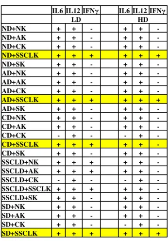
4.2 Delineation of synergistic immunostimulatory effects of liposomal D-‐ and K-‐ type CpG ODNs in Mice and in Man ... 39
4.2.1. Studies with Mouse Spleen cells ... 39
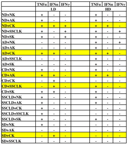
4.2.2 Studies with human PBMCs ... 43
4.3 Establishing the benefit of selected liposome combinations in healthy donors ... 48
4.4.Stimulation of PBMC with different D-‐ and K-‐ type ODNs sequences retain liposome based benefits ... 53
4.5 Intracellular Detection of IFNα and TNFα upon PBMC stimulation ... 55
4.5.1 Antigen presentation ability of PBMCs when simultaneously treated with liposomes encapsulating D and K type CpG ODNs ... 63
4.6 Stimulation of Splenocytes does not depend on mice strain ... 65
4.7 Ex-‐vivo activity of labeled liposomes in with C57/BL6 mice ... 67
4.7.1 Internalization of labeled liposomes by immune cells ... 70
4.8 Screening the trajectory of labeled liposomes on mouse cell line by fluorescence microscope ... 72
5.Discussion ... 74
6.APPENDIX ... 85
List of Figures
Figure 1.1: Toll-like receptors and their ligands.. ... 4
Figure 1.2 TLR signaling pathways.. ... 8
Figure 1.3: Delivery of D- and K-type CpG ODN into late and early endosomes ... 11
Figure 1.4: Y-shaped CpG ODN. ... 12
Figure 1.5:Nanoparticle drug delivery systems ... 15
Figure 1.6: Represantative diagram of liposomes.. ... 16
Figure 4 1 A-F :Dose dependent IL6 secretion of different liposome formulations and alone forms of candidate CpG ODNs from mouse spleen cells.. ... 33
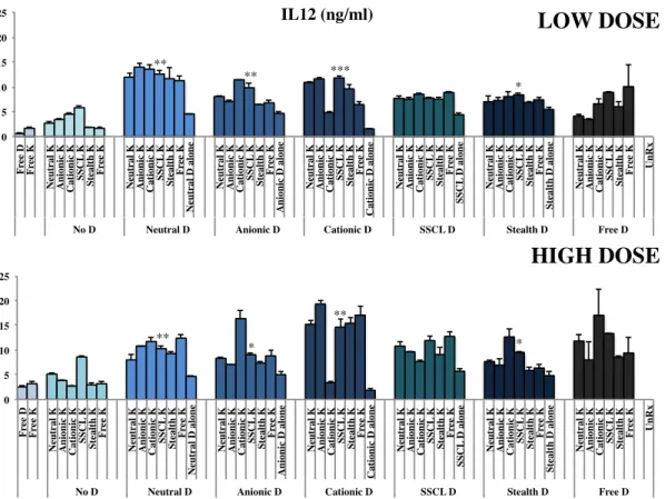
Figure 4 2 A-F: Dose dependent IL12 secretion of different liposome formulations and alone forms of candidate CpG ODNs from mouse spleen cells. ... 34
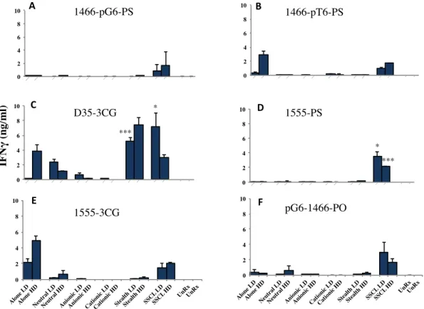
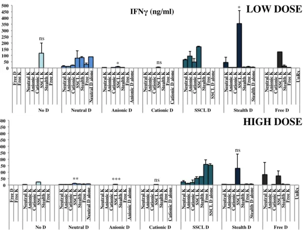
Figure 4 3 A-F: Dose dependent IFNγ secretion of different liposome formulations and alone forms of candidate CpG ODNs from mouse spleen cells in the repeat experiments. ... 35
Figure 4 4 A-F: Dose dependent IFNγ secretion levels from stimulated PBMC supernatants, after incubation of 24h were assessed by cytokine ELISA. ... 37
Figure 4 5A-F:Dose dependent TNFα secretion levels from stimulated PBMC supernatants, after incubation of 24h were assessed by cytokine ELISA. ... 38
Figure 4 6 : D35-3CG was the only liposomal and alone form of CpG ODN that showed detectable IFNα secretion from stimulated PBMC in a dose dependent manner ... 38
Figure 4 7: IL6 cytokine ELISA results for low and high doses of simulatenous stimulation of liposomal D- and K- type ODN with splenocytes.. ... 40
Figure 4 8: IL12 cytokine ELISA results for low and high doses of simultaneous stimulation of liposomal D- and K- type ODN with splenocytes.. ... 41
Figure 4 9: IFNγ cytokine ELISA results for low and high doses of simultaneous stimulation of liposomal D- and K- type ODN with splenocytes. ... 42
Figure 4 10: IFNα cytokine ELISA results for low and high doses of simultaneous stimulation of liposomal D- and K- type ODN with PBMC. ... 45
Figure 4 11: TNFα cytokine ELISA results for low and high doses of simultaneous stimulation of liposomal D- and K- type ODN with PBMC ... 46
Figure 4 12: IFNγ cytokine ELISA results for low and high doses of simultaneous stimulation of liposomal D- and K- type ODN with PBMC. ... 47 Figure 4 13: Representative cytokine ELISA results for TNFα, IFNα, IFNγ, IP10 with chosen 5 different liposomal formulations for 2 different donors out of 10.). ... 50 Figure 4 14 A-B: Fold Inductions of TNFα and IFNα for 10 healthy donor ... 51 Figure 4 15: Fold Inductions of IFNa secretion obtained from cytokine ELISA results.
... 54 Figure 4 16: Fold Inductions of TNFa secretion obtained from cytokine ELISA results. ... 54 Figure 4 17: Fold induction of TNFα secretion from pDC.. ... 56 Figure 4 18: Fold induction of IFNα secretion from pDC.. ... 57 Figure 4 19:FACS analysis of IFNα secretion from PBMC isolated from healthy donor 1. ... 58 Figure 4 20: FACS analysis of IFNα secretion from PBMC isolated from healthy donor 2.. ... 59 Figure 4 21: Fold induction of IFNα secretion from 2 healthy donors pDCs.. ... 60 Figure 4 22: FACS analysis of TNFα secretion from PBMC isolated from healthy donor 1. ... 61 Figure 4 23: FACS analysis of TNFα secretion from PBMC isolated from healthy donor 2. ... 62 Figure 4 24: Fold induction of TNFα secretion from 2 healthy donors pDCs. pDCs were defined by CD123-FITC positive cell gating. ... 62 Figure 4 25: FACS results for CD80-FITC and HLA-DQ-PE expressions from PBMC. ... 64 Figure 4 26: Fold inductions of HLA-DQ-PE and CD80-FITC double positive cells.
... 64 Figure 4 27: IL6 cytokine ELISA results C57/BL6 mice. ... 66 Figure 4 28: IL12 cytokine ELISA results for C57/BL6 mice.. ... 67 Figure 4 29: IL6 cytokine ELISA results of ex-vivo, experiment was run in duplicates. Cell supernatants were incubated for 24 hours. ... 68 Figure 4 30: IL12 cytokine ELISA results of ex-vivo, ... 69
Figure 4 31: IFNγ cytokine ELISA results of ex-vivo ... 69 Figure 4 32: PCR products of cytokine-related gene transcripts obtained from total RNA ... 70 Figure 4 33: FACS analysis of labeled liposomes from PEC. ... 72 Figure 4 34: Fluorescent microscope (Nikon Eclipse Ti) Images 40X ... 73
List of Tables
Table 1.1 Innate vs Adaptive immunity ... 2 Table 1.2: Pattern recognition receptors in mammals and their ligands. ... 3 Table 1 3: Comparasion of different types of ODN. ... 10 Table 3.1: Sequences of ODN used. Upper case letters: Phosphorothoiate linkage between bases, lower case letters: Phosphorodiester linkage between bases ... 19 Table 3.2: Lipid Compositions of different liposome types ... 24 Table 3.3 : concentrations of coating antibodies for mouse and human cytokine ELISA experiments (50ul/well transferred) ... 25 Table 3.4: Starting concentrations and detection limits for each recombinant standard cytokines (each cytokine was diluted by 11X by 1/2 dilutions) ... 25 Table 3.5: Mouse primer sequences and their expected product sizes. ... 27 Table 3. 6: PCR conditions for mouse primers. ... 28 Table 4.1: General outlook of formulation dependent cytokine specific upregulation for splenocytes. ... 43 Table 4.2: General outlook of formulation dependent cytokine specific upregulation for PBMC. ... 48 Table 4.3 : Overall response of 10 healthy donors to selected liposome combinations loaded with D and K-Type CpG ODNs ... 52 Table 4 4 : Overall patient-to-patient immune response to liposomal CpG formulations ... 52
Abbreviations
A Anionic liposome
APC Antigen presenting cell
Bp Base pairs
cDNA Complementary deoxyribonucleic acid
C Cationic liposome
CpG Unmethylated cytosine-phosphate-guaniosine motifs
CXCL CXC-chemokine ligand
DC Dendritic cell
DNA Deoxyrobinucleic acid
dsRNA Double-stranded RNA
ELISA Enzyme linked-immunosorbent assay
FBS Fetal bovine serum
Ig Immunoglobulin
IκK Inhibitor kappa b kinase
IL Interleukin
IFN Interferon
IP Interferon induced protein
IRAK IL1 receptor-associated kinase
IRF Interferon-regulatory factor
LPS xii Lipopolysaccharide
MHC Major histocompatibility complex
MIP Macrophage inflammatory protein
MyD88 Myleoid differentiation primary response gene 88
N Neutral liposome
NF-κB Nuclear factor-kappa b
NK Natural killer cell
NLR Nucleotide-binding oligomerization domain like proteins or
receptors
NOD Nucleotide-binding oligomerization domain
PAMP Pathogen associated molecular patterns
PBS Phosphate buffer solution
PCR Polymerase chain reaction
pDC Pasmacytoid dendritic cell
pIC Polyionosinic acid:cytidylic acid
PNPP Para-nitrophenyl pyro phosphate
PRR Pathogen recognition receptor
RIG Retinoic acid inducible gene
RNA Ribonucleic acid
RPMI Roswell Park Memorial Institute
RT Reverse transcriptase
S Stealth Liposome
SA-AKP Streptavidin Alkaline-phosphatase
SSCL Sterically Stabilized Cationic Liposome
ssRNA Single-stranded RNA
TIR Toll/IL1 receptor
TIRAP Toll/IL1 receptor-associated protein
TLR Toll-like receptor
TNF Tumor necrosis factor
TRAF TNF-associated factor
TRAM TRIF-related adaptor molecule
CHAPTER 1
1. Introduction
1.1. The Immune System
A powerful and efficient defense mechanism is operational for the discrimination between self and infectious/pathogenic non-self among organisms of different genera. The system that has highly effective weapons awaiting any intruders is called the immune system [1]. The immune system consists of effector cells and molecules, which protects the body from infectious agents and their damages. Immunity can be divided into two main categories; innate and adaptive immunity.
Adaptive (acquired) immunity is a specific immune response against pathogens, which provides recognition and antigen production, and is developed throughout the lifetime of an individual [2]. Adaptive immune system is only present in vertebrates and cartilaginous fish.
The innate immune system on the other hand, is the first line of defense against pathogens [3]. Innate immunity consists of a fixed number of germ-line encoded receptors that recognizes components of microorganisms and viruses [1]. Phagocytic cells and complement system mediate innate immune system. Upon infection, cellular mediators of innate immunity; macrophages, neutrophils and natural killer cells migrate towards to the infection leading to the activation of inflammatory signaling cascades.
It is accepted that innate immune signals play an important role for the activation of adaptive immunity. Upon the activation of innate immunity, dendritic cells are maturated and migrated in to lymph nodes where they are capable of
activating naive CD4+
phenotypes depending on the pathogen. Th1 cells support the cell-mediated immunity whereas Th2 cells shape humoral immunity. [1], [4].
Table 1.1 Innate vs Adaptive immunity (Adapted from Albertz et al., 2002)
Innate Immune System Adaptive Immune System
Response is non-specific Pathogen and antigen specific response
Exposure leads to immediate Lag time between exposure and
maximal response maximal response
Cell mediated and humoral Cell mediated and humoral
components components
No immunological memory Exposure leads to immunological memory
Found in nearly all forms of life Found only in jawed vertebrates
1.2. Innate Immunity
1.2.1.Pathogen Recognition Receptors
The innate immune system has a limited specific recognition system for pathogens, which relies on germline-encoded-pathogen-recognition receptors (PRRs) [3]. PRRs recognize typical repeating patterns that are expressed by microorganisms. These molecular structures are called pathogen-associated molecular patterns (PAMPs) [2]. Recently, it was revealed that, PRRs also recognize endogenous molecules released from damaged cells, called damage/pathogen associated molecular patterns (D/PAMPs).
PRRs have common features. They recognize PAMPs, that are important for the microorganism survival, and because of that it is difficult for microorganism to alter. Secondly, PRRs are expressed in the host regardless of their life-cycle stage. Thirdly, PRRs are germline encoded, nonclonal and expressed in all cells without a memory. Different PRRs recognize distinct PAMPs, and activate different signaling pathways, which leads to specific immune responses. Mechanisms of recognition are highly conserved from plants and fruit flies to mammals [3].
PRRs can be divided into 2 groups on basis; cytoplasmic and signaling PRRs. Cytoplasmic PRRs can be grouped into 3 families; interferon (IFN)-inducible proteins, caspase-recruiting domain (CARD) helicases, and nucleotide-binding
oligomerization domain (NOD)-like receptors (NLRs).Toll-like receptors (TLRs) and C-type lectin receptors belong to signaling PRR family [6] (Table 2).
Table 1.2: Pattern recognition receptors in mammals and their ligands (Adapted from Lee,2007a).
Various non-professional immune cells express these PRRs including macrophages and DCs. PRRs upregulate the transcription of genes involved in many inflammatory responses by PAMPs or DAMPs [7]. Innate immunity is activated through these of proinflammatory molecules, which are triggered by PRRs [8],[67].
1.2.2. Toll-like Receptor Family
Toll-like receptors are major and most studied class of PRRs, which are conserved through the worm Caenorhabditis elegans to mammals [9], [10], [11], [12]
TLRs are integral glycoproteins, containing leucine-rich repeat (LRR) motifs that mediate the recognition of PAMPs and intracellular Toll-interleukin 1 (IL-1) receptor (TIR) domains which activate the downstream signaling pathways by using adaptor molecules such as MyD88, TIRAP, TRIF, and TRAM [13],[74]. 10 TLRs have been identified in human that recognize different PAMPs derived from viruses, bacteria and fungi [8].
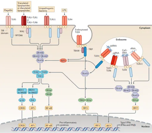
divided into two subgroups [13]. TLR1, TLR2, TLR4, TLR5, and TLR6 are found on the cell surface and typically recognize ligands that are usually bacterial in origin. Endosomal TLRs; TLR3, TLR7, TLR8, and TLR9 are responsible for recognizing nucleic acids including double-stranded (ds) RNA and CpG DNA [14] (Figure 1.1).
Figure 1.1: Toll-like receptors and their ligands (Adapted from Rakoff-Nahoum, 2009).
1.2.2.1. TLR1, TLR2 and TLR 6
TLR2 recognizes a wide spectrum of microbial components such as; lipoproteins, lipopeptides, peptidoglycan and lipoteichoic acid from Gram-positive bacteria, lipoarabinomannan from mycobacteria, glycosylphospatidylionsitol anchors from Trypanosoma cruzi, a phenol-soluble modulin from Straphylococcus epidermis, zymosan from fungi and glycolipids from Treponema maltophilum [9]. In general,
TLR2 forms heterodimers with TLR1 and TLR6. By heterodimerization TLR2 gains the ability to recognize different microbial components. TLR2- TLR1 heterodimer recognizes triacylated lipopeptides from Gram-negative bacteria and mycoplasma, where as TLR2-TLR6 heterodimer recognizes diacylated lipopeptides from Gram-positive bacteria and mycoplasma. Studies have proved that the structure caused by heterodimerization provides recognition of different PAMPs by TLR2 [13]. Upon recognition, TLR2 activates the adaptor molecules TRAP and MyD88 , which initiates a MyD88 dependent signaling pathway leading inflammatory cytokine production [6].
1.2.2.2.TLR3
TLR3 recognizes, polyinosinic-polycytidylic acid (poly (I:C)), which is a synthetic analog of double stranded RNA (dsRNA) and mimics viral infection by inducing antiviral immune responses. Type I interferon and inflammatory cytokine productions are promoted by TLR3 activation through TRIF-dependent pathway [13],[6] deficient mice are impaired in their response do dsRNA but not abolished completely. Which indicates that TLR3 is not the only receptor for dsRNA, there are additional sensors exist such as; CARD helicases [16].
1.2.2.3. TLR4
TLR 4 is essential for protection against gram-negative bacteria in both human and mice. Lipopolysaccharide (LPS) is the major component of the outer layer of gram-negative bacteria [17]. Recognition of LPS by TLR4 activates two major intracellular signaling pathways; MyD88- dependent and TRIF- dependent [18]. TLR4 is the only TLR which can initiate both MyD88 and TRIF-dependent pathways by using all four adaptors [13]. The MyD88-dependent pathway is activated by the recruitment of MyD88 to cytoplasmic TIR domain of TLR4, TRIF-dependent pathway is activated similarly by the recruitment of TRIF to cytoplasmic TIR domain of TLR4 on the plasma membrane [18]. Inflammatory cytokine expressions such as; IL6, IL12 and TNFa are induced by the MyD88-dependent pathway, whereas type I IFN expressions are upregulated by the TRIF-dependent pathway. TLR4 can also
activate other signaling molecules including phosphatidylinositol-3 kinase (PI-3K) and MAP3Ks such as MEKK3, TPL2, and ASK1A. Recently it was reported that CD14, a co-receptor, helps the diversification of TLR4 ligand repertoire and activation of downstream signaling pathways [6]. Additionally TLR4 recognizes various substances from viruses, fungi and mycoplasma. Danger associated molecular patterns (DAMPs) also activates TLR4 [19].
1.2.2.4. TLR5
TLR5 is responsible for the recognition of flagellin, a protein component of bacterial flagella. Mostly epithelial cells, monocytes and immature DCs especially in the small intestine express TLR5. Mouse TLR11, a relative of TLR5, expression is high in the kidney and bladder. Uropathogenic bacterial components are recognized by TLR11. Additionally TLR5 is highly expressed in lungs and they are important in the defense against respiratory tract pathogens [13],[3].
1.2.2.5. TLR7 and TLR8
TLR7 and TLR8 are phylogenetically and structurally closely related subgroup of TLR superfamily. Upon recognition of ssRNA by TLR7 and TLR8, DCs are activated. TLR7 is mostly expressed in plasmascytoid DCs (pDCs) myeloid DCs (mDCs) expression of TLR7 is still controversial [75]. Mostly TLR7 expression is not detected in human monocyte-derived DCs [20]. TLR7 and TLR8 activate innate immune responses upon viral infections; ssRNA sequences containing GU-rich or poly-U sequences can stimulate both receptors. Recently synthetic compounds of imidazoquinoline have been identified as an agonist as well [21]. Recognition activates MyD88-dependent signaling pathway and leads to recruitment of interleukin-1 receptor-associated kinase family [78]. This results in the activation of the transcription factor nuclear factor NF-κB, which controls the expression of proinflammatory cytokine genes [22],[80].
1.2.2.6. TLR9
caused by the presence of unmethylated CpG dinucleotides [3].The frequency of CpG motifs are highly reduced and the cytosine are mostly methylated which leads to abrogation of the immunostimulatory activity [9]. Synthetic oligonucleotides that contain CpG motifs mimic bacterial DNAs immunostimulatory activity [3],[73]. There are two types of CpG DNA; A/D-type and B/K-type, which have different immune responses. B/K-type CpG DNA induces inflammatory cytokines such as; IL12 and TNFα, whereas A/D-type CpG DNA stimulates greatly pDCs to secrete IFNα but have less ability to induce IL12. TLR9 is essential for both types of CpG DNA recognition by the activation of MyD88-dependent signaling pathway. Upon recognition DCs, macrophages and B cells are directly activated and a strong Th1 immune response is observed [9],[70]. TLR9 also recognizes insoluble crystal hemozoin, a byproduct of the detoxification process after digestion of host hemoglobin by Plasmodium falciparum [13],[69].
1.2.2.7. TLR Signaling Pathways
Recognition of PAMPs and DAMPs by TLRs triggers the activation of several signaling cascades, leading to upregulation of related gene transcriptions. Dimerization and conformational changes of TLRs are required for TIR-domain containing adaptor molecule recruitment to the TLR [3]. The TIR domain is conserved through all TLRs except for TLR3 [23],[65]. There are four adaptor molecules namely MyD88, TIR-associated protein (TIRAP)/MyD88- adaptor-like (MAL), TIR-domain-containing adaptor protein-inducing IFN-β (TRIF)/TIR-domain-containing molecule 1 (TICAM1) [3]. TLR signaling is divided into 2 distinct pathways on the usage of the adaptor molecules; MyD88 and TRIF [7]. Activation of distinct signaling pathways, leads to production of different pro-inflammatory cytokines and type IFNs (Figure 1.2) [7],[72].
Figure 1.2 TLR signaling pathways (Adapted from O'Neill, 2013).
1.2.2.7.1. MyD88 Dependent Pathway
MyD88 was first defined as a myeloid differentiation primary response gene in 1990 [77]. Afterwards, the homology between MyD88 and the cytoplasmic domains of Drosophila Toll and mammalian IL1 receptors were discovered, MyD88 was defined as a signal transduction molecule that has an important function in the activation of the immune system [25], [26]. MyD88 is critical for signaling from all TLRs [23]. Upon stimulation, MyD88 recruits IL1 receptor associated kinases; IRAK4, IRAK1, IRAK2 and IRAKM [13]. IRAK4 mediates phosphorylation of IRAK1, which leads to association of TRAF6 and activates 2 distinct signaling pathways. First pathway activates AP-1 transcription factors through MAP kinases. And the other pathway activates TKA1/TAB complex that leads to enhanced activation of IκB kinase (IKK) complex and finally transcription factor NF-κB is activated. Other structures related to MyD88 also are essential for the TLR signaling
pathways such as; TIR domain contacting molecule TIRAP. TIRAP/Mal plays an important role for the activation of TLR2 and TLR4 via the MyD88-depent pathway [9].
1.2.2.7.2. MyD88 Independent/Trif Dependent Pathway
TLR3 and TLR4, upon recognition, activate another pathway by recruitment of another adaptor protein TRIF [7]. In MyD88- deficient macrophages, stimulation with TLR4 ligands activates NF-KB however production of inflammatory cytokines are not induced, which indicates that production of inflammatory cytokines through TLR4 depends on MyD88-dependent pathway, whereas NF-κB does not. Studies have showed that, TLR4 stimulation activates IRF-3 and leads to NF-κB activation by TRIF dependent pathway. The TRIF-dependent pathway activates booth IRF3 and NF-κB, by the recruitment of TRAF6 and the activation of TAK1 for NF-κB. RIF forms a complex with TRAF6, TRADD, Pellino-1 and RIP for the activation of TAK1 and leads to NF-κB and MAPK pathway activation [13]. Hence, TLR3 and TLR4 signal through the MyD88-independent pathway to induce IFN-β [9].
1.3. Synthetic CpG ODNs
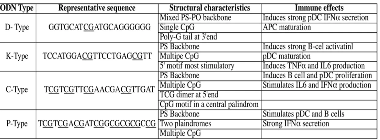
TLR9 recognizes bacterial DNA that contains unmethylated CG dinucleotides and triggers immune responses. The immunostimulatory effects of bacterial DNA are mimicked by synthetic oligodeoxynucleotide (ODN) sequences expressing unmethylated CpGs [27]. Immunostimulatory effects of these ODNs were first reported in 1992 [28]. Minimum ODN length for stimulatory effect has been shown as 8 bp. Optimally, stimulatory ODN contain an unmethylated CpG dinucleotide flanked by two 5’ purines (Pu) and two 3’ pyrimidines (Py) in mice [79]. However, immunostimulatory effects of CpG ODN differ between species. Human PBMC’s immune responses to ODN that are highly stimulatory in mice are poor [29]. TLR9 is highly expressed on professional antigen presenting cells in human such as; B cells and plasmacytoid dendritic cells (pDCs) [30]. Four classes of synthetic CpG ODN have been described (Table 1.3), that have different structural properties leading to different types of cytokine and chemokine releases through different pathways
[27].CpG ODN boost up the efficacy of vaccines against bacterial, viral and parasitic pathogens in both mice and human [31].
Table 1 3: Comparasion of different types of ODN. Adapted from Bode et al., 2011.
1.3.1. D or A Type ODN
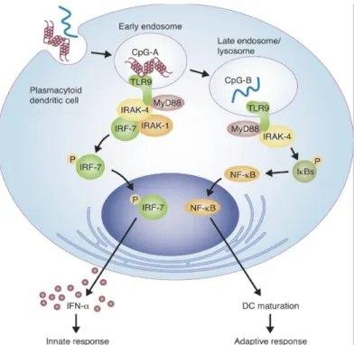
D-type ODNs (also referred to as A type) have a mixed, phosphodiester/phosphorothioate backbone, contain a single CpG and have poly G tails at the 3’ ends. The poly G tails interact with each other, forming ‘G tetrads’ and ODN clusters [32]. This type of ODN interacts with CXCL16 that is expressed on pDCs but not on B cells. D-type ODN activates pDCs to secrete IFNα, however it has weak affects on B cells [33]. They are retained in the early endosome for long time of period, where they interact with MyD88/IRF-7 complexes. This interaction triggers a signaling pathway leading to IFNα production eventually [27].
1.3.2. K or B Type ODN
Typical K/B-type ODNs have a complete phosphothioate backbone, which decreases its susceptibility to DNAse digestion, resulting in a longer in vivo half-life than ODN with phosphodiester backbone [32]. Unlike D/A-type ODN, it does not form higher ordered structures and also lacks poly-G tails. K/B-type ODN is used in most studies with CpG ODNs, because of its stable structure [33], [31]. K/B-type ODN expresses multiple CpG motifs [34]. The K/Btype ODN also promotes the maturation and activation of pDCs, however it does not induce IFNα secretion but stimulates TNFα production because this type of CpG ODN is rapidly transported
into late endosomes through early endosomes (Figure 1.3). It stimulates IL6 secretion, induces B cells to proliferate and secrete IgM [29].
Figure 1.3: Subcellular location of D- and K-type CpG ODNs into late and early endosomes in pDCs (adapted from Williams, 2006).
1.3.3. C-type CpG ODN
The immunostimulatory effects of C-type ODNs are intermediate between D and K classes [33]. It also have an entire phopsphorothioate backbone and expresses TCGTCG at the 5’end, it resembles to D-type ODN because of its palindromic CpG motifs [32]. This class of CpG ODN stimulates B cells to secrete IL6 and induces IFNα production from pDCs. It is found in both early and late endosome, thus expresses both D and K- type properties [27].
1.3.4. Other Types of CpG ODNs
There is several more CpG ODNs, which do not fit to any other class described above. One of them is, P-type ODN, they contain two palindromic
sequences that enable them to form higher ordered structures. P-Type ODNs, stimulate B cells and pDCs to secrete IFNα [27].
Another type is Y-shaped DNA (Figure 1.4), they are composed of using 3 30-base ODNs. Y-type ODN induces TNFα and IL6 [35].
.
Figure 1.4: Y-shaped CpG ODN (Adapted from Nishikawa, 2008).
1.3.5. The Dichotomy of D-and K-Type ODNs
The CpG ODN classes described above have different responses in human pDCs as mentioned. The response mechanisms of these ODNs are poorly understood and still being investigated. Studies revealed that both types of ODNs enter the same immune cells, however, their localization differs leading to different immune responses [30].
In mice, it was established that D- and K- type ODNs trigger different signaling pathways that involves IRF-7 or NF-κB. Depending on the physical forms of the molecules one is localized to early endosomes, where the other to late endosomes. Sequence and backbone structures are important for their localization. Localization differentiation causes the activation of different signaling pathways [36]. K-type ODNs have been shown to localize in early endosome where it interacts with TLR9 directly, activating a signaling cascade that involves serine kinase interleukin-1 receptor-associated kinase interaction with the adaptor protein tumor necrosis
receptor-associated factor 6. NF-κB signaling pathway is activated as well as activated protein-1 transcription family members Jun and Fos, causing the transcription of immune response genes such as IL6 [30]. However there is no evidence that D-type ODN interacts with TLR9. It has been established that, D-type ODNs bind to the chemokine and scavenger receptor CXCL16 that pDCs express on their surfaces. This binding leads D-type ODN to accumulate in early endosome for much longer period, where TLR9-MyD88-IRF7 signaling pathway is activated. Eventually IFNα secretion is induced from pDCs [37].
Moreover, to their different immune responses, when these two types of ODNs, D- and K-type, are introduced simultaneously to human monocytes, K-type cross inhibits D-type ODN’s response. Studies showed that D-type ODN was unable to trigger monocytes to differentiate and secret IFNα in the presence of K-type ODN. This dichotomy is not caused because of any competing uptake mechanisms, they do not block one another’s entry to cell, moreover they are found in different locations in the cell; D-type in endosome and K-type in lysosomes [30]. An explanation to their dichotomy is; lysosomal TLR9 signaling by K-type ODN inhibition to the induction
of IFNα secretion by endosomal TLR9 [38].
To overcome this dichotomy, new CpG ODNs were described or modifications were made to K-type ODN in order to stimulate pDCs to secrete IFNα. A third CpG ODN described was the C-type ODN, as mentioned above, that has the ability of both B-cell activation leading IL6 secretion and IFNα induction from pDCs [69].K-type ODNs are the only CpG ODN that are being used in clinical trials. Due to their ability to from G quadruplex based structures that complicated their manufacturing made it difficult for the D-type ODNs to progress into the clinic.
Another developed strategy for using IFNα inducer CpG in clinical trials, is converting a conventional K-type ODN into a IFNα inducer CpG, by multimerizing them. Multimerized CpG ODN is retained in early endosome causing the activation of MyD88/IRF-7 signaling pathway, which leads to IFNα secretion. In a recent study, a nanoring with Tat peptide and a K-type ODN was formed, which induced IFNα production similar to D-type ODN did [34]. Another study was done with K3 and
Schizophyllan (SPG), a soluble B-glucan derived from Schizophyllum commune. K3 forms a nanostructure with SPG that shows characteristics of both K-and D-type ODNs at the same time. It has been reported that K3-SPG induces PBMC into secreting large amounts of both type I and II IFN. By nanostructure formation with SPG, K-type ODN does not lose its K-type activity rather it gains D-type activity at the mean time [39].
1.4. Drug Delivery with Nanoparticles
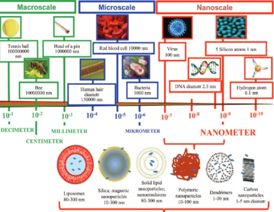
There are many issues facing delivery of drugs to target sites, such as; high toxicity, poor solubility, aggregation, non-specific targeting, instability in vivo and short half –lives. A proposed method to overcome many physical and degradative barriers is encapsulating drugs into nanoparticles. Encapsulation of drugs into nanoparticles, protects the drug from degradation and improves cellular uptake, in many cases reduces toxicity [40]. Solid lipid nanoparticles, dendrimers, polymers, silicon or carbon materials, magnetic nanoparticles and liposomes are the examples of nanoparticles for drug delivery systems (Figure 1.5) [41]. Liposomes has been the most successful candidate in clinical applications, there are many currently approved liposomal drugs are being used and numerous in late-stage clinical development in the United States [42], [43]. Doxorubicin encapsulated Sterically stabilized PEG`ylated
liposome, DoxilTM
, is approved for the treating of metastatic breast cancer, ovarian cancer, and Kaposi’s sarcoma. Chitosan nanoparticles are also effective against tumor
growth [44]. Poly(lactic-co-glycolic acid) (PLGA) NPs are being used in cancer
therapy, causing enhanced binding due to their surface modifications leading to more
efficient anti-tumoral effect [45]. In addition to synthetic particles, natural polymers
Figure 1.5: Nanoparticle drug delivery systems (adapted from Wilczweska, 2012). Liposomes are small vesicles, which are made of cholesterol and natural non-toxic phospholipids. There are different types of vesicles; large multilamellar vesiscles, (LMV) ranges in size between 500nm to 5um, small unilamellar vesicles (SUV) they are formed by single bilayer and around 100nm in size, large unilamellar vesicles are formed by single bilayer and range in size between 200nm to 800 nm [46].
Late Alec Bangham and colleagues published the first description of a swollen phospholipid system in 1965. Later, Gregory Gregoriadis, one of the early pioneers in liposome field, first established the concept of liposomes. Gregoriadis and colleagues were the first to entrap drugs into liposomes and describing a novel drug delivery system in 1971 [47], [68]. The name liposome, was derived from two Greek words; ‘lipos’ means fats and ‘soma’ means body [48].
Liposomes (Figure 1.6) are promising delivery systems, due their sizes, their hydrophobic and hydrophilic character. Moreover, most drug toxicities decrease when encapsulated into liposomes and drugs that are loaded into liposomes are protected from degradation leading to longer circulation time of the drug through the body [46]. Additionally, with liposomal loading, drugs can be targeted to the cells of interest by surface functionalization of the carriers. The most common surface modification used is polyethylene glycol (PEG), by which liposome gains immunogenicity along with prolonged half time and accumulation to the preferential target tissue by enhanced permeation retention (EPR) effect [49]. Both amphiphilc and lipophilic molecules can be loaded into them, due to their phospholipid bilayers [50]. According to their lipid composition, surface charge, size and the method of preparation, they have different properties. Their bilayer components determine the rigidity and fluidity. Liposomes are greatly used in cosmetic and pharmaceutical industries [47].
Figure 1.6: Representative diagram of liposomes (Adapted from Dhandapani, 2013).
1.4.1. Delivery of CpG ODN in liposomes
Even though, nucleic acids have been shown to have great immunostimulatory effects, their in vivo and in vitro applications require delivery vehicles [43]. The uses of free CpG ODNs have several disadvantages. Free ODN lacks specifity for target cells; poor cellular uptake and most importantly, they are sensible to nuclease degradation [52].
as targeting, simplicity and protection like described above [53]. Liposomes can be administered in vivo by different ways, including intravenous, intraperitoneal, aerosol or direct injection [54]. Moreover, encapsulation of CpG ODN into liposomes enhances delivery to target cells when compared with free forms[71]. Most importantly, liposomes protect ODNs from nuclease degradation thus allowing a prolonged circulation time. Therefore, liposomal delivery systems enhance immunostimulatory activity of CpG ODNs [52].
Chapter 2
2. Aim of this study
CpG ODNs are promising candidates to be used as adjuvants in the vaccine development. However, they also have some disadvantages like mentioned before. Studies have been still going on to overcome these disadvantages. D- and K- type ODNs have different immune responses. K-type ODN, which has progressed into clinical trials as adjuvants, strongly stimulates B-cells and pDC that leads to secretion of IL6 and TNFα, as an antibacterial response. Whereas, D-type ODN induces pDCs to secrete IFNα causing an antiviral immune response. Moreover manufacturing process of D-type ODN is complicated due to its quadruplex structures, and D-type ODN shows batch-to-batch variation, which makes it difficult for D-type ODN to enter any clinical trial. Researchers are still keen to develop a system to combine D- and K- effects in a single CpG ODN or in a multimerized system.
Liposomes are extensively used as drug delivery systems. Along with many features it provides to the drug, most importantly it brings protection. CpG ODNs are highly sensitive to DNase degradation, and encapsulation in liposomes protects CpG from degradation, thus provides a longer circulation time. Additionally, when D- and K- type ODN are introduced to cell in their free forms, K-type ODN blocks D-type response, and only K-type specific response is observed.
To overcome all of these disadvantages and to develop a system that will combine both CpG ODN effect in a universal system, we will encapsulate both of these CpG ODNs to different kinds of liposomes, and introduce them to human PBMC and murine splenocyte to observe whether we will see a combined effect. The main aim of this study is to overcome the dichotomy of D- and K- type ODN by encapsulating them into liposomes and to identify proper liposome combinations encapsulating D- and K- that leads to expression of both D- and K- superior activity.
Chapter 3
3. Materials and Methods
3.1. Materials
3.1.1.ELISA Reagents
Mouse cytokine ELISA reagents, including monoclonal antibodies for cytokines, recombinant antibodies for IL6 and IL12 were purchased from Biolegend (USA), whereas reagents for IFNg and streptavidine-alkaline phosphatase (SA-AKP) were purchased from MabTech (USA). P-nitrophenyl phosphate disodium salt (PNPP)-substrate for alkaline phosphates was purchased from Thermo (USA).Human cytokine ELISA reagents for TNFa and IFNg were purchased from BioLegend (USA) , IP10 reagents were from BD and reagents for pan IFN-a were from MabTech (USA).
3.1.2.TLR Ligands
D- and K-Type CpG Oligodeoxynucleotides (ODNs) (TLR9 ligand) and their CG dinucleotides flip forms (i.e. control ODNs) were synthesized either by Alpha
DNA (Montreal, Canada) or on an in-house machine (Mermade6
, ODN Synthesizer, USA). their sizes and base sequences are listed in Table 3.1.
Table 3.1: Sequences of ODN used. Upper case letters: Phosphorothoiate linkage
between bases, lower case letters: Phosphorodiester linkage between bases
K-type ODN
K3(20mer)
5’- ATCGACTCTCGAGCGTTCTC-3’
K23(12mer)
5’-TCGAGCGTTCTC-3’
1555’(15mer)
5’-GCTAGACGTTAGCGT-3'
1555-3CG(20mer)
5’-GACGTTGACGTTGACGTTGG-3'
1466-pG6-PS(16mer)
5’-CAACGTTGAGGGGGG-3'
1466-pT6-PS(16mer)
5’-TCAACGTTGATTTTT-3'
3.1.3. Lipids
Cholesterol was purchased from Sigma Aldrich (USA).
L-a-Phosphatdylcholine (PC), 3a-[N-(N’,N’-Dimethylaminoethane)-carbamoyl]
Cholesterol Hydrochloride (DC-Chol),
1,2-Dioleoyl-sn-GLycero-3-Phosphoethanolamine-N-[MEthoxy(Polyethyleneglycol)-2000] (Ammonium Salt)
(PEG-PE), 1,2-Dioleoyl-sn-Glycero-3-Phosphoethanolamine (DOPE),
L-a-Phosphatdylserine (PS) were all purchased from Avanti Polar Lipids (Alabama, USA).
3.2. Methods
3.2.1. Cell Culture
3.2.1.1. Spleen and Lymph Node Single Cell Suspension
Preparation
Mice (C57/BL6 or BALB/C strains) were sacrificed by cervical dislocation, their spleens and Lymph nodes were removed and transferred into 3 ml of 2% FBS supplemented complete RPMI-1640 media. Single cell suspensions were obtained, by smashing the spleens with back of a sterile syringe plunger with circular movements. Media was collected into 15 ml falcons, leaving the tissue clumps and connective tissues in the Petri plate, and washed twice with 10 ml of fresh 2% FBS supplemented complete RPMI-1640 media followed by centrifugation at 540 g for 10 minutes. At
D-type ODN
D35 (20mer)
5’-GGtgcatcgatgcaggggGG-3'
D35-3CG (20mer)
5’-GGtcgatcgatcgaggggGG-3'
pG6-1466-PO
5’-ggggggtcaacgttga-3'
Control ODNs
1471-PS
5'-TCAAGCTTGA-3'
1466-Acore-PS
5'-TCAACGGTTGAGGGGGG-3'
1466-Acore-PO
5'-tcaacggttgattcaaa-3'
1466-Acore-MB
5'-tcaacggttgattcaaa-3'
the end of the last washing, cells were re-suspended in 1ml of 5% FBS supplemented complete RPMI-1640 for counting by Flow Cytometry.
3.2.1.2. Peritoneal Exudate Cell Preparation
Mice were injected intraperitoneally (ip) with 10 ml of 2% FBS supplemented complete RPMI-1640 and peritoneal exudate cells (PECs) containing macrophages were collected back into the syringe (usually 8-9ml was recovered). Cells were washed twice with fresh media followed by centrifugation at 540 g for 10 minutes. After the washing steps, cells were resuspended in 1 ml of 5% FBS supplemented complete RPMI-1640 for counting by Flow Cytometry.
3.2.1.3. Human Peripheral Blood Mononuclear Cell (PBMC)
Isolation
Blood samples were collected from healthy donors into vacutainer tubes that contained EDTA as anti-coagulant (BD Vacutainer, purple cap). 22.5 ml blood was slowly layered onto, 15 ml of ficoll (Lymphocyte Separation Medium, Lonza). Samples were centrifuged at 540 g for 30 minutes with the break set to off for keeping layers separated. The cloudy layer that was settled over the ficoll, (which contained PBMCs), was collected carefully using a sterile Pasteur pipette. Recovered PBMCs were washed with fresh media twice followed by centrifugation at 540 g for 10 minutes. After the washing steps, the pellet was re-suspended in 1 ml of 5% FBS containing complete RPMI-1640 and counted by Flow Cytometry.
3.2.2. Cell Lines
3.2.2.1. RAW 264.7
RAW 264.7 (Mus musculus), macrophage/monocyte cells (ATCC) were cultured with 10 % FBS supplemented complete RPMI-1640. Cells were passaged every 3-4 days when they reached 80-90% confluency.
3.2.3. Time Lapse detection of Labeled Liposome Uptake
within RAW 264.7 cells
Differential internalization of D-and K-Type liposomes within cells were followed by labeling two different populations of liposomes with two different dyes that excitates at different wavelengths by Nikon Fluorescent (Nikon Eclipse Ti)
microscope with an attachments of CO2 chamber and live camera recording.
While D-type ODN were encapsulated in Neutral Liposome (labeled with FM1-43 dye), K-type ODN were encapsulated in Cationic Liposome (labeled with SPDiOC Dye). Initial experiments were conducted first with RAW cells that were incubated with one type of liposome. Images, as well as live video recordings were recorded for 15 mins, with 15-second intervals. Next, D- and K-Type CpG ODN loaded two different liposome populations were co-incubated with RAW 264.7 cells
(2x10⁵
cells/well) and early stage simultaneous uptake/internalization kinetics of liposomes by cells were recorded by fluorescence microscope (Nikon Eclipse Ti). As done before, images were recorded for 15 mins, with 15-second intervals. Later proper images were converted into a video file.
3.2.4. Cell Counting by Flow Cytometer
20 μl of single cell suspensions, which were prepared in 1 ml (dilution factor 1ml/20μl ; 1000/20=50), were added into 10 ml of isotonic solution . 20 μl from the prepared 10 ml solutions (dilution factor 10ml/20μl; 10000/20=500) were counted in Accuri C6 Flow Cytometer. Live and nucleated cells were gated, apoptotic, dead or non-nucleated blood cells were ignored. Counted number was multiplied with 25000 (multiplication of dilution factors; 50x500=25000).
3.2.5. In Vitro Stimulation of Murine Cells
For stimulation in 96-well plates, from cell culture stock (2x10⁶
cells/ml, 150
μl fractions) of i) lymph node and ii) PECs were layered (3x10⁵
cells/well). D-and K-Type CpG ODNs as stimulants in either free form (0.2-3.0 µM) or different liposome
loaded form were added in 50 μl RPMI-1640 media to adjust the final well volumes to 250 μl. Stimulations were done in duplicate wells for each treatment groups. Supernatants were collected after 24 hours. Each experiment, at least repeated with 3-5 independent mice done on at least 2-3 separate occasions.
3.2.6. In Vitro Stimulation of PBMCs
PBMCs are isolated and counted as described above (Section 3.2.2).
Afterwards, 3x10⁵
cells in 150 μl were layered to 96-well cell culture plates. Stimulants (as mentioned in Section 3.2.3.) were added in 50 μl of 5% FBS supplemented RPMI-1640, each well were prepared as duplicates and the final volume was set to 250 μl. Supernatants were collected after 24 hours of stimulation.
3.2.7. Liposome Preparation
All phospholipid and cholesterol stock solutions were prepared as 10 mg/ml or 20 mg/ml in chloroform and stored at -20°C. Phospholipids and cholesterol were mixed according to the ratios in Table 3.2 to obtain different liposomes with different physicochemical characteristics. Chloroform was evaporated with rotary evaporator (ILVAC, Germany) and a thin uniform film was formed. Nitrogen gas was streamed through the round bottom flasks to prevent remaining oxygen initiate peroxidation of the lipids. Next, onto lipid film, 1 ml of PBS was added and to obtain large multilamellar vesicles, films were disrupted by adding glass beads and shaking the flasks rotationally, which resulted in a milky solution. In order to generate small unilamellar vesicles (SUVs), liposomes were sonicated 10 times for 30s durations with amplitude of 50% (VibraCell, SONICS, USA). 150 mg of ODN was added to each 4,5 μmol of liposomes. The mixture was snap frozen in liquid nitrogen and lyophilized overnight (Benchtop K, Virtis, USA) to obtain lipid/ODN powder.
Encapsulation of ODNs within liposomes was achieved by controlled
re-hydration method; 20 μl of DNase RNase free ddH20 was added to dehydrated
liposomes and vortexed for 15s every 5 minutes for 25 minutes. Same amount of PBS was added to the mixture and vortexed for a few seconds and let stand on the bench
for another 10mins. Finally, 110 μl of PBS was added to the mixture to have a final volume of 150 μl bringing the ODN concentration within liposomes to 1µg/µl of lipozomes. Liposome formulations were kept at 4°C until for further use.
Table 3.2: Lipid Compositions of different liposome types
Liposome Compostion of Lipids (molar ratio)
Neutral PC:Chol (1:1)
Anionic PC:DOPE:PS (1:0.5:0.25)
Cationic DC-Chol:PC:DOPE (4:6:0.06)
Stealth Chol:DOPE:PEG-PE (4:6:0.06)
Cationic-Stealth (SSCL) DC-Chol:DOPE:PEG-PE (4:6:0.06)
3.2.7.1. Labeling Liposomes with SP-DiOC and FM1-43
Cationic liposomes were labeled with 1uM of SP-DiOC, anionic and neutral liposomes were labeled with 200 μg/ml of FM-143. Labeled liposomes were kept at dark for 30 minutes at RT, washed with 1 ml of PBS by centrifugation at 15000g for 30 minutes (Eppendorf Minifuge; Model: 5415R). The supernatant was discarded and the pellet was resuspended at 150 μl of PBS.
3.2.8. Cytokine Enzyme Linked Immunosorbent Assay (ELISA)
After 24 hours of stimulation, 96 well plates were centrifuged at 540 g for 5 minutes. Approximately 230 μl of supernatants were collected and transferred into a new 96 well plate for immediate use or storing at -20°C for further use. 2HB plates were coated with monoclonal antibodies against mouse or human cytokines (50 μl/well) according to working concentrations listed in Table 3.3 and incubated overnight at +4°C. Wells were blocked with Blocking Buffer (200 μl, 5% w/v BSA-1X PBS, 0.025% Tween-20) at RT for 2 hours after coating antibody solutions were discarded. Plates were washed with washing buffer (PBS-Tween 20) for 3 minutes 5 times. Cell Supernatants and appropriately diluted standard recombinant cytokine antibodies (Table 3.4) were added (50 μl/well) and incubated overnight at +4°C.
Plates were washed as previously mentioned. Biotinylated anti-cytokine antibodies (50µl/well) were added to wells, in T-cell buffer with 1:1000 dilutions and incubated for 2 hours at RT. Streptavidin alkaline-phosphatase (SA-AKP) 1:1000 diluted was added again as 50 μl/well portions, following the washing steps and incubated for 1 hour at RT. For SA-AKP detection PNPP substrate was prepared according to the product`s protocol sheet and pipetted over each well at 50 µl portions. Yellow color formation was followed by reading the plates in automated ELISA Reader (Molecular Devices, USA) at OD set to 405nm.Concentrations of the culture supernatants were calculated from the S-Shaped curve of each standard recombinant cytokine.
Table 3.3 : concentrations of coating antibodies for mouse and human cytokine ELISA experiments (50ul/well transferred)
Anti mouse IL6 2 μg/ml
Anti mouse IL12 4 μg/ml
Anti mouse IFNg 1 μg/ml
Anti human TNFa 2 μg/ml
Anti human IFNa 4 μg/ml
Anti human IFNg 4 μg/ml
Anti human IP10 2 μg/ml
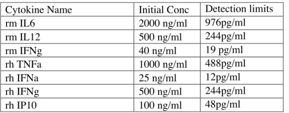
Table 3.4: Starting concentrations and detection limits for each recombinant standard cytokines (each cytokine was diluted by 11X by 1/2 dilutions)
Cytokine Name Initial Conc Detection limits
rm IL6 2000 ng/ml 976pg/ml rm IL12 500 ng/ml 244pg/ml rm IFNg 40 ng/ml 19 pg/ml rh TNFa 1000 ng/ml 488pg/ml rh IFNa 25 ng/ml 12pg/ml rh IFNg 500 ng/ml 244pg/ml rh IP10 100 ng/ml 48pg/ml
3.2.9. Ex-Vivo Experiments
C57/BL6 mice were injected i.p. either with free 20 μg D + 20 μg K-Type CpG ODN, or 20 μg of FM1-43 labeled D ODN neutral liposome + SP-DiOC labeled
K ODN loaded SSCL,. In another experiment, Neutral liposome was replaced by anionic liposome and mixed with K-Type CpG ODN loaded SSCL. Also labeled stealth liposome that was loaded with D ODN was again mixed and injected with SP-DiOC labeled K ODN loaded SSCL. All formulations were suspended in 200 μl of 1X PBS. 6 hours later mice were sacrificed. Spleen, lymph nodes and PEC were collected. Single cell suspensions were prepared as described previously for cytokine ELISA. Cells were also used to assess surface marker expression levels by FACS analyses. Additionally, LN, PEC and splenocyte cells were used to purify total RNA, and study various cytokine and chemokine gene expression levels by PCR.
3.2.10. Determination of the Gene Expression
3.2.10.1. Total RNA Isolation
Untreated or ODN treated cells (5-10x105
/ml) from sacrificed mice were centrifuged at 540xg for 10 minutes, supernatants were discarded and pellets were resuspended in 1 ml of Trizol (Invitrogen, USA). Samples were either stored at -80°C for further use or in some cases directly continued to extract total RNA as described below. Of note, all steps during RNA isolation including centrifugations were performed on ice and at +4°C.
Samples were transferred into Eppendorf tubes and 200 μl chloroform was added into each. After the tubes were shaken vigorously for 10-15 seconds, samples were centrifuged at maximum speed for 18 minutes (Eppendorf Minifuge; Model: 5415R, @14000 rpm). At the end of centrifugation, 450-500 μl of upper clear phase was collected into new Eppendorf tubes. 500 μl isopropanol was added in order to precipitate RNA, incubated at RT for 10 minutes and centrifuged again @ 14000rpm for 13 minutes. The pellet was washed with the addition of 1 ml 75 % EtOH and centrifuging at 8000 rpm for 8 minutes. Washing step was repeated with 99,8 % EtOH. The pellet was left under laminar flow hood to air dry. Purified RNA was
resuspended in 20 μl of DNase/RNase free H2O. The OD of the samples were
Technologies, USA). The absorption ratios of the samples at 260/280 were expected to be in 1.8-2.0. RNA samples were then either stored at -80°C for further use or continued for PCR protocols.
3.2.10.2. cDNA Synthesis
cDNA synthesis kit (NEB, USA) ProtoScript First Strans cDNA was used to transcribe total RNA samples.1 μg of total RNA was mixed with 100 ng of Oligo (dT)
15 mer promer (1 μl) and each sample was completed with DNase/RNase free H2O to
8 μl. Samples were pre-denatured at 65°C for 5 minutes. 10 μl RT Buffer ( dNTP mix
and 10mM MgCl2) and 2 μl M-MuLV RNase H+ reverse transcriptase ( includes
RNase inhibitor) were added to samples. After spinning down the samples quickly, were incubated at 42° C for one hour and at 85° C for 5 minutes. cDNA samples were stored at -20°C for further use.
3.2.10.3. PCR
All primers (Table 3.5) except mIL18 were adopted from Guiletti et al 2001,
mIL18 was designed using Primer3 Input 0.4.0 program
(http://frodo.wi.mit.edu/primer3/input.htm). PCR reaction was prepared with Quick-load Taq 2X Master Mix (NEB, USA). Final volume of the reaction was 25 μl, containing 12.5 μl Master Mix, 2 μl cDNA, 1 μl of each primer (forward and reverse)
and 8.5 μl dd H2O (Table 3.6).
Table 3.5: Mouse primer sequences and their expected product sizes.
Primer Direction Sequence
Product Size
mIP10 Forward 5'-GCCGTCATTTTCTGCCTCAT-3 127bp
Reverse 5'-GCTTCCCTATGGCCCTCATT-3'
mIL18 Forward 5'-GATCAAAGTGCCAGTGAACC-3' 491bp
Reverse 5'-ACAAACCCTCCCGACCTAAC-3'
mIL15 Forward 5'-CATCCATCTCGTGCTACTTGTGTT-3' 126bp
Reverse 5'-CATCTATCCAGTTGGCCTCTGT-3'
mCD40 Forward 5'-GTCATCTGTGGTTTAAAGTCCCG-3' 91bp
mGAPDH Forward 5'-AGCTCATTTCCTGGTATGACA-3' 128bp
Reverse 5'-CTCTCTTGCTCAGTGTCCTT-3'
Table 3. 6: PCR conditions for mouse primers.
Gene Names (mouse)
PCR Steps (Temp;Time) gapdh ip10, il18, il15, cd40
Initial Denaturation 94° C; 5' 94° C; 5' Denaturation 94° C; 30'' 94° C; 30'' Annealing 60° C; 30'' 55° C; 30'' Extension 72° C; 40'' 72° C; 40'' Cycle # 30 35 Final Extension 72° C; 5' 72° C; 5'
3.2.10.4. Agarose Gel Electrophoresis
2 % agarose gel was prepared (w/v) with 150 ml of 1X TAE buffer. Ethidium bromide was added as 1µg/ml. Each lane was loaded with 10 μl PCR product and 5μl of 100 or 50 bp DNA ladders (NEBiolabs, USA) as markers. Gels were run at 100V for 30 minutes and visualized under UV transilluminator (Vilber Lourmat, France). The gel images of different samples were then pooled and transcript level comparison was done on combined gel image.
3.2.11. Flow Cytometry
3.2.11.1. Fixation of Cells
After single cell preparations and treatments of cells, they were centrifuged and pellets were resuspended in 50 μl of fixation solution (4% paraformaldehyde Invitrogen, USA) while vortexing. Cells were incubated at RT for 15 min and 20x more PBS-BSA Azide (1ml, FACS Buffer) was added and cells were centrifuged at 1800 rpm for 10 minutes. Supernatants were discarded and the cells were resuspended in 1ml of FACS Buffer and stored at +4°C for further use.
3.2.11.2. Ex-vivo FACS Analyses of cells to determine
Liposomal CpG ODN Internalization
Six hours after injection with labeled D- or K-Type CpG ODN loaded liposomal formulations, animals were sacrificed. Spleen, lymph node and PEC single cells were recovered fixed after washing and centrifugation as described previously and analyzed by BD Accuri C6 Flow Cytometer. From FL1 channel for SP-DiOC signal and FL2 channel for FM-143 signal two different liposome population`s level of internalization were assessed.
3.2.11.3. Intracellular Cytokine Staining (ICCS) of IFNα and
TNFα
3.2.11.3.1. IFNα Secretion Assay
PBMCs were stimulated with different free or liposomal CpG ODN formulations for 5 hours. To detect secreted IFNα, an IFN-α Secretion Assay Kit was used and protocol from the product data sheet was followed (Cell Enrichment and Detection Kit (PE) MACS, Miltenyi Biotec). The experiment was done on ice and centrifuges were set to 4°C. After the washing steps, cells were incubated with IFNα catch reagent on ice for 10 minutes before the addition of cell supernatant medium and further incubated at 37°C for additional 20 minutes for achieving the capture of the secreted form of IFNα to the supernatants. IFNα which were bound by pDCs were stained with i) anti-IFN-α detection (PE-labeled), ii) anti-BDCA-2 (FITC labeled, Biolegend, USA) and iii) anti-CD123 (PE/Cy5 labeled, BD, USA) for 30 minutes at dark. FACS analyses were performed after washing and re-suspending cells in PBS.
3.2.11.3.2. TNFα ICCS
For the detection of TNFα, cells were stimulated for 4 hours in the presence of Brefeldin-A (in order to store the secreted TNFα inside the cells). After stimulation, cells were washed fixed as described above Section and stained with
anti-CD123-FITC (Biolegend) and and anti-TNFα-PE/Cy7 (Biolegend) for 30 minutes at dark. FACS analyses were performed after washing and re-suspending cells in PBS.
3.2.12. Statistical Analysis
Statistical significance between untreated (or control) and treated groups was evaluated using Student's t-test. p<0.05 set as statistically significance between the analyzed groups.