Antioxidant Vitamin E/Cyclodextrin Inclusion Complex Electrospun
Nano
fibers: Enhanced Water Solubility, Prolonged Shelf Life, and
Photostability of Vitamin E
Asli Celebioglu
†and Tamer Uyar
*
,††Institute of Materials Science & Nanotechnology, UNAM-National Nanotechnology Research Center, Bilkent University, Ankara
06800, Turkey
*
S Supporting InformationABSTRACT: Here, we demonstrated the electrospinning of polymer-free nanofibrous webs from inclusion complex (IC) between hydroxypropyl-β-cyclodextrin (HPβCD) and Vitamin E (Vitamin E/HPβCD-IC NF). The inclusion complexation between HPβCD and Vitamin E was prepared by using two different molar ratios (Vitamin E/HPβCD; 1:2 and 1:1), which correspond to theoretical value of ∼13% (w/w) and 26% (w/w) loading of Vitamin E in the nanofiber (NF) matrix. After electrospinning and storage, a very high loading of Vitamin E (up to∼11% w/w, with respect to fiber matrix) was preserved in Vitamin E/HPβCD-IC NF. Because of the cyclodextrin inclusion complexation, only a minimal weight loss (only ∼2% w/w) was observed. While pure Vitamin E is insoluble in water, Vitamin E/HPβCD-IC NF web has displayed fast-dissolving behavior. Because of the greatly enhanced water-solubility of Vitamin E, Vitamin E/HPβCD-IC NF web has shown effective antioxidant activity. Additionally, Vitamin E/HPβCD-IC NF web has provided enhanced photostability for the sensitive Vitamin E by the inclusion complexation in which Vitamin E/HPβCD-IC NF still kept its antioxidant activity even after exposure to UV-light. Moreover, a 3 year-old Vitamin E/HPβCD-IC NF sample has shown very similar antioxidant efficiency when compared with freshly prepared Vitamin E/HPβCD-IC NF indicating that long-term stability was achieved for Vitamin E in the CD−IC fiber matrix. In brief, our results suggested that polymer-free electrospun Vitamin E/HPβCD-IC nanofibrous webs could have potential applications in food, pharmaceuticals, and healthcare thanks to its efficient antioxidant activity along with enhanced water-solubility, prolonged shelf life, and high photostability of Vitamin E.
KEYWORDS: electrospinning, nanofibers, Vitamin E (alpha-tocopherol), cyclodextrins, inclusion complex, antioxidant activity, photostability, water-solubility
■
INTRODUCTIONThe electrospinning of nanofibers/nanowebs is becoming promising approach for encapsulation of active agents such as drugs1 and food additives.2,3 Electrospinning technique has versatility and simplicity for the production of functional nanofibers/nanowebs from a wide range of synthetic and natural materials.4 The unique characteristics of electrospun nanofibers/nanowebs such as high surface-to-volume ratio, highly porous structure, and adjustable and modifiable properties provide great potential from food, biomedical, filtration, and energy to agriculture.4−6
General aspect of electrospinning based on the production of polymeric nano-fibers by using organic solvents causes concerns in terms of cost and toxicity for healthcare and food applications. On the other hand, water-soluble polymers can be substituted as an alternative to eliminate the disadvantages originating from organic solvents. The starch-based oligosaccharides, cyclo-dextrins (CDs), are also water-soluble, and as we have demonstrated in our previous studies, possibilities exist to obtain nanofibers from these biorenewable material without using a carrier polymeric matrix.7−11Practically, it might be a challenge to obtain bead-free and uniform fibers for the electrospinning process; hence, tedious work needs to be done for the optimization of the parameters. Here, another challenge with the nonpolymeric systems is the extra efforts needed to
obtain uniform fibers by electrospinning. On the other hand, having high concentration of CD systems enables us to perform electrospinning of bead-free and uniform fibers without the break of electrospinning jet and without the need of using polymeric carrier matrix.7−17 CDs are attractive oligo-saccharide types owing to their cyclic and truncated supra-molecular structures. CDs are produced by the enzymatic degradation process of starch, and the positioning of hydro-phobic carbon backbones through the interior part of CD leads to formation of relatively hydrophobic cavity.18,19The unique characteristic of CD is due to its hydrophobic cavity, which makes noncovalent host−guest inclusion complexation (IC) with variety of active compounds.18,19Besides, their edible and nontoxic nature makes them attractive for numerous formulations for food, pharmaceutical, and cosmetic prod-ucts.18,19 Depending on the subunit quantity, there are three native CD typesα-CD, β-CD, and γ-CD composed of 6, 7, and 8 glucopyranose, respectively.19Apart from these native CDs, chemically modified CDs (hydroxypropyl and methylated CDs) are also synthesized to increase the water-solubility of
Received: April 5, 2017
Revised: June 8, 2017
Accepted: June 13, 2017
Published: June 13, 2017
guest molecules and provide better stability to complexed guest molecules against light and oxygen when compared to native CDs.20
In our recent studies, we have produced polymeric electrospun nanofibers incorporating CD-IC of various active compounds like essential oils and fragrance/flavors.21−25 However, the weight loading of these compounds was generally limited up to 5% (w/w) (with respect to polymer matrix) because the incorporation of higher amount of CD-IC affects the electrospinning to produce uniform nanofibers. Therefore, the electrospinning of polymer-free CD-IC nanofibers provides much higher loading capacity of guest molecules (up to∼10− 15%, w/w).12−17Besides, much higherfiber throughput can be achieved for the electrospinning of polymer-free CD-IC systems when compare to polymeric systems. For example, for polymeric systems, typically 5−25% (w/v) polymer solution is used for electrospinning,21−25whereas, for polymer-free CD-IC systems, typically 120−160% (w/v) CD-IC solution is used for electrospinning.12−17Hence, we can produce much higher amount of electrospun nanofibrous materials in the case of electrospinning of polymer-free CD-IC systems.
Vitamin E is commonly used in drug delivery and wound dressing applications especially due its antioxidant property, and α-tocopherol is the most biologically active form of Vitamin E.26,27 However, the poor water solubility and sensitivity to oxygen, light, and alkali pH create limitations during the practices of Vitamin E.28On the other hand, CDs have drawn interest to parry the limitations of Vitamin E by inclusion complexation. As reported in the literature, CD-IC of Vitamin E can be utilized to enhance the solubility and oxidation stability of this active agent.29−31Even in the related studies, which were performed by our group, CD-ICs of Vitamin E were incorporated into polymeric nanofibers such polylactic acid32and polycaprolactone33for potential applica-tions in food packaging and wound healing, where we have achieved enhanced release and effective antioxidant profile along with the improved water-solubility and UV-light stability for Vitamin E.
In the present study, electrospinning of inclusion complex (IC) between hydroxypropyl-β-cyclodextrin (HPβCD) and Vitamin E was performed without using a polymeric matrix to produce Vitamin E/HPβCD-IC nanofibers (NF). The inclusion complexation between HPβCD and Vitamin E was prepared by using two different molar ratios (Vitamin E/ HPβCD; 1:2 and 1:1, which correspond to theoretical value of ∼13% (w/w) and ∼26% (w/w) loading of Vitamin E in the fiber matrix, respectively). The morphological characterization of the free-standing electrospun nanofibrous webs of Vitamin E/HPβCD-IC NF was performed by scanning electron microscopy (SEM) imaging. The detailed structural and thermal characterization of Vitamin E/HPβCD-IC NF was carried out by proton nuclear magnetic resonance (1H NMR), Fourier transform infrared (FTIR), differential scanning calorimetry (DSC), and X-ray diffraction (XRD). The fast-dissolving characteristic of Vitamin E/HPβCD-IC NF in water was tested. The antioxidant property of Vitamin E/HPβCD-IC NF was also investigated along with a comparison tests by using 3 year-old Vitamin E/HPβCD-IC NF to study the long-term stability. In addition, Vitamin E/HPβCD-IC NF samples were exposed UV-light to investigate the photostability of Vitamin E in CD-IC NF.
■
MATERIALS AND METHODSMaterials. Vitamin E ((alpha-tocopherol), Sigma, > 96% (HPLC)) was obtained commercially. The hydroxypropyl-β-cyclodextrin (HPβCD) (degree of substitution:∼ 0.6, CavasolW7 HP Pharma) was kindly donated by Wacker Chemie AG (Germany). Potassium bromide (KBr, 99%, FTIR grade, Sigma-Aldrich), deuterated dimethyl sulfoxide (d6-DMSO, deuteration degree min. 99.8% for NMR spectroscopy, Merck), ethanol (99.8%, Sigma-Aldrich), methanol (extra pure, Sigma-Aldrich), and 2,2-diphenyl-1-picrylhydrazyl (DPPH, Sigma-Aldrich) were used in this study. The water used was from a Millipore Milli-Q ultrapure water system. The materials were used as-received without any further purification process.
Preparation of Vitamin E/HPβCD-IC and Electrospinning of Nanofibers. The formation of inclusion complexes (IC) of HPβCD with Vitamin E (Vitamin E/HPβCD-IC) was achieved by using two different molar ratios, 1:1 and 1:2 of Vitamin E/HPβCD. In the case of 1:1 molar ratio of Vitamin E/HPβCD, it is anticipated that excess amount of guest molecule (Vitamin E) was used since Vitamin E is a long molecule and it is likely that two HPβCD molecules are required to be included fully in the CD cavity. For Vitamin E/HPβCD-IC, first, Vitamin E was dissolved in ethanol and HPβCD (160% (w/v) with respect to solvent) was dissolved in water separately, then Vitamin E solution was drop-wised added into HPβCD solution slowly. During the inclusion complex preparation, ethanol/water ratio was kept as 1:9 (v/v). Clear aqueous solution of HPβCD turned into turbid and the solution was stirred overnight to obtain Vitamin E/HPβCD-IC. As a control sample, homogeneous and clear aqueous solution of HPβCD was prepared by dissolving HPβCD (160% (w/v)) in water by stirring for 1 h at 50°C; thereafter, it was cooled down to room temperature prior to electrospinning. The clear HPβCD solution and the turbid Vitamin E/HPβCD-IC solutions having 1:2 and 1:1 molar ratios were separately electrospun into the form of nanofibers. The electro-spinning was performed in a horizontal position by using plastic syringefitted with a metallic needle of 0.45 mm of inner diameter. The syringe pump (KD Scientific, KDS-101) was used to control the solution feed rate at 0.5 mL/h. The electrode was clamped to the metal needle tip and the collector. The voltage of 15 kV was set by the high voltage power supply (Matsusada Precision, AU Series, Japan). Electrospun nanofibers were deposited on a grounded stationary cylindrical metal collector covered by a piece of aluminum foil at a distance of 10 cm. The electrospinning apparatus was enclosed in Plexiglas box, and the electrospinning was carried out at 24°C at 30% relative humidity. For comparison, the physical mixture of Vitamin E and HPβCD was also prepared by simple blending of two components as to have 1:2 molar ratio (Vitamin E/HPβCD).
Measurements and Characterization. The viscosity of the HPβCD and Vitamin E/HPβCD-IC solutions was measured by rheometer (Anton Paar Physica MCR 301) equipped with a cone/ plate accessory (spindle type CP40−2) at a constant shear rate of 100 Hz at 22°C. The conductivity of the solutions was measured with Multiparameter meter (InoLab Multi 720, WTW) at RT. Scanning electron microscope (FEI-Quanta 200 FEG) was used for the morphological analyses of the nanofibers. To avoid charging and better imaging, Au/Pd was sputtered onto the samples prior to SEM imaging. The averagefiber diameter (AFD) was measured from the SEM images, and around 100 fibers were counted from various locations of the samples. The molar ratio between Vitamin E/HPβCD was determined by using proton nuclear magnetic resonance (1H NMR, Bruker D PX-400) system. The electrospun Vitamin E/ HPβCD-IC NF and Vitamin E were dissolved in d6-DMSO at the 20 g/L concentration. The spectra were recorded at 400 MHz and at 16 total scans. Integration of the chemical shifts (δ) given in parts per million (ppm) of the samples was calculated by using Mestrenova software. The XRD data were recorded for the nanofibers of HPβCD and Vitamin E/HPβCD-IC NF by using PANalytical X’Pert Powder diffractometer with Cu Kα radiation in a range of 2θ = 5°−30°. Thermal properties of the samples were analyzed by DSC (TA Q2000). DSC analyses were carried out in modulated mode under N2 atmosphere; initially, samples were equilibrated at−90 °C and then
heated to room temperature at a heating rate of 10 °C/min. The infrared spectra of the samples were taken by using FTIR spectrometer (Bruker-VERTEX 70). The samples were mixed with potassium bromide (KBr) and pressed as pellets, and 64 scans were recorded between 4000 and 400 cm−1at a resolution of 4 cm−1. For the visual water-solubility test, a piece of Vitamin E/HPβCD-IC NF and Vitamin E (the same amount in the CD-IC NF) were put into Petri dishes, and water was poured onto the samples (Movie S1). Both the photographs and video were recorded during this test.
Antioxidant activity of the samples was examined by 2,2-diphenyl-1-picrylhydrazyl (DPPH) radical scavenging method. For this, DPPH solution in methanol (75μM) was prepared freshly, and 2.3 mL of this solution was mixed with 200μL of aqueous solutions of Vitamin E/ HPβCD-IC NF and Vitamin E. While ultimate solution concentration of Vitamin E/HPβCD-IC NF was adjusted to 0.4 mg/mL, Vitamin E concentration in nanofibers was determined from the Vitamin E content amount (∼11%, w/w with respect to total sample amount), which was determined from1H NMR measurement. The Vitamin E/ HPβCD-IC NF sample was dissolved immediately in water; however, in case of pure Vitamin E system, we also had tofilter solution to eliminate undissolved part. The ultimate solutions were incubated in dark at RT for 30 min. The DPPH solution has maximum absorption at 517 nm and along with reduction by antioxidant compound, this dominant absorbance disappears. Thus, after incubation time, the reduction at the absorbance intensity (517 nm) was recorded by using UV−vis spectroscopy (Varian, Carry 100). To investigate the long-term stability and photostability of Vitamin E/HPβCD-IC NF, the antioxidant tests were also performed for the 3 year-old Vitamin E/ HPβCD-IC NF (stored in refrigerator at 4 °C) and freshly prepared Vitamin E/HPβCD-IC NF, which are exposed to UV light (300 W, Osram Ultra-Vitalux, E27/ES) from a distance of 30 cm for 120 min. Videos were also recorded for the antioxidant tests for UV-light treated Vitamin E/HPβCD-IC NF to show the scavenging rate change before (Movie S2) and after the UV-light exposure (Movie S3). For video record, methanol solution of DPPH (2.3 mL) was poured into the aqueous solution of Vitamin E/HPβCD-IC NF (200 μL). These
experiments were conducted in triplicate. The antioxidant activities (%) were calculated according to the following equation:
= A −A A ×
Antioxidant activity (%) ( control sample)/ control 100
■
RESULTS AND DISCUSSIONElectrospinning of Vitamin E/HPβCD-IC NF. Here, solutions of inclusion complexes between Vitamin E and HPβCD (Vitamin E/HPβCD-IC) having two different molar ratio (1:2 and 1:1 of Vitamin E/HPβCD) were prepared, and then these highly concentrated solutions (160% (w/v) HPβCD) were electrospun to produce nanofibrous webs (Figure 1). As reported in the literature, Vitamin E molecules intend to form inclusion complexes with HPβCD by 1:2 molar ratio.34 The 1:2 molar ratio (Vitamin E/HPβCD) can be optimal because the long chain structure of Vitamin E molecule might induce steric effect during the complexation and so at least two HPβCD molecules needed for the full molecular coverage of Vitamin E in HPβCD cavities (Figure 1). Even so, along with 1:2 molar ratio of Vitamin E/HPβCD, we have also prepared Vitamin E/HPβCD-IC with 1:1 molar ratio for the electrospinning to have higher loading (∼26% (w/w, respect to totalfiber matrix) of Vitamin E in the nanofiber matrix. Yet, the electrospinning of Vitamin E/HPβCD-IC (1:1) system resulted in some beaded nanofiber morphology as discussed below. It is worth to mention that, much higher loading of Vitamin E can be achieved since Vitamin E/HPβCD-IC was electrospun into nanofibers by itself without the addition of any carrier polymeric matrix. The initial loading of Vitamin E in the fiber matrix is ∼26% (w/w) and ∼13% (w/w, respect to total fiber matrix) for Vitamin E/HPβCD-IC (1:1) NF and Vitamin E/HPβCD-IC (1:2) NF, respectively, whereas in our previous studies, generally we were able to load ∼5% (w/w) active agents21−25when fully complexed guest molecules with CD-IC
Figure 1. Chemical structure of (a) HPβCD and (b) Vitamin E. (c) Schematic illustration of the Vitamin E/HPβCD IC formation and electrospinning of Vitamin E/HPβCD-IC NF; photographs of the Vitamin E/HPβCD-IC nanofibrous webs having free-standing and flexible characteristic.
was incorporated into polymeric electrospun matrix. More importantly, unlike the usual CD-IC, which are mostly in solutions35or powder forms31,36here, we successfully produced nanofibrous webs from Vitamin E/HPβCD-IC by using electrospinning technique as a free-standing material (Figure 1).
The optimized conditions/parameters for the electrospinning of nanofibers from HPβCD and Vitamin E/HPβCD-IC were summarized in the Materials and Methods. Electrospun nanofibers from HPβCD without containing Vitamin E were also produced as a control sample in this study. The morphology of the electrospun nanofibrous web samples was examined by SEM imaging. The representative SEM images of the electrospun nanofibers of HPβCD, Vitamin E/HPβCD-IC (1:1), and Vitamin E/HPβCD-IC (1:2) are shown in Figure 2a−c. By using the optimized electrospinning conditions/
parameters, we were able to electrospun bead-free nanofibers from pure HPβCD and Vitamin E/HPβCD-IC systems having 1:2 molar ratio. On the other hand, very few beads were observed in case of Vitamin E/HPβCD-IC systems having 1:1 molar ratio. The AFDs of pure HPβCD, Vitamin E/HPβCD-IC (1:1), and Vitamin E/HPβCD-IC (1:2) electrospun nanofibers were 745 ± 370 nm, 630 ± 285 nm, and 735 ± 345 nm, respectively. The same concentration of HPβCD (160% (w/v)) was used for the electrospinning and the AFD of each sample was more or less similar, yet, the slight variation observed was possibly due to the differences in conductivity and viscosity of
the electrospinning solutions (Table 1). The presence of Vitamin E causes a decrease in solution conductivity, but, at the same time, the viscosity of the solution increases. As shown in our previous studies,8 the sufficient aggregation and inter-molecular interactions between the HPβCD molecules in their highly concentrated (160% (w/v)) aqueous solutions of HPβCD and Vitamin E/HPβCD-IC ensure the uniform nanofibers formation during the electrospinning. On the other hand, a very few beads were observed in the Vitamin E/HPβCD-IC NF (1:1), most probably because of the lower solution conductivity, which could limit the full stretching of the electrospinning jet at all time.37 It is known that urea disturbs the self-aggregation of the HPβCD molecules in water by interfering the hydrogen bonds between the HPβCD molecules.8For instance, the addition urea (20% (w/w), with respect to HPβCD) to the Vitamin E/HPβCD-IC (1:2) solution resulted in significant decrease in viscosity of the system in which it was decreased from 0.166 Pa s to 0.0539 Pa s (Table 1). The attempt of formingfibers by electrospinning of Vitamin E/HPβCD-IC (1:2) solution with addition of urea was not successful due to frequent splashing, and thus, no fibers were formed (Figure S1). This is because of the break up the electrospinning jet due to the lack of sufficient HPβCD aggregates as disturbed by the presence of urea.12
The detailed structural characterizations and antioxidant tests were performed for the Vitamin E/HPβCD-IC NF (1:2) having uniform and bead-free morphology. The character-izations were carried out by 1H NMR, XRD, FTIR spectros-copy, and DSC. Pure Vitamin E and pristine electrospun HPβCD NF were also analyzed for comparison. The DSC experiment was also performed for Vitamin E/HPβCD (1:2) physical mixture.
The presence and the molar ratio of Vitamin E in the electrospun Vitamin E/HPβCD-IC NF were confirmed by1H NMR (Figure 3). Here, Vitamin E/HPβCD-IC NF were dissolved in d6-DMSO for1H NMR measurements; then the integration peak of HPβCD at 5.5−5.8 range and the integration peak of Vitamin E at 1.9−2.1 ppm were taken into account to calculate the molar ratio of Vitamin E/HPβCD in the Vitamin E/HPβCD-IC NF (Figure 3). The molar ratio of Vitamin E:CD was found as 0.80:2.00 in Vitamin E/ HPβCD-IC NF (1:2), and this value is little bit lower compared to the initial molar ratio (Vitamin E/CD = 1:2). The loss of Vitamin E is possibly due to evaporation of Vitamin E during the electrospinning process; however, it is still obvious that the considerable amount of Vitamin E (∼11% (w/w), which corresponds to 83% of the initial amount (∼13% (w/w)) was preserved in Vitamin E/HPβCD-IC NF after the electro-spinning process and storage.
The FTIR spectroscopy analyses of Vitamin E/HPβCD-IC NF were carried out to investigate the presence of Vitamin E in
Figure 2.Representative SEM images of (a) HPβCD NF, (b) Vitamin E/HPβCD-IC NF (1:2), and (c) Vitamin E/HPβCD-IC NF (1:1).
Table 1. Properties of Electrospinning Solutions and Resulting Electrospun Nanofibers
solution % HβCD(w/v)a Vitamin E/HβCD molarratio
viscosity (Pa s)
conductivity
(μS/cm) fiber morphology fiber diameter(nm) HPβCD 160 0.117 222.0 bead-free nanofibers 745± 370 Vitamin E/HPβCD-IC 160 1:1 0.146 124.9 nanofibers with few
beads
630± 285 Vitamin E/HPβCD-IC 160 1:2 0.166 173.6 bead-free nanofibers 735± 345 Vitamin E/HPβCD-IC + 20% (w/w)
Urea
160 1:2 0.054 151.2 nofiber formation
aWith respect to solvent (solvents systems are water and water/ethanol (9:1, v/v) for HPβCD and Vitamin E/HPβCD-IC, respectively.
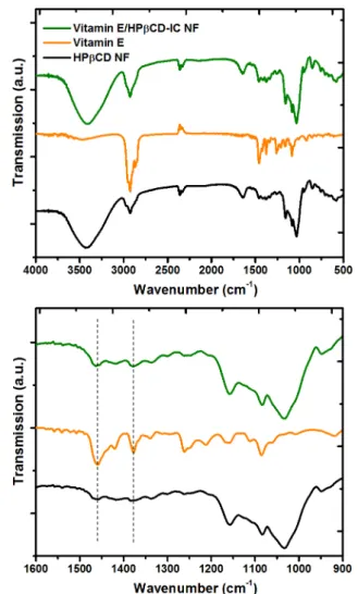
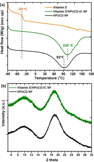
the nanofibrous Vitamin E/HPβCD-IC NF sample (Figure 4). For Vitamin E/HPβCD-IC NF and the pristine HPβCD NF, the major absorption peaks were recorded at around 1020, 1070, and 1150 cm−1corresponding to the coupled C−C/C− O stretching vibrations and the antisymmetric stretching vibration of the C−O−C glycosidic bridge of HPβCD molecules (Figure 4).14 While the characteristic FTIR absorption bands of Vitamin E at 1377 cm−1 correspond to the phenyl skeletal and the overlap of asymmetrical methyl bending and methylene scissoring vibration, the band at 1458 cm−1 is owing to symmetrical methyl bending.31 The overlapping of IR absorption peaks of HPβCD and Vitamin E makes the FTIR spectrum of Vitamin E/HPβCD-IC NF somehow difficult to identify the presence of Vitamin E. However, the absorption bands at 1377 and 1458 cm−1were observed in more intense and sharp manner in case of Vitamin E/HPβCD-IC NF, which confirms the presence of Vitamin E guest molecules in the Vitamin E/HPβCD-IC NF web samples. DSC is a useful technique to study the inclusion complex-ation between the guest molecules and host CD molecules.14 For instance, the absence of thermal transitions such as melting point (Tm) or glass transition temperature (Tg) of a guest molecule in a CD-IC is generally taken as an evidence of the proper inclusion complexation. In DSC thermogram of the pure Vitamin E, the Tg around −40 °C corresponds to the transition from glassy state to a supercooled liquid (Figure 5a).31 While the same transition is obvious for Vitamin E/ HPβCD physical mixture (Figure S2), there is no Tg recorded for Vitamin E/HPβCD-IC NF (Figure 5a), suggesting that the Vitamin E in the nanofiber matrix was completely complexed
within the HPβCD cavity.14,31 In addition, the total enthalpy change (ΔH in J/g) formed in the DSC thermograms of CD NF is due to the dehydration of CD molecules, and theΔH differences between pure CD and CD-IC can demonstrate the complex formation.14,38,39 As it is known, guest molecules compete with water molecules, which exist in the CD cavity during the complexation; therefore, ΔH value obtained as a replacement of water molecules will be lower for CD-IC structure compared to pure CD form. Here, ΔH and peak temperature were determined in the range of 0−165 °C for the samples by performing calculation using TA-Instruments Software program. While the ΔH values were calculated as 374 J/g and 195 J/g, the peak values were determined as 92 and 103 °C for HPβCD and Vitamin E/HPβCD-IC NF, respectively. The apparent decrease forΔH value and increase for peak temperature supported the inclusion complex formation between Vitamin E and CD molecules for Vitamin E/HPβCD-IC NF sample.
XRD patterns of electrospun HPβCD NF and Vitamin E/ HPβCD-IC NF are shown inFigure 5b. The pure Vitamin E is in liquid state at room temperature, and therefore, the XRD was not recorded. It is known that modified CD such as HPβCD are amorphous.14As anticipated, the XRD data of the pristine HPβCD NF have shown a broad halo pattern confirming its amorphous structures. Similarly, the broad XRD patterns were
Figure 3.1H NMR spectra of (a) pure Vitamin E and (b) Vitamin E/ HPβCD-IC NF dissolved in d6-DMSO.
Figure 4.FTIR spectra of HPβCD NF, pure Vitamin E, and Vitamin E/HPβCD-IC NF in full range (4000−500 cm−1) and expanded range (1600−900 cm−1).
also recorded for Vitamin E/HPβCD-IC NF. In the case of inclusion complexation, the guest molecules are isolated from
each other by the CD cavities and therefore cannot form crystals.12,14For Vitamin E/HPβCD-IC NF, the XRD did not show any crystalline peak suggesting that there is no crystal formation by inclusion complexation between Vitamin E and HPβCD molecules.
Vitamin E has a wide range of use as food supplements and in pharmaceuticals; however, its poor solubility is a drawback for its use and processing. Inclusion complexation with CD molecules enables to overcome this challenge and enhance the water solubility of Vitamin E compounds.29−33Here, to show the fast dissolution of Vitamin E/HPβCD-IC NF and to show the enhanced water-solubility of Vitamin E in Vitamin E/ HPβCD-IC NF, a piece of Vitamin E/HPβCD-IC NF web sample (approximately 4 cm × 4 cm) and a drop of pure Vitamin E (at the similar amount presence in the nanofiber) put into separate Petri-dishes and water was poured onto the samples. As apparent fromFigure 6and theMovie S1, Vitamin E/HPβCD-IC NF web was dissolved immediately in water, whereas pure Vitamin E remained in the Petri dishes without dissolving. This is a clear indication that water-insoluble Vitamin E becomes readily water-soluble when electrospun into Vitamin E/HPβCD-IC NF due to the inclusion complex-ation with HPβCD along with very high surface area of nanofiber matrix.
α-Tocopherol is one of the most abundant forms of Vitamin E, and it is a chain-breaking antioxidant and its activity based on the scavenging the free-radicals. α-Tocopherol influentially transfers a hydrogen atom to a free radical and gives nonradical product and α-tocopheroxyl radical. Once α-tocopheroxyl radical forms, it reacts with the second free radical, and this reaction continues until the free-radical chain reaction is terminated.40Because of this property, Vitamin E can be used as an effective inhibitor for the lipid peroxidation in foods and living cells.40In this study, we have investigated the antioxidant property of Vitamin E/HPβCD-IC NF and we have anticipated to obtain enhanced antioxidant efficiency compared to pure Vitamin E compound due to improved solubility of Vitamin E as a result of inclusion complexation. For this, antioxidant properties of Vitamin E/HPβCD-IC NF and pure Vitamin E were tested by a model organic radical molecule (DPPH)
Figure 5. (a) DSC thermograms of HPβCD NF, Vitamin E, and Vitamin E/HPβCD-IC NF. (b) XRD patterns of HPβCD NF and Vitamin E/HPβCD-IC NF.
Figure 6.Visual presentation of the water-solubility behavior of (a) Vitamin E/HPβCD-IC NF and (b) Vitamin E. The pictures are taken after a very few seconds of water contact with the samples.
(Figure 7). For comparison, the antioxidant test was also carried out for 3-year old Vitamin E/HPβCD-IC NF, which was stored in refrigerator at 4°C and still preserve Vitamin E without any loss by ∼0.80:2.00 molar ratio of Vitamin E/ HPβCD (Figure S3). Here, DPPH molecule is reduced by a hydrogen donor such as free radical scavenging antioxidant. During the DPPH reduction, a decline occurred at strongest absorption (517 nm) band and the purple color of solution turns into yellowish at the end of the reaction (Figure 7c). For antioxidant tests, 0.4 mg/mL concentrated clear Vitamin E/ HPβCD-IC NF solution and ∼11% (w/w) concentrated pure Vitamin E system was used. The antioxidant test of pure Vitamin E was also performed after the undissolved part of the system wasfiltered. UV−vis absorption graphs were recorded after incubating all systems in dark at RT for 30 min. UV−vis measurement results indicated that DPPH radical scavenging was completed for Vitamin E/HPβCD-IC NF samples after 30 min of incubation time (100.0 ± 0.0% antioxidant activity) (Figure 7a). In case of unfiltered Vitamin E system, we have observed a 60.4± 1.3% scavenging of DPPH, since methanol used for the solubility of DPPH induced dissolution of Vitamin E molecules in the test system (Figure 7a). The significant antioxidant efficiency of Vitamin E/HPβCD-IC NF is intrinsi-cally originated from the inclusion complexation, which increases the water-solubility. Therefore, Vitamin E in CD-IC can take part in the scavenging process, yetfiltered Vitamin E system prepared with the same amount of CD-IC NF cannot show any antioxidant activity since there is hardly any presence of Vitamin E in this filtered solution (Figure 7a). Regarding Vitamin E/HPβCD-IC NF, the antioxidant test results also proved that the antioxidant activity of Vitamin E can be effectively sustained in case of CD-IC NF sample without a shielding on the scavenging performance. The inclusion complexation with CD molecules also provided a prolonged
shelf life stability for Vitamin E molecules. 1H NMR measurement (Figure S3) demonstrated that there is no structural change observed for Vitamin E molecule, which was encapsulated in CD-IC NF and stored for 3 years, and there was no loss of Vitamin E/HPβCD-IC NF (∼0.80:2.00 molar ratio for Vitamin E/HPβCD, Figure S3), which still exhibited the similar high antioxidant property as the freshly prepared electrospun Vitamin E/HPβCD-IC NF sample.
As reported previously, UV-light can diminish the activity of Vitamin E by degrading the structure of compound.41 Therefore, the photostability of Vitamin E is crucial issue for its use as antioxidant. The encapsulation of Vitamin E into CD cavity might be a promising alternative approach to enhance stability against photodegradation since it is known fact that, the effect of UV-light or heat on active compound can be taken under control by inclusion complexation with CD mole-cules.28,33 Here, antioxidant tests were also performed for Vitamin E/HPβCD-IC NF samples, which were exposed to UV-light for 2 h. First, Vitamin E/HPβCD-IC NF samples still kept their web structure without deformation after the UV light treatment (Figure 7c-vii and viii). Additionally, as it is observed in Figure 7b, even after UV-light treatment, that Vitamin E/ HPβCD-IC NF still has shown a very effective antioxidant activity, which enables the full radical scavenging under the same experimental conditions. The apolar environment and screening effect of CD cavity protect Vitamin E against degradation under UV light. Therefore, to detect the possible antioxidant activity differences between UV-light treated and untreated samples, a video was recorded during the antioxidant tests. Movie S2 shows that the radical scavenging reaction completed in∼35 s for unexposed Vitamin E/HPβCD-IC NF sample; on the other hand, the scavenging reaction of UV-light exposed Vitamin E/HPβCD-IC NF sample took a slightly longer time (>90 s) (Movie S3). This is possibly because of a
Figure 7.UV−vis absorption graphs indicating the DPPH scavenging efficiency of (a) pure and filtered Vitamin E systems and freshly prepared Vitamin E/HPβCD-IC NF and 3-year old Vitamin E/HPβCD-IC NF samples. (b) DPPH scavenging efficiency of freshly prepared Vitamin E/ HPβCD-IC NF after UV light exposure. (c) Photographs of the solution (i) DPPH, (ii) Vitamin E, (iii) Vitamin E (filtered), (iv) Vitamin E/ HPβCD-IC NF, (v) Vitamin E/HPβCD-IC NF (3-year old), (vi) UV-light exposed Vitamin E/HPβCD-IC NF and the photographs of Vitamin E/ HPβCD-IC NF web (vii) before and (viii) after UV light exposure.
small amount of Vitamin E in CD-IC NF might be degraded by UV-light; however, the remaining Vitamin E still has effective antioxidant property. Hence, it is clear that Vitamin E/HP βCD-IC NF could be employed as an effective antioxidant material due to its high water-solubility, prolonged stability, and enhanced photostability of Vitamin E.
In conclusion, we have demonstrated the fabrication of nanofibers from Vitamin E/HPβCD inclusion complex in the form of free-standing nanofibrous webs without using any polymeric carrier matrix by electrospinning technique. The electrospinning was performed for two different molar ratios; 1:1 and 1:2 molar ratios of Vitamin E/HPβCD and Vitamin E/ HPβCD-IC NF (1:2) yielded bead-free nanofiber morphology under the used electrospinning parameters. The presence of Vitamin E and inclusion complexation between Vitamin E and HPβCD in electrospun Vitamin E/HPβCD-IC NF sample was confirmed by1H NMR, XRD, FTIR, and DSC techniques. The Vitamin E/HPβCD-IC NF (1:2) facilitated the loading of Vitamin E up to∼11% (w/w, with respect to fiber matrix), and ∼83% of Vitamin E was still protected for Vitamin E/HPβCD-IC NF suggesting that inclusion complexation between Vitamin E and HPβCD prevents the loss of Vitamin E during electrospinning and even after the electrospinning during storage. The rapid dissolution of Vitamin E/HPβCD-IC NF sample in water was achieved due to the high surface area nanofibrous morphology; more importantly, the water-solubility of Vitamin E was greatly enhanced for Vitamin E/ HPβCD-IC NF sample due to the inclusion complexation. Vitamin E/HPβCD-IC NF has shown an effective antioxidant activity due to presence of Vitamin E and its high water-solubility. The photostability test under UV-light and the prolonged shelf life stability for 3-year storage of Vitamin E/ HPβCD-IC NF confirmed that Vitamin E was structurally stable and kept its antioxidant activity due to the inclusion complexation with CD molecules. Apart from our previous reports on polymer-free CD-IC NF systems,14−17here we have successfully obtained CD-IC NF of Vitamin E, which consists of long hydrophobic chain in its molecular structure. Moreover, we could load Vitamin E into electrospun nanofibers matrix with an excess amount (1:1 molar ratio). Our result suggests that it might be also possible to produce CD-IC NF from big/ long molecules or oligomers and polymers as well. In brief, using our results suggested that polymer-free electrospun Vitamin E/HPβCD-IC nanofibrous webs could have potential applications in food, pharmaceuticals, and healthcare owing to enhanced water-solubility, prolonged shelf life, and high photostability of Vitamin E along with its efficient antioxidant activity.
■
ASSOCIATED CONTENT*
S Supporting InformationThe Supporting Information is available free of charge on the ACS Publications websiteat DOI:10.1021/acs.jafc.7b01562.
SEM image of Vitamin E/HPβCD-IC NF (1:2) including 20% urea; DSC thermograms of Vitamin E and Vitamin E/HPβCD-IC phy. mix;1H NMR spectra of pure Vitamin E and Vitamin E/HPβCD-IC NF (3-year old) dissolved in d6-DMSO (PDF)
Water-solubility of Vitamin E/HPβCD-IC NF and Vitamin E (AVI)
Antioxidant scavenging performance of Vitamin E/ HPβCD-IC NF before UV-light exposure (AVI)
Antioxidant scavenging performance of Vitamin E/ HPβCD-IC NF after UV-light exposure (AVI)
■
AUTHOR INFORMATIONCorresponding Author
*E-mail:[email protected]. Phone: +90-3122908987. ORCID
Tamer Uyar:0000-0002-3989-4481 Funding
T.U. acknowledges The Scientific and Technological Research Council of Turkey (TUBITAK)-Turkey (Project No. 213M185) for funding this research. T.U. also acknowledge partial support from The Turkish Academy of Sciences -Outstanding Young Scientists Award Program (TUBA-GEBIP)-Turkey. A.C. thanks TUBITAK-BIDEB for the Ph.D. scholarship.
Notes
The authors declare no competingfinancial interest.
■
REFERENCES(1) Ramakrishna, S.; Zamani, M.; Prabhakaran, M. P. Advances in drug delivery via electrospun and electrosprayed nanomaterials. Int. J. Nanomed. 2013, 8, 2997−3017.
(2) Bhushani, J. A.; Anandharamakrishnan, C. Electrospinning and electrospraying techniques: Potential food based applications. Trends Food Sci. Technol. 2014, 38 (1), 21−33.
(3) Noruzi, M. Electrospun nanofibres in agriculture and the food industry: a review. J. Sci. Food Agric. 2016, 96 (14), 4663−4678.
(4) Wendorff, J. H.; Agarwal, S.; Greiner, A. Electrospinning: Materials, Processing, and Applications; John Wiley & Sons: Weinheim, Germany, 2012.
(5) Ramakrishna, S.; Fujihara, K.; Teo, W.; Lim, T.; Ma, Z. An Introduction to Electrospinning and Nanofibers; World Scientific Publishing Company, 2005.
(6) Uyar, T.; Kny, E. Electrospun Materials for Tissue Engineering and Biomedical Applications: Research, Design, and Commercialization; Elsevier, Woodhead Publishing Series in Biomaterials, 2017.
(7) Celebioglu, A.; Uyar, T. Cyclodextrin nanofibers by electro-spinning. Chem. Commun. 2010, 46 (37), 6903−5.
(8) Celebioglu, A.; Uyar, T. Electrospinning of nanofibers from non-polymeric systems: polymer-free nanofibers from cyclodextrin derivatives. Nanoscale 2012, 4, 621−631.
(9) Celebioglu, A.; Uyar, T. Electrospinning of nanofibers from non-polymeric systems: Electrospun nanofibers from native cyclodextrins. J. Colloid Interface Sci. 2013, 404, 1−7.
(10) Celebioglu, A.; Uyar, T. Electrospun gamma-cyclodextrin (γ-CD) nanofibers for the entrapment of volatile organic compounds. RSC Adv. 2013, 3, 22891−22895.
(11) Celebioglu, A.; Uyar, T. Green and one-step synthesis of gold nanoparticles incorporated into electrospun cyclodextrin nanofibers. RSC Adv. 2013, 3, 10197−10201.
(12) Celebioglu, A.; Uyar, T. Electrospinning of polymer-free nanofibers from cyclodextrin inclusion complexes. Langmuir 2011, 27 (10), 6218−26.
(13) Celebioglu, A.; Umu, O. C. O.; Tekinay, T.; Uyar, T. Antibacterial electrospun nanofibers from triclosan/cyclodextrin inclusion complexes. Colloids Surf., B 2014, 116, 612−619.
(14) Celebioglu, A.; Kayaci-Senirmak, F.; İpek, S.; Durgun, E.; Uyar, T. Polymer-free nanofibers from vanillin/cyclodextrin inclusion complexes: high thermal stability, enhanced solubility and antioxidant property. Food Funct. 2016, 7, 3141−3153.
(15) Aytac, Z.; Yildiz, Z. I.; Kayaci-Senirmak, F.; San Keskin, N. O.; Kusku, S. I.; Durgun, E.; Tekinay, T.; Uyar, T. Fast-Dissolving, Prolonged Release, and Antibacterial Cyclodextrin/Limonene-Inclu-sion Complex Nanofibrous Webs via Polymer-Free Electrospinning. J. Agric. Food Chem. 2016, 64, 7325−7334.
(16) Aytac, Z.; Yildiz, Z. I.; Kayaci-Senirmak, F.; San Keskin, N. O.; Tekinay, T.; Uyar, T. Electrospinning of polymer-free cyclodextrin/ geraniol−inclusion complex nanofibers: enhanced shelf-life of geraniol with antibacterial and antioxidant properties. RSC Adv. 2016, 6, 46089−46099.
(17) Aytac, Z.; Yildiz, Z. I.; Kayaci-Senirmak, F.; Tekinay, T.; Uyar, T. Electrospinning of cyclodextrin/linalool-inclusion complex nano-fibers: Fast-dissolving nanofibrous web with prolonged release and antibacterial activity. Food Chem. 2017, 231, 192−201.
(18) Del Valle, E. M. M. Cyclodextrins and their uses: a review. Process Biochem. 2004, 39 (9), 1033−1046.
(19) Szejtli, J. Introduction and general overview of cyclodextrin chemistry. Chem. Rev. 1998, 98 (97), 1743−1753.
(20) Szente, L.; Szejtli, J. Highly soluble cyclodextrin derivatives: chemistry, properties, and trends in development. Adv. Drug Delivery Rev. 1999, 36, 17−28.
(21) Kayaci, F.; Ertas, Y.; Uyar, T. Enhanced thermal stability of eugenol by cyclodextrin inclusion complex encapsulated in electrospun polymeric nanofibers. J. Agric. Food Chem. 2013, 61 (34), 8156−8165. (22) Kayaci, F.; Sen, H. S.; Durgun, E.; Uyar, T. Functional electrospun polymeric nanofibers incorporating geraniol-cyclodextrin inclusion complexes: High thermal stability and enhanced durability of geraniol. Food Res. Int. 2014, 62, 424−431.
(23) Kayaci, F.; Uyar, T. Encapsulation of vanillin/cyclodextrin inclusion complex in electrospun polyvinyl alcohol (PVA) nano-webs:Prolonged shelf-life and high temperature stability of vanillin. Food Chem. 2012, 133, 641−649.
(24) Uyar, T.; Hacaloglu, J.; Besenbacher, F. Electrospun poly-ethylene oxide (PEO) nanofibers containing cyclodextrin inclusion complex. J. Nanosci. Nanotechnol. 2011, 11, 3949−3958.
(25) Aytac, Z.; Kusku, S. I.; Durgun, E.; Uyar, T. Quercetin/β-cyclodextrin inclusion complex embedded nanofibres: Slow release and high solubility. Food Chem. 2016, 197, 864−871.
(26) Taepaiboon, P.; Rungsardthong, U.; Supaphol, P. Vitamin-loaded electrospun cellulose acetate nanofiber mats as transdermal and dermal therapeutic agents of vitamin A acid and vitamin E. Eur. Eur. J. Pharm. Biopharm. 2007, 67, 387−397.
(27) Sheng, X.; Fan, L.; He, C.; Zhang, K.; Mo, X.; Wang, H. Vitamin E-loaded silk fibroin nanofibrous mats fabricated by green process for skin care application. Int. J. Biol. Macromol. 2013, 56, 49−56.
(28) Bramley, P.; Elmadfa, I.; Kafatos, A.; Kelly, F. J.; Manios, Y.; Roxborough, H. E.; Schuch, W.; Sheehy, P. J. A.; Wagner, K. H. Vitamin E. J. Sci. Food Agric. 2000, 80, 913−938.
(29) Celik, S. E.; Ozyurek, M.; Guclu, K.; Apak, R. CUPRAC total antioxidant capacity assay of lipophilic antioxidants in combination with hydrophilic antioxidants using the macrocyclic oligosaccharide methylβ-cyclodextrin as the solubility enhancer. React. Funct. Polym. 2007, 67, 1548−1560.
(30) Sueishi, Y.; Hori, M.; Inazumi, N. Characterization of inclusion complex of vitamin E compound with 2, 6-di-O-methylated β-cyclodextrin as the solubility enhancer and its kinetic determination for radical scavenging ability. J. Inclusion Phenom. Mol. Recognit. Chem. 2012, 72, 467−472.
(31) Koontz, J. L.; Marcy, J. E.; O’Keefe, S. F.; Duncan, S. E. Cyclodextrin inclusion complex formation and solid-state character-ization of the natural antioxidantsα-tocopherol and quercetin. J. Agric. Food Chem. 2009, 57, 1162−1171.
(32) Aytac, Z.; Keskin, N. O. S.; Tekinay, T.; Uyar, T. Antioxidanta-tocopherol/c-cyclodextrin−inclusion complex encapsulated poly(lactic acid) electrospun nanofibrous web for food packaging. J. Appl. Polym. Sci. 2017, 134 (21), 44858.
(33) Aytac, Z.; Uyar, T. Antioxidant activity and photostability of a-tocopherol/ b-cyclodextrin inclusion complex encapsulated electro-spun polycaprolactone nanofibers. Eur. Polym. J. 2016, 79, 140−149.
(34) Motoyama, K.; Nagatomo, K.; Elazim, S. O.; Hirayama, F.; Uekama, J.; Arima, H. Potential Use of 2-Hydroxypropyl-b-cyclo-dextrin for Preparation of Orally Disintegrating Tablets Containing dl-a-Tocopheryl Acetate, an Oily Drug. Chem. Pharm. Bull. 2009, 57 (11), 1206−1212.
(35) Kimura, M.; Ooya, T. Enhanced solubilization of alpha-tocopherol by hyperbranched polyglycerol-modified beta-cyclodextin. J. Drug Delivery Sci. Technol. 2016, 35, 30−33.
(36) Lange, K.; Gierlach-hladon, T. Solid state characterization of α-tocopherol in inclusion complexes with cyclodextrins. Acta Polym. Pharm. ACTA 2015, 72, 21−30.
(37) Uyar, T.; Besenbacher, F. Electrospinning of uniform polystyrene fibers: The effect of solvent conductivity. Polymer 2008, 49, 5336−5343.
(38) Veiga, M.; Merino, M.; Fernandez, D.; Lozano, R. Character-ization of some cyclodextrin derivatives by thermal analysis. J. Therm. Anal. Calorim. 2002, 68, 511−516.
(39) Cloudy, P.; Letoffe, J.; Germain, P.; Bastide, J.; Bayol, A.; Blasquez, S.; Rao, R.; Gonzalez, B. J. Therm. Anal. 1991, 37, 2497− 2506.
(40) Tian, F.; Decker, E. A.; Goddard, J. M. Controlling lipid oxidation of food by active packaging technologies. Food Funct. 2013, 4, 669−680.
(41) Iaconinoto, A.; Chicca, M.; Pinamonti, S.; Casolari, A.; Bianchi, A.; Scalia, S. Influence of cyclodextrin complexation on the photodegradation and antioxidant activity ofa-tocopherol. Pharmazie 2004, 59 (1), 30−33.