INVITED AUTHOR
Complete Root Coverage for Miller Class I and II – Recession Type Defects: Meta-Analysis of CAF+ EMD Versus CAF + FCTG
Rüdiger JUNKER, Andreas LEINER, Daniela ZITSCH, Thomas ULRICH, Wilhelm FRANK, Adrian KASAJ ORIGINAL ARTICLE
The Effect of Bleaching Treatment and Tea on Color Stability of Two Different Resin Composites Funda Öztürk BOZKURT, Tuğba Toz AKALIN, Burcu GÖZETİCİ, Gencay GENÇ, Harika GÖZÜKARA BAĞ
The Effect of Smoking on Postoperative Period of Extraction of Impacted Mandibular Third Molars Seda YILMAZ, Hatice HOŞGÖR, Özlem Akbelen KAYA, Burcu BAŞ, Bora ÖZDEN
CASE REPORT
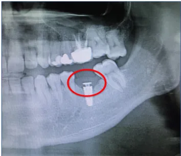
The Closure Screw Loosening Complication of Single-Molar Implant Mimicking Peri-Implant Mucositis: A Case Report
Sibel DİKİCİER, Emre DİKİCİER, Jülide ÖZEN
Oroantral Fistula Associated with Destructive Periodontitis
Feyza OTAN ÖZDEN, Bora ÖZDEN, H.İlyas KÖSE, Esra DEMİR, Peruze ÇELENK
Veillonella Spp. Infection As a Rare Cause for Early Multiple Dental Implant Failures: A Case Report
Mustafa TUNALI, Cenker Zeki KOYUNCUOGLU, Burak SELEK, Bayhan BEKTORE, Mustafa OZYURT, Rana ÇULHAOĞLU
Immediate Prosthetic Treatment of an Edentulous Patient With “All-On-4®” Concept
Alper UYAR, Simel AYYILDIZ, Bülent PİŞKİN, Can DURMAZ REVIEW
Orthodontic and Surgical Approach: Accelerated Osteogenic Orthodontics
Orhan AKSOY, Fatma YILDIRIM, M. İrfan KARADEDE
AYDIN DENTAL
Year 2 Number 2 - 2016
ISTANBUL AYDIN UNIVERSITY
JOURNAL OF FACULTY OF DENTISTRY
JOURNAL OF FACULTY OF DENTISTRY
AYDIN DENTAL
Ahu URAZ Gazi University, Turkey Ali ZAIMOGLU Istanbul Aydın University, Turkey Arzu ATAY Gulhane Military Medical Academy, Turkey Aylin BAYSAN The London School of Medicine and Dentistry, London, U.K.
Ayşen NEKORA AZAK Istanbul Aydın University, Turkey Behçet EROL Istanbul Aydın University, Turkey
Berna TARIM, Istanbul University, Turkey Bilgin GİRAY Istanbul Aydın University, Turkey Bora ÖZDEN Ondokuz Mayıs University, Turkey
Can DÖRTER Istanbul University, Turkey Cansu ALPASLAN Gazi University, Turkey
Cem TANYEL Istanbul University, Turkey Cemal ERONAT Ege University, Izmir, Turkey Didem ÖNER ÖZDAŞ Istanbul Aydın University, Turkey Elif KALYONCUOĞLU Ondokuz Mayıs University, Turkey
Engin Fırat CAKAN, Istanbul Aydın University, Turkey Erman BULENT TUNCER Istanbul Aydın University, Turkey Ersin YILDIRIM Gulhane Military Medical Academy, Turkey
Esra SOMTÜRK Istanbul Aydın University, Turkey Feyza OTAN ÖZDEN Ondokuz Mayıs University, Turkey Fulya TOKSOY TOPÇU Gulhane Military Medical Academy, Turkey
Gülce ALP Istanbul Aydın University, Turkey
Günseli GÜVEN POLAT Gülhane Military Medical Academy, Turkey Hakan ÖZBAŞ Istanbul University, Turkey
Handan ERSEV Istanbul University, Turkey Kadriye DEMİRKAYA Gulhane Military Medical Academy, Turkey
Kemal SÜBAY Istanbul Aydın University, Turkey Korkud DEMİREL Istanbul University, Turkey Leyla KURU Marmara University, Istanbul, Turkey Mehmet CUDİ BALKAYA Istanbul Aydın University, Turkey Işıl Kaya BÜYÜKBAYRAM, Istanbul Aydın University, Turkey
Raif ERİŞEN Istanbul University, Turkey Rezzan ÖZER Dicle University, Turkey Rüdiger JUNKER Danube Private University, Austria
Sedat ÇETİNER Gazi University, Turkey Sema BELLİ Selçuk University, Turkey Sema ÇELENK Dicle University, Turkey Semih BERKSUN Ankara University, Turkey Serap KARAKIŞ Istanbul Aydın University, Turkey
Serdar CİNTAN Istanbul University, Turkey Simel AYYILDIZ Gulhane Military Medical Academy, Turkey
Şeniz KARAÇAY Gulhane Military Medical Academy, Turkey Ümit KARAÇAYLI Gulhane Military Medical Academy, Turkey
Vesela STEFANOVA Medical University of Plovdiv, Bulgaria Proprietor - Sahibi
Mustafa Aydın
Editor-in-Chief - Yazı İşleri Müdürü Nigar Çelik
Editor - Editör
Jülide Özen Editorial Board - Yayın Kurulu Jülide Özen
Sercan Küçükkurt
Academic Studies Coordination Office (ASCO) Akademik Çalışmalar Koordinasyon Ofisi
İdari Koordinatör - Administrative Coordinator Nazan Özgür
Technical Editor - Teknik Editör Hakan Terzi
Language - Dili English - Türkçe
Publication Period - Yayın Periyodu Published twice a year - Yılda iki kere yayınlanır April and October - Nisan ve Ekim
Correspondence Address - Yazışma Adresi Beşyol Mahallesi, İnönü Caddesi, No: 38 Sefaköy, 34295 Küçükçekmece/İstanbul Tel: 0212 4441428 - Fax: 0212 425 57 97 web: www.aydin.edu.tr - e-mail: [email protected] Printed by - Baskı
Veritas Baskı Merkezi
İstanbul Tuzla Kimya Sanayicileri Organize Sanayi Bölgesi
Melek Aras Bulvarı, Analitik Caddesi, PK 34959 No: 46 Tuzla – İstanbul
Tel: 444 1 303 - Faks: 0 216 290 27 20
ISSN: 2149-5572
Scientific Board
İstanbul Aydın Üniversitesi, Diş Hekimliği Fakültesi, Aydın Dental Dergisi özgün bilimsel araştırmalar ile uygulama çalışmalarına yer veren ve bu niteliği ile hem araştırmacılara hem de uygulamadaki akademisyenlere seslenmeyi amaçlayan hakem sistemini kullanan bir dergidir.
INVITED AUTHOR
Complete Root Coverage for Miller Class I and II – Recession Type Defects: Meta-Analysis of CAF+ EMD Versus CAF+ FCTG
Rüdiger JUNKER, Andreas LEINER, Daniela ZITSCH, Thomas ULRICH, Wilhelm FRANK, Adrian KASAJ... 1
ORIGINAL ARTICLE
The Effect of Bleaching Treatment and Tea on Color Stability of Two Different Resin Composites
Funda Öztürk BOZKURT, Tuğba Toz AKALIN, Burcu GÖZETİCİ, Gencay GENÇ, Harika GÖZÜKARA BAĞ... 23
The Effect of Smoking on Postoperative Period of Extraction of Impacted Mandibular Third Molars
Seda YILMAZ, Hatice HOŞGÖR, Özlem Akbelen KAYA, Burcu BAŞ, Bora ÖZDEN... 31
CASE REPORT
The Closure Screw Loosening Complication of Single-Molar Implant Mimicking Peri-Implant Mucositis: A Case Report
Sibel DİKİCİER, Emre DİKİCİER, Jülide ÖZEN... 39
Oroantral Fistula Associated with Destructive Periodontitis
Feyza OTAN ÖZDEN, Bora ÖZDEN, H.İlyas KÖSE, Esra DEMİR, Peruze ÇELENK... 45
Veillonella Spp. Infection As a Rare Cause for Early Multiple Dental Implant Failures: A Case Report
Mustafa TUNALI, Cenker Zeki KOYUNCUOGLU, Burak SELEK, Bayhan BEKTORE, Mustafa OZYURT, Rana ÇULHAOĞLU... 51
Immediate Prosthetic Treatment of an Edentulous Patient With “All-On-4®” Concept
Alper UYAR, Simel AYYILDIZ, Bülent PİŞKİN, Can DURMAZ... 59
REVIEW
Orthodontic and Surgical Approach: Accelerated Osteogenic Orthodontics
Dear Readers of Journal of Aydin Dental (JAD),
After publishing the first issue in 2015, we are proud of publishing the second JAD now.
The JAD has already been added into the “TUBITAK DERGIPARK” (http://dergipark.ulakbim. gov.tr/adj/ ) within considerably short timeframe of six months.
In the upcoming period, we intend to indexed in ULAKBİM database and to make it commonly well known and looked for as a reference journal among the scientific circles because of its high academic reliability.
I would like to express my special thanks and appreciations to Prof. Dr. Rudiger Junker and all other academic colleagues because of their invaluable contributions to the second issue of JAD.
Best regards, Prof. Dr. Jülide ÖZEN Editor of Aydin Dental Journal
Editörden Mesaj;
Sayın Aydın Dental Journal Okurları,
İlk sayısını 2015 yılında yayınladığımız dergimizin 2. Sayısını basmaktan gurur duyuyoruz. Dergimiz 6 ay gibi çok kısa bir sürede Tübitak Dergipark’a (http://dergipark.ulakbim.gov.tr/adj/ ) girmiş bulunmaktadır.
Hedefimiz ilerleyen süreçte ULAKBİM veritabanı listesine de girerek, dergimizin adını daha geniş kitlelere duyurarak seçkin, aranan ve akademik güvenilirliği yüksek bir duruma getirmektir. Bu sayımızda değerli katkılarıyla beni ve dergimizi onurlandıran sayın hocam ve aynı zamanda çok değerli bir bilim adamı Prof. Dr. Rudiger Junker’e ve bilimsel yazılarıyla dergimize katkıda bulunan bütün akademisyen arkadaşlarıma teşekkür etmek istiyorum.
Saygılarımla, Prof. Dr. Jülide ÖZEN Aydın Dental Journal Editörü
Complete Root Coverage for Miller Class I and II – Recession
Type Defects: Meta-Analysis of CAF+EMD Versus CAF+FCTG
Rüdiger JUNKER1, Andreas LEINER1, Daniela ZITSCH1, Thomas ULRICH1, Wilhelm FRANK2,
Adrian KASAJ3 ABSTRACT
Background: To the best of our knowledge, the equality of CAF + EMD and CAF + FCTG regarding complete root coverage in Miller class I and II recession-type defects is still uncertain.
Aim: The aim of the current paper is to compare the effect of CAF + EMD versus CAF + FCTG regarding complete root recession coverage. Thereby, equality on the longer term of both therapeutic options is hypothesized (H0- hypothesis). Materials and methods: Three reviewers searched independently within the electronic database Pubmed/Medline. Only RCTs reporting quantitative data for the outcome variable percentage complete root coverage (%CRC) for the therapeutic options CAF + EMD or CAF + FCTG were considered. Additionally a manual search in the reference lists of all included publications was accomplished.
Results: After electronical and manual search for relevant studies, the three independent reviewers (DZ, TU, AL) screened 552 titles, resulting in 102 abstracts and 41 full-texts. Eventually, twenty-five papers could be included for meta-analysis. By comprehensively comparing data from RCTs for the outcome variable “percentage complete root coverage”, statistically significant weighted mean differences in favor of CAF + FCTG were found at 6, 12 and 24 months.
Conclusion: Regarding percentage complete root coverage, CAF + EMD is not as effective as CAF + FCTG for Miller class I and II recession- type defects.
Keywords: Miller class I and II recession-type defects, root coverage, coronally advanced flap, free connective tissue graft, enamel matrix derivative
1 Department of Prosthodontics and Biomaterials, Danube Private University (DPU), Krems, Austria
2 Department of Health management, - economics, and - system research, Danube Private University (DPU), Krems, Austria 3 Department of Operative Dentistry and Periodontology, University of Mainz, Germany
Introduction
Gingival recession is defined as displacement of the gingival margin apical to the cemento-enamel junction with root surface exposure (Wennström 1996). Recession-type defects can be seen in subjects of all ages with suboptimal as well as with excellent oral hygiene (Sagnes & Gjermo 1976). Possible causes are for example periodontal disease, traumatic tooth brushing, as well as malposition of teeth or orthodontic tooth movement out of the bony envelope ( Gorman 1967; Boyd 1978; Miller & Allen 1996).
Main indications for root coverage procedures are esthetic / cosmetic demands, root sensitivity as well as changing the topography of the marginal soft tissue to facilitate plaque control (Wennström, et al. 2008; Zuhr & Hürzeler 2011). Interestingly, Miller (1985) created a classification system for recession-type defects to predict treatment success. Thereby, it is presumed that for Class I and II recession–type defects (i.e. recession within the attached gingiva and recession up to the mucogingival margin, respectively) complete root coverage can be achieved.
Up to now, the so-called coronally advanced flap (CAF) combined with a free connective tissue graft (FCTG) is considered the “gold standard” for root coverage therapy (Cairo, et al. 2014; Chambrone & Tatakis 2015). Consequently, alternative techniques are generally compared to CAF + FCTG and evaluated according to their ability to reduce recession and achieve root coverage (Oates, et al. 2003, Academy-Report 2005, Chambrone, et al. 2009). During the last decades, an alternative method avoiding a second surgery for the FCTG, the „Coronally Positioned Flap” in combination with “Enamel Matrix
(Modica et al. 2000) However, to the best of our knowledge, the equality of CAF + FCTG and CAF + EMD regarding complete root coverage in Miller class I and II recession-type defects is still uncertain. Therefore, the aim of the current paper is to compare the effect of CAF + EMD versus CAF + FCTG regarding complete root recession coverage. Thereby, equality on the longer term of both therapeutic options is hypothesized (H0- hypothesis).
Material and Methods Search Strategy
Three reviewers (Daniela Zitsch (DZ), Thomas Ulrich (TU), Andreas Leiner (AL)) searched independently within the electronic database PubMed/Medline, US National Library of Medicine, National Institute of Health (http:// www.ncbi.nlm.nih.gov/pubmed) for relevant publications.
The typewritten search strategy for „Coronally Advanced Flap” in combination with a “free connective tissue graft“ (CAF + FCTG) was: (Recession coverage OR root coverage OR plastic periodontal surgery) AND (FCTG OR free connective tissue graft OR connective tissue graft OR subepithelial graft)
Thereafter, that search strategy for CAF + FCTG was translated by the search engine of Pubmed/Medline to:
((Recession[All Fields] AND (“AHIP Cover”[Journal] OR “coverage”[All Fields])) OR ((“plant roots”[MeSH Terms] OR (“plant”[All Fields] AND “roots”[All Fields]) OR “plant roots”[All Fields] OR “root”[All Fields]) AND (“AHIP Cover”[Journal] OR “coverage”[All Fields])) OR ((“plastics”[MeSH Terms] OR “plastics”[All Fields] OR “plastic”[All
(“surgery”[Subheading] OR “surgery”[All Fields] OR “surgical procedures, operative”[MeSH Terms] OR (“surgical”[All Fields] AND “procedures”[All Fields] AND “operative”[All Fields]) OR “operative surgical procedures”[All Fields] OR “surgery”[All Fields] OR “general surgery”[MeSH Terms] OR (“general”[All Fields] AND “surgery”[All Fields]) OR “general surgery”[All Fields]))) AND (FCTG[All Fields] OR (free[All Fields] AND (“connective tissue”[MeSH Terms] OR (“connective”[All Fields] AND “tissue”[All Fields]) OR “connective tissue”[All Fields]) AND (“transplants”[MeSH Terms] OR “transplants”[All Fields] OR “graft”[All Fields])) OR ((“connective tissue”[MeSH Terms] OR (“connective”[All Fields] AND “tissue”[All Fields]) OR “connective tissue”[All Fields]) AND (“transplants”[MeSH Terms] OR “transplants”[All Fields] OR “graft”[All Fields])) OR (subepithelial[All Fields] AND (“transplants”[MeSH Terms] OR “transplants”[All Fields] OR “graft”[All Fields])))
In analogue, the typewritten search strategy for “Coronally Advanced Flap” in combination with “Enamel matrix Derivative” (CAF + EMD) was: (Recession coverage OR root coverage OR plastic periodontal surgery) AND (Emdogain OR EMD OR Amelogenin OR enamel proteins OR growth factor)
Thereafter, that search strategy for CAF + EMD was translated by the search engine of PubMed/Medline to:
((Recession[All Fields] AND (“AHIP Cover”[Journal] OR “coverage”[All Fields])) OR ((“plant roots”[MeSH Terms] OR (“plant”[All Fields] AND “roots”[All Fields]) OR “plant roots”[All Fields] OR “root”[All Fields]) AND (“AHIP Cover”[Journal] OR “coverage”[All
Fields])) OR ((“plastics”[MeSH Terms] OR “plastics”[All Fields] OR “plastic”[All Fields]) AND periodontal[All Fields] AND (“surgery”[Subheading] OR “surgery”[All Fields] OR “surgical procedures, operative”[MeSH Terms] OR (“surgical”[All Fields] AND “procedures”[All Fields] AND “operative”[All Fields]) OR “operative surgical procedures”[All Fields] OR “surgery”[All Fields] OR “general surgery”[MeSH Terms] OR (“general”[All Fields] AND “surgery”[All Fields]) OR “general surgery”[All Fields]))) AND ((“enamel matrix proteins”[Supplementary Concept] OR “enamel matrix proteins”[All Fields] OR “emdogain”[All Fields]) OR EMD[All Fields] OR (“amelogenin”[MeSH Terms] OR “amelogenin”[All Fields]) OR ((“dental enamel”[MeSH Terms] OR (“dental”[All Fields] AND “enamel”[All Fields]) OR “dental enamel”[All Fields] OR “enamel”[All Fields]) AND (“proteins”[MeSH Terms] OR “proteins”[All Fields])) OR (“intercellular signaling peptides and proteins”[MeSH Terms] OR (“intercellular”[All Fields] AND “signaling”[All Fields] AND “peptides”[All Fields] AND “proteins”[All Fields]) OR “intercellular signaling peptides and proteins”[All Fields] OR (“growth”[All Fields] AND “factor”[All Fields]) OR “growth factor”[All Fields])
Screening Process
The three independent reviewers (DZ, TU, AL) searched titles and evaluated abstracts as well as fulltexts for meta-analytical inclusion. Any difference between the reviewers was resolved by discussion. Eventually, for papers selected for meta-analysis, data extraction sheets were developed, tested and operated (Table 1). Thereafter, data for meta-analysis were synopsized in an excel-sheet.
Manual Search
Additionally to the search in the electronic database PubMed/Medline, a manual search for relevant studies was performed independently by the three reviewers (DZ, TU, AL). Thereby, the references of the studies included for meta- analysis were screened according to the above detailed process (i.e. title-, abstract-, and full text-analysis).
Outcome Variable
In this paper the outcome variable percentage complete root coverage (%CRC) is meta-analyzed.
Thereby, the formula for the outcome variable was:
Inclusion- / Exclusion Criteria
To test the hypothesis of equality on the longer term of both therapeutic options only randomized controlled clinical trials (RCTs) were considered for the current systematic review / meta- analysis. Further, studies with a publishing date before the year 2000 were excluded. Moreover, only RCTs with at least 10 patients per treatment protocol were included. Thereby, only reports on Miller class I and II recession- type defects were considered. Studies with Miller Class III and IV recession– type defects were excluded. It goes without saying that only studies using either FCTG or EMD in combination with a “Coronally Advanced Flap” were included. For comparison, the included studies should present quantitative data at 6, 12, 18 and/or 24 months after surgery.
Quality Assessment
Risk of bias assessment of the included articles was accomplished. It was done according to
(http://handbook.cochrane.org/chapter _ 8 / 8 _ 5the _ coch- rane_collaborations_tool_for_ assessing_risk_of_bias-htm).Thereby, five main criteria were analyzed: random sequence generation (RSG), allocation concealment (ALC), blinding of outcome assessment (BOA), incomplete outcome (ICD), selective reporting (SLR). Accordingly, by judging a paper for all five criteria as being associated with a low risk of bias, the publication was judged as being associated with a low risk of bias. Further, by judging a paper for three to four criteria as being associated with a low risk of bias, the paper itself was judged as being associated with a moderate risk of bias. Whereas, by judging a paper for less than three criteria as being associated with a low risk of bias, the publication itsef was judged as being associated with a high risk of bias (Higgins & Green 2011).
Cochrane Review Manager / Meta-analytic Approach
The so-called „Cochrane Review Manager (RevMan)” (current version 5.3.5; http:// community.cochrane.org / editorial-and- publishing-policy-resource/review-manager-revman) is a software program providing guidance to write systematic reviews and / or meta-analyses. All relevant data - the authors agreed to analyze all parameters on tooth level and not on patient level - at 6, 12, 18 and / or 24 months of all included studies were conveyed to RevMan version 5.3.5 and meta-analyzed. Thus, for comprehensive comparison at different time points, „weighted means“ for the outcome variable percentage complete root coverage (%CRC) were determined by the calculator program of RevMan version 5.3.5. Further, RCTs evaluating CAF + EMD versus CAF + FCTG per protocol were
with RevMan version 5.3.5 is only possible, if standard deviations are given. It goes without saying that for the variable %CRC no standard deviations could be retrieved from the included RCTs. Therefore, for all %CRC-data a standard deviation tending to zero and thereby not effecting the meta-analysis (i.e. 0.001) was operated. Further, for directly compared data statistical heterogeneity was tested. As a result, either fixed- or random-statistical models were operated. A significance level of 0.05 was chosen.Additionally, forest plots were generated.
Heterogeneity
Heterogeneity was defined and tested according to the „Cochrane Handbook“ for systematic reviews (http://handbook. cochrane .org / chapter_9/9_5_2_ identifying _ and _ measuring_heterogeneity.htm). Thereby, RevMan version 5.3.5 is operating a chi-squared test (χ2, or Chi2) to determine
heterogeneity and its impact on the meta-analysis is represented with I2. The test
evaluates whether observed differences in results are compatible with chance alone. A low p-value (p < 0.1 or a large chi-squared statistic relative to its degree of freedom) provides evidence of heterogeneity of intervention effects (i.e. variation in effect estimates beyond chance). The interpretation of I2 can be done as follows: 0% to 40% might
not be important, 30% to 60% may represent moderate heterogeneity, 50% to 90% may represent substantial heterogeneity and 75% to 100% might be seen as considerably heterogeneity (Higgins & Green 2011).
Detailed Hypotheses
As stated above, the aim of the current meta-analysis is to compare the effects of CAF + EMD and CAF + FCTG regarding root recession coverage. Thereby, equality on
the longer term of both therapeutic options is hypothesized (H0- hypothesis). More in detail, equality for the outcome variable percentage complete root coverage (%CRC) is hypothesized.
Results
Selection of the Studies
After electronical and manual search for relevant studies, the three independent reviewers (DZ, TU, AL) screened 552 titles, resulting in 102 abstracts and 41 full-texts. Eventually, twenty-five papers could be included for the meta-analysis (Fig. 1). After fulltext reading excluded papers are listed in Table 3. Reasons for exclusion are given.
Included studies
In the end the following studies could be included:
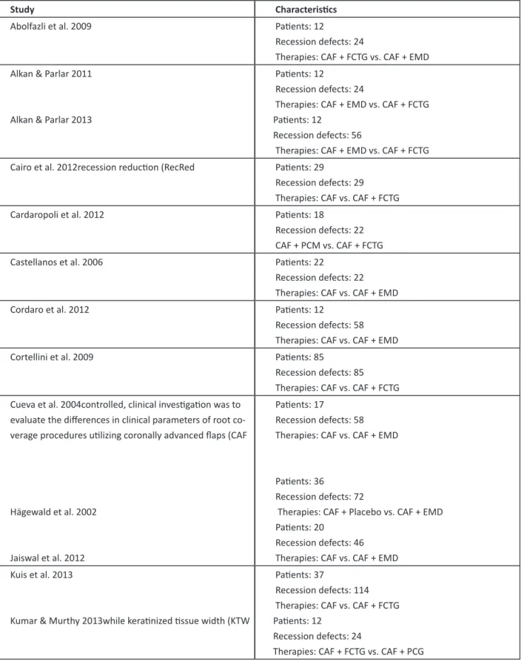
Table 2. Included Studies
Study Characteristics
Abolfazli et al. 2009 Patients: 12
Recession defects: 24
Therapies: CAF + FCTG vs. CAF + EMD Alkan & Parlar 2011
Alkan & Parlar 2013
Patients: 12
Recession defects: 24
Therapies: CAF + EMD vs. CAF + FCTG Patients: 12
Recession defects: 56
Therapies: CAF + EMD vs. CAF + FCTG Cairo et al. 2012recession reduction (RecRed Patients: 29
Recession defects: 29 Therapies: CAF vs. CAF + FCTG
Cardaropoli et al. 2012 Patients: 18
Recession defects: 22 CAF + PCM vs. CAF + FCTG
Castellanos et al. 2006 Patients: 22
Recession defects: 22 Therapies: CAF vs. CAF + EMD
Cordaro et al. 2012 Patients: 12
Recession defects: 58 Therapies: CAF vs. CAF + EMD
Cortellini et al. 2009 Patients: 85
Recession defects: 85 Therapies: CAF vs. CAF + FCTG Cueva et al. 2004controlled, clinical investigation was to
evaluate the differences in clinical parameters of root co-verage procedures utilizing coronally advanced flaps (CAF
Hägewald et al. 2002
Jaiswal et al. 2012
Patients: 17
Recession defects: 58 Therapies: CAF vs. CAF + EMD
Patients: 36
Recession defects: 72
Therapies: CAF + Placebo vs. CAF + EMD Patients: 20
Recession defects: 46 Therapies: CAF vs. CAF + EMD Kuis et al. 2013
Kumar & Murthy 2013while keratinized tissue width (KTW
Patients: 37
Recession defects: 114 Therapies: CAF vs. CAF + FCTG Patients: 12
Recession defects: 24
McGuire & Nunn 2003
McGuire & Scheyer 2010
Patients: 20
Recession defects: 40
Therapies: CAF + EMD vs. CAF + FCTG Patients: 25
Recession defects: 50
Therapies: CAF + FCTG vs. CAF + CM
McGuire et al. 2014 Patients: 30
Recession defects: 60
Therapies: CAF + FCTG vs. CAF + HPDGF + β-TCP Modica et al. 2000one site was randomly assigned to the
test group and the contralateral site to the control group. The treatment consisted of a CAF procedure with (test
Patients: 12
Recession defects: 28 Therapies: CAF vs. CAF + EMD
Del Pizzo et al. 2005 Patients: 15
Recession defects: 30 Therapies: CAF vs. CAF + EMD
Roman et al. 2013 Patients: 42
Recession defects: 68
Therapies: CAF + FCTG vs. CAF + FCTG + EMD Salhi et al. 2014
Sayar et al. 2013
Patients: 40
Recession defects: 40
Therapies: CAF + FCTG vs. FCTG + Pouch Technique Patients: 13
Recession defects: 40
Therapies: CAF + FCTG vs. CAF + EMD
da Silva et al. 2004 Patients: 11
Recession defects: 22 Therapies: CAF vs. CAF + FCTG
Spahr et al. 2005 Patients: 30
Recession defects: 60
Therapies: CAF + Placebo vs. CAF + EMD Zucchelli, Marzadori, et al. 2014 Patients: 50
Recession defects: 50
Therapies: CAF + FCTG vs. CAF + FCTG + removed LST Zucchelli, Mounssif, et al. 2014 Patients: 50
Recession defects: 149 Therapies: CAF vs. CAF + FCTG
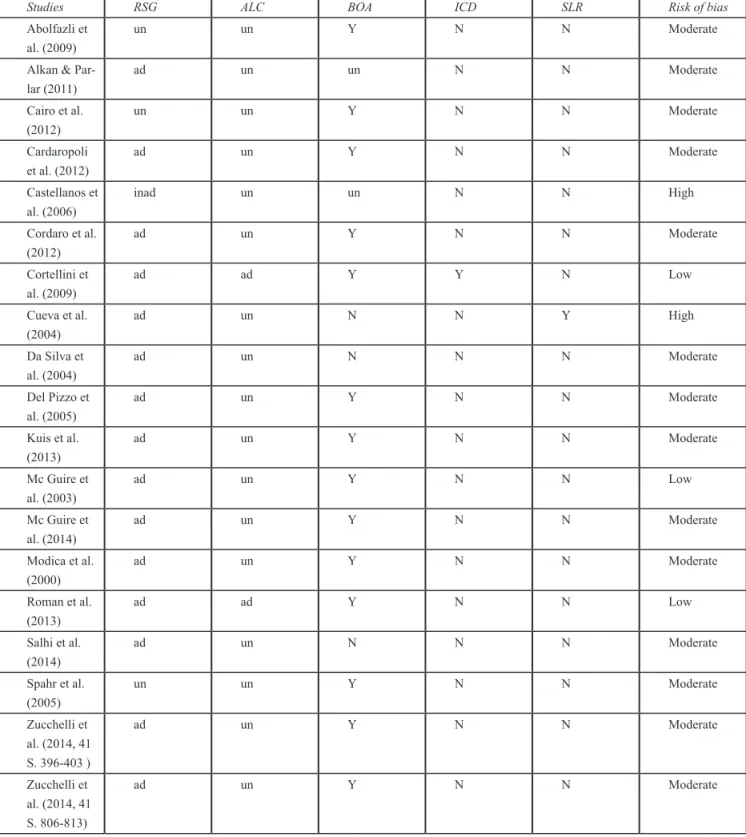
Risk of Bias
In the present systematic review / meta- analysis of the current literature, three included studies were judged as being associated with a low risk of bias (Cortellini et al. 2009, McGuire & Nunn 2003 and Roman et al. 2013), whereas twenty studies were judged as being associated with a moderate risk of bias (Abolfazli et al. 2009, Alkan & Parlar 2011, Alkan & Parlar 2013, Cairo et al. 2012recession reduction (RecRed, Cardaropoli et al. 2012, Cordaro et al. 2012, da Silva et al. 2004, del Pizzo et al. 2005, Hägewald et al. 2002, Jaiswal et al. 2012, Kuis et al. 2013, Kumar & Murthy 2013while keratinized tissue width (KTW, McGuire & Scheyer 2010, McGuire et al. 2014, Modica et al. 2000one site was randomly assigned to the test group and the contralateral site to the control group. The treatment consisted of a CAF procedure with (test, Salhi et al. 2014, Sayar et al. 2013, Spahr et al. 2005, Zucchelli Mounssif, et al. 2014 and Zucchelli, Marzadori, et al. 2014), and two studies were judged as being associated with a high risk of bias (Castellanos et al. 2006 and Cueva et al. 2004)(Tab. 4).
Percentage Complete Root Coverage (%CRC)
RCTs comprehensively compared at six months
For meta- analysis of percentage complete root coverage (%CRC) at six months the studies of Cordaro et al. (2012); Cueva et al. (2004); Modica et al. (2000with the adjunct of EMD for test sites, was performed. Clinical measurements (recession length, keratinized tissue, probing depth, and clinical attachment level) for CAF + EMD and the studies of Cairo et al. (2012); Cortellini et al. (2009); Kuis et al. (2013); McGuire et al. (2014); Roman et al. (2013); Salhi et al. (2014); da Silva et al.
(2014recession reduction (RecRed) for CAF + FCTG could be included.
At six months after root coverage surgery a weighted mean percentage complete root coverage of 54.1% (SD: 19.3%) for CAF + EMD was calculated. For CAF + FCTG a weighted mean percentage complete root coverage of 78.8% (SD: 18.2%) was found. Thereby, the calculated weighted mean difference of 24.7% (95% CI [19.8%, 29.7%]) in favor of CAF + FCTG was statistically significant (Fig. 3).
However, it should be kept in mind that only two of the included studies were judged as being associated with a low risk of bias (Cortellini et al. 2009; Roman et al. 2013), eight studies were judged as being associated with a moderate risk of bias (Cairo et al. 2012; Cordaro et al. 2012; Kuis et al. 2013; McGuire et al. 2014; Modica et al. 2000; Salhi et al. 2014; da Silva et al. 2004; Zucchelli, Mounssif, et al. 2014)recession reduction (RecRed and one study as being associated with a high risk of bias (Cueva et al. 2004)controlled, clinical investigation was to evaluate the differences in clinical parameters of root coverage procedures utilizing coronally advanced flaps (CAF.
RCTs directly compared at six months
At six months, no publication reported the effect difference for the outcome variable %CRC directly.
RCTs comprehensively compared at twelve months
For meta- analysis at twelve months the studies of Abolfazli et al. (2009); Alkan & Parlar (2011); Castellanos et al. (2006); McGuire &
of Abolfazli et al. (2009); Alkan & Parlar (2011); Cardaropoli et al. (2012); Kuis et al. (2013); McGuire & Nunn (2003); Roman et al. (2013); Zucchelli, Marzadori, et al. (2014); Zucchelli, Mounssif, et al. (2014) for CAF + FCTG could be included.
At twelve months after root coverage surgery a weighted mean percentage complete root coverage of 67.0 % (SD: 16.2%) for CAF + EMD was calculated. For CAF + FCTG a weighted mean percentage complete root coverage of 78.4% (SD: 14.7%) was found. Thereby, the calculated weighted mean difference of 11.5% (95% CI [7.1%, 15.9%]) in favor of CAF + FCTG was statistically significant (Fig.3).
However, it should be understood that only two studies were judged as being associated with a low risk of bias (McGuire & Nunn 2003; Roman et al. 2013), six studies were judged as being associated with a moderate
risk of bias (Abolfazli et al. 2009; Alkan & Parlar 2011; Cardaropoli et al. 2012; Kuis et al. 2013; Zucchelli, Marzadori, et al. 2014; Zucchelli, Mounssif, et al. 2014) and one study as being associated with a high risk of bias (Castellanos et al. 2006).
RCTs directly compared at twelve months
At twelve months after plastic periodontal surgery three papers comparing percentage complete root coverage for CAF + EMD versus CAF + FCTG per protocol, i.e. directly, were eventually included in the current meta-analysis (Abolfazli et al. 2009; Alkan & Parlar 2011; McGuire & Nunn 2003) (Fig.4).Thereby, Abolfazli et al. (2009) found a percentage of complete root coverage of 50% for CAF + EMD and 58.3% for CAF + FCTG. The mean difference was 8.0% in favor of CAF + FCTG. This difference was statistically significant (Fig. 4). In contrast, McGuire & Nunn (2003) found a percentage of complete root coverage of 89.5% for CAF + EMD and 79% for CAF Fig. 2. Percentage complete root coverage at six months
+ FCTG. The mean difference was 10.5% in favor of CAF + EMD. Again, this difference was statistically significant (Fig. 4). Further, Alkan & Parlar (2011) reported a percentage of complete root coverage of 75% for CAF + EMD and 58.3% for CAF + FCTG. The mean difference was 16.7% in favor of CAF + EMD. This difference was statistically significant (Fig. 4). In total, the weighted mean difference between CAF + EMD and CAF + FCTG of all three studies together was 6.3% (95% CI [-7.7%, 20.3%]). This difference was not statistically significant (Fig. 4).
Additionally, it should be mentioned that statistical heterogeneity across the studies could be found (Heterogeneity: Chi² = 2068926160.98, df = 2 (P < 0.00001); I² = 100%). Moreover, it should be kept in mind that only one study was judged as being associated with a low risk of bias (McGuire & Nunn 2003), whereas two studies were judged
bias (Abolfazli et al. 2009; Alkan & Parlar 2011).
Comparing comprehensively versus directly at twelve months
The weighted mean difference of comprehensively compared data was statistically significant in favor of CAF + FCTG (11.6%; 95% CI [7.1%, 15.9%]), whereas the weighted mean difference of directly compared data (6.3% in favor of CAF + EMD; 95% CI [-7.7%, 20.3%]) was not statistically significant.
RCTs comprehensively compared at 24 months
For meta- analysis at twenty-four months the studies of Abolfazli et al. (2009); Cordaro et al. (2012); Del Pizzo et al. (2005); Spahr et al. (2005) for CAF + EMD and the studies of Abolfazli et al. (2009); Kuis et al. (2013) for CAF + FCTG could be included.
At twenty-four months after root coverage surgery a weighted mean percentage complete root coverage of 40.5% (SD: 21.6%) for CAF + EMD was calculated. For CAF + FCTG a weighted mean percentage complete root coverage of 85.5% (SD: 8.7%) was found. Thereby, the calculated weighted mean difference of 45.0% (95% CI [40.0%, 50.0%]) in favor of CAF + FCTG was statistically significant (Fig. 5).
Further, all five studies were judged as being associated with a moderate risk of bias (Abolfazli et al. 2009; Cordaro et al. 2012; Kuis et al. 2013; Del Pizzo et al. 2005; Spahr et al. 2005).
RCTs directly compared at twenty-four months
At twenty-four months only one study comparing CAF + FCTG versus CAF + EMD
per protocol, i.e. directly, was eventually included in the current paper (Abolfazli et al. 2009). For CAF + EMD 25% complete root coverage and for CAF + FCTG 66.6% complete root coverage was found. This difference was statistically significant in favor of CAF + FCTG.
Discussion
Briefly, the current meta-analysis aimed to compare the effects of CAF + EMD versus CAF + FCTG regarding root recession coverage. Thereby, it was hypothesized (H0- hypothesis) that for the outcome variable “percentage complete root coverage” the results achieved by CAF + EMD do not differ statistically significant from CAF + FCTG on the longer term.
By comprehensively comparing data from RCTs for the outcome variable “percentage Fig. 4. Percentage complete root coverage at twelve months (directly compared)
complete root coverage”, statistically significant weighted mean differences of 24.7% (95% CI [29.7%, 19.8%]), 11.5% (95% CI [7.1%, 15.9%]) and 45.0% (95% CI [40.0%, 50.0%]) in favor of CAF + FCTG were found at 6, 12 and 24 months, respectively. At 6 months there were no studies reporting mean differences between CAF + EMD and CAF + FCTG directly. In contrast to comprehensively compared data from RCTs, for RCTs directly comparing CAF + EMD versus CAF + FCTG, no statistically significant difference was found at twelve months. However, at 24 months a statistically significant difference in favor of CAF + FCTG was found. Thereby, it should be kept in mind that most of the studies were not judged as having a low risk of bias and statistical heterogeneity was found. Therefore, we tend to reject the H0 – hypothesis of no difference and we have the tendency to accept a superiority of CAF + FCTG regarding the outcome variable percentage complete root coverage on the longer term. It should be kept in mind that comprehensively comparing RCTs resulted in weighted mean percentages of complete root coverage of 78.8% (SD: 18.2%), 78.4% (SD: 14.7%), and 85.5% (SD: 8.7%) for CAF + FCTG in Miller class I and II recession-type defects at six, twelve, and twenty-four months after plastic periodontal surgery, respectively. Thus, as presumed by Miller (1985) in general complete root coverage can be achieved in Class I and II recession–type defects and obviously the mean percentages of complete root coverage increase over time. However, it should be understood that even with the so-called “gold standard” prediction of complete root coverage is not possible.
Somehow in contrast, comprehensively comparing RCTs resulted in weighted mean
54.1% (SD: 19.3%), 67.0 % (SD: 16.2%), and 40.5% (SD: 21.6%) for CAF + EMD in Miller class I and II recession-type defects at six, twelve, and twenty-four months after plastic periodontal surgery, respectively. It goes without saying that with this method the clinician cannot predict complete root coverage at all.
Moreover, conclusions of earlier reports that CAF + EMD resulted in root coverage similar to CAF + FCTG but without the morbidity and potential clinical difficulties associated with the donor site surgery (McGuire and Nunn 2003; Alkan and Parlar 2011, 2013; Sayer et al. 2013) must be - at least regarding percentage of complete root coverage - interpreted with caution.
Conclusion
Within the limits of the current meta-analysis of the literature regarding plastic periodontal surgery of Miller class I and II recession- type defects it is concluded that CAF + EMD is not as effective as CAF + FCTG as regards percentage complete root coverage.
Appendix
Publication
(authors, title, journal, date) Abstract
Earlier reports of same study
Study design Study design Treatment test group Treatment control group Split mouth
Study duration Funding
Methodological quality Allocation concealment Surgeon blinding Examiner blinding Sequence generation Sample size calculation Dropouts
Intervention Type surgery
FCTG from tuberosity / palate Pre-surgical treatment of site (scaling/ rootplaning, reducing root convexity, AB)
Healing / complications Treatment of complications Supportive Periodontal Therapy
Inclusion criteria patients / sites
Number of patients (M/F/Age) Other inclusion criteria patients Number of sites (= teeth) Miller class
Other inclusion criteria sites Smokers
Single / multiple recessions Upper / lower jaw
Type of teeth Outcome variable
Frequency of complete root-coverage (%CRC) 6 / 12/ 24 months Control variable for oral hygiene
API [%] PBI [%]
Table 3. Excluded studies (RCTs) after fulltext analysis with reason
Publication
electronical search
Intervention Test group Control group Reason(s) for exclusion
Berlucchi et al. 2002 Recession coverage Miller Class I and II
CAF + EMD CAF + FCTG + EMD no RCT Berlucchi et al. 2005 Recession coverage
Miller Class I and II
CAF + EMD no no RCT
Cheng et al. 2007 Recession coverage Miller Class I and II
CAF + EMD CAF or CAF + CRSC no RCT Cheng et al. 2014 Recession coverage
Miller Class I, II and III
CAF + EMD or CAF + FCTG + EMD
CAF or CAF + FCTG no RCT and Miller Class III recessions Lafzi et al. 2007 Recession coverage
Miller Class I and II
CAF + FCTG (P- flap) CAF + FCTG (P- teeth) only 8 patients Martorelli de Lima et al. 2006 Recession coverage Miller Class I and II
CAF + FCTG no no RCT (no control
group) Mc Guire et al. 2012 Recession coverage
Miller Class I and II
CAF + EMD CAF + FCTG only 9 patients Nart et al. 2012 Recession coverage
Miller Class II and III
CAF + FCTG no no RCT and Miller
Class III recessions Nemcovsky et al.
2004
Recession coverage Miller Class I and II
CAF + EMD CAF + FCTG no RCT
Pilloni et al. 2006 Recession coverage Miller Class I and II
CAF + EMD no single study with
outcomes only at 18 months, outcomes are not comparable Pini- Prato et al. 2010 Recession coverage
Miller Class I, II and III
CAF + FCTG CAF Miller Class III
recession Tatakis et al. 2015 Recession coverage
Miller Class I and II
CAF + EMD or CAF + ADMG
CAF + FCTG no RCT Tonetti et al. 2014 Recession coverage
Miller Class I, II and III
CAF + EMD or CAF + GTR
CAF + FCTG no RCT
Publication
manual search
Intervention Test group Control group Reason(s) for exclusion
Buti et al. 2013 Recession coverage Miller Class I and II
CAF + EMD or CAF + CM
CAF + FCTG no RCT Montebugnoli et al.
2012
Recession coverage Miller Class I and II
BT LMCAF other surgical
therapies Zucchelli et al. 2005 Recession coverage
Miller Class I and II
Table 4. Quality assessment (risk of bias)
Studies RSG ALC BOA ICD SLR Risk of bias
Abolfazli et al. (2009)
un un Y N N Moderate
Alkan & Par-lar (2011) ad un un N N Moderate Cairo et al. (2012) un un Y N N Moderate Cardaropoli et al. (2012) ad un Y N N Moderate Castellanos et al. (2006) inad un un N N High Cordaro et al. (2012) ad un Y N N Moderate Cortellini et al. (2009) ad ad Y Y N Low Cueva et al. (2004) ad un N N Y High Da Silva et al. (2004) ad un N N N Moderate Del Pizzo et al. (2005) ad un Y N N Moderate Kuis et al. (2013) ad un Y N N Moderate Mc Guire et al. (2003) ad un Y N N Low Mc Guire et al. (2014) ad un Y N N Moderate Modica et al. (2000) ad un Y N N Moderate Roman et al. (2013) ad ad Y N N Low Salhi et al. (2014) ad un N N N Moderate Spahr et al. (2005) un un Y N N Moderate Zucchelli et al. (2014, 41 S. 396-403 ) ad un Y N N Moderate Zucchelli et al. (2014, 41 S. 806-813) ad un Y N N Moderate
ad: adequate; inad: inadequate; y: yes, n: no, un: unclear;
REFERENCES
[1] Abolfazli N, Saleh-Saber F, Eskandari A, Lafzi A. A comparative study of the long term results of root coverage with connective tissue graft or enamel matrix protein: 24-month results. Medicina oral, patología oral y cirugía bucal 2009;14;304–309. [2] Academy Report. Oral reconstrucitve
and corrective considerations in periodontal therapy. J Periodontol 2005;76;1588-1600.
[3] Alkan EA, Parlar A. EMD or subepithelial connective tissue graft for the treatment of single gingival recessions: A pilot study. J Periodontol Res 2011; 46;637–642.
[4] Alkan EA, Parlar A. Enamel matrix derivative (emdogain) or subepithelial connective tissue graft for the
treatment of adjacent multiple gingival recessions: a pilot study. Int J Perio Rest Dent 2013;33;619–25.
[5] Allen E, Miller P. Coronal positioning of existing Gingiva : Short term results in the treatment of shallow marginal tissue recession. J Periodontol 1989;60;316–319.
[6] Aroca S, Molnár B, Windisch P, Gera I, Salvi GE, Nikolidakis D, Sculean A. Treatment of multiple adjacent Miller class I and II gingival recessions with a Modified Coronally Advanced Tunnel (MCAT) technique and a collagen matrix or palatal connective tissue graft: A randomized, controlled clinical trial. J Clin Periodontol 2013;40;713–720.
[7] Boyd RL. Mucogingival considerations and their relationship to orthodontics. J Periodontol 1978;49;67–76. [8] Cairo F, Cortellini P, Tonetti M, Nieri
M, Mervelt J, Cincinelli S,
Pini-[9] Coronally advanced flap with and without connective tissue graft for the treatment of single maxillary gingival recession with loss of interdental attachment. A randomized controlled clinical trial. J Clin Periodontol, 2012;39;760–768. [10] Cairo F, Nieri M, Pagliaro U. Efficacy
of periodontal plastic surgery
procedures in the treatment of localized facial gingival recessions. A systematic review. J Clin Periodontol
2014;41;44–62.
[11] Cairo F, Pagliaro U, Nieri M. Treatment of gingival recession with coronally advanced flap procedures: A systematic review. J Clin Periodontol 2008; 35 Suppl. 8;136–162.
[12] Cardaropoli D, Tamagnone L, Roffredo A, Gaveglio L. Treatment of gingival recession defects using coronally advanced flap with a porcine collagen matrix compared to coronally advanced flap with connective tissue graft: A randomized controlled clinical trial. J Periodontol 2012;83;321–328. [13] Castellanos A, de la Rosa M, de
la Garza M, Caffesse RG. Enamel matrix derivative and coronal flaps to cover marginal tissue recessions. J Periodontol 2006;77;7–14. [14] Chambrone L, Sukekava F, Araújo
M, Pustiglioni F, Chambrone L, Lima, L. Root coverage procedures for the treatment of localized recession-type defects: A Cochrane Systematic Review. J Periodontol
2009;81;452–78.
[15] Chambrone L, Tatakis DN. Periodontal soft tissue root coverage procedures: a systematic review from the AAP Regeneration Workshop. J Periodontol 2015;86;8-51.
[16] Cheng GL, Fu E, Tu YK, Shen EC, Chiu HC, Huang RJ, Yuh DY, Chiang CY. 2014. Root coverage by coronally advanced flap with connective tissue graft and/or enamel matrix derivative : a meta-analysis. J Periodont Res 2014;501-11.
[17] Cordaro L, di Torresanto VM, Torsello F. Split-mouth comparison of a coronally advanced flap with or without enamel matrix derivative for coverage of multiple gingival recession defects: 6- and 24-month follow-up. Int J Perio Rest Dent 2012;32;10–20.
[18] Cortellini P, Tonetti M, Baldi C, Francetti L, Rasperini G, Rotundo R, Nieri M, Franceschi D, Labriola A, Pini Prato G. Does placement of a connective tissue graft improve the outcomes of coronally advanced flap for coverage of single gingival recessions in upper anterior teeth? A multi-centre, randomized, double-blind, clinical trial. J Clin Periodontol 2009;36;68–79.
[19] Cueva M, Boltchi FE, Hallmon WW, Nunn ME, Rivera-Hidalgo F, Rees T. A comparative study of coronally advanced flaps with and without the addition of enamel matrix derivative in the treatment of marginal tissue recession. J Periodontol 2004;75;949–956.
[20] Gorman W. Prevalence and etiology of ginigival recession. J Periodontol 1967; 38;316–322.
[21] Grupe H, Warren R. Repair of gingival defects by a sliding flap operation. J Periodontol
1956;27;92–95.
[22] Hägewald S, Spahr A, Rompola E, Haller B, Heijl L, Bernimoulin JP. Comparative study of Emdogain and coronally advanced flap technique in
the treatment of human gingival recessions. A prospective controlled clinical study. J Clin Periodontol 2002;29;35–41.
[23] Higgins JP, Green S. 2011. Cochrane Handbook at http://handbook. cochrane.org/.
[24] Hofmänner P, Alessandri R,
Laugisch O, Sculean A. Predictability of surgical techniques used for coverage of multiple adjacent ginigval recessions. A systematic review. Quintessence International 2012;43;545–554.
[25] Jaiswal G, Khatri P, Bhongade M, Kumar R, Jaiswal S. The effectiveness of enamel matrix protein (Emdogain®)
in combination with coronally advanced flap in the treatment of multiple marginal tissue recession: A clinical study. Journal of Indian Society of Periodontol
2012;16;224-230.
[26] Kuis D, Sciran I, Lajnert V, Snjaric D, Prpic J, Pezelj-Ribaric S, Bosnjak A. Coronally advanced flap alone or with connective tissue graft in the treatment of single gingival recession defects: a long-term randomized clinical trial. J Periodontol 2013;84;1576–85.
[27] Kumar GNV, Murthy KRV. A comparative evaluation of
subepithelial connective tissue graft (SCTG) versus platelet concentrate graft (PCG) in the treatment of gingival recession using coronally advanced flap technique: A 12-month study. Journal of Indian Society of Periodontology 2013;17;771–776. [28] Langer B, Langer L. Subepithelial connective tissue graft technique for root coverage. J Periodontol 1985;56;715–720.
[29] McGuire MK, Scheyer ET. Xenogeneic collagen matrix with coronally advanced flap compared to connective tissue with coronally advanced flap for the treatment of dehiscence-type recession defects. J Periodontol 2010;81;1108–1117. [30] McGuire MK, Scheyer ET, Snyder
MB. Evaluation of recession defects treated with coronally advanced flaps and either recombinant human platelet-derived growth factor-BB plus Beta Tricalcium Phosphate or Connective Tissue: Comparison of clinical parameters at 5 Years. J Periodontol 2014;85;1361-1370. [31] McGuire, Nunn. Evaluation of human
recession defects treated with
coronally advanced flaps. J Periodontol 2003;74;1126–1135.
[32] Miller PD. A classification of marginal tissue recession. Int J Periodont Rest Dent 1985;5;8–13.
[33] Miller PD, Allen EP. The development of periodontal plastic surgery. J Periodontol 1996;11;7-17.
[34] Modica F, Del Pizzo M, Roccuzzo M, Romagnoli R. Coronally advanced flap for the treatment of buccal gingival recessions with and without enamel matrix derivative. A split-mouth study. J Periodontol 2000;71;1693–1698. [35] Oates TW, Robinson M, Gunsolley
GC. Surgical therapies for the treatment of gingival recessions: A systematic review. Ann Periodontol 2003;8;303-320.
[36] Pini-Prato G, Nieri M, Pagliaro U, Giorgi TS, La Marca M, Franceschi D, Buti J, Giani M, Weiss JH, Padeletti L, Cortellini P, Chambrone L, Barzagli L, Defraia E, Rotundo R. Surgical treatment of single gingival recessions: clinical guidelines. European journal
[37] Del Pizzo M, Zucchelli G, Modica F, Villa R, Debernardi C. Coronally advanced flap with or without enamel matrix derivative for root coverage: A 2-year study. J Clin Periodontol 2005;32;1181–1187.
[38] Roccuzzo M, Bunino M, Needleman I, Sanz M. Periodontal plastic surgery for treatment of localized gingival recessions: a systematic review. J Clin Periodontol 2002a; 29 Suppl
3;178–196.
[39] Roccuzzo M, Bunino M, Needleman I, Sanz M. Periodontal plastic surgery for treatment of localized ginigval recessions: a systematic review. J Clin Periodontol 2002b;29;178–194. [40] Roman, Soancǎ, Kasaj, Stratul SI.
2013. Subepithelial connective tissue graft with or without enamel matrix derivative for the treatment of Miller class I and II gingival recessions: A controlled randomized clinical trial. J Periodontol Res 2013;48;563–572. [41] Sagnes G, Gjermo P. Prevalence
of oral soft and hard tissue lesions related to mechanical tooth cleaning procedures. Community Dent Oral Epidemiol 1976;4;77–83.
[42] Salhi L, Lecloux G, Seidel L, Rompen E, Lambert F. Coronally advanced flap versus the pouch technique combined with a connective tissuegraft to treat Miller’s class I gingival recession: A randomized controlled trial. J Clini Periodontol
2014;41;387–395.
[43] Sayar F, Akhundi N, Gholami S. Connective tissue graft vs. emdogain: A new approach to compare the outcomes. J Dent Res 2013;10;38–45. [44] Sculean A, Cosgarea R, Stähli A,
Katsaros C, Arweiler N, Brecx M, Deppe H. The modified coronally
enamel matrix derivative and subepithelial connective tissue graft for the treatment of isolated mandibular Miller Class I and II gingival
recessions: A report of 16 cases. Quintessence International 2014;10;829–835.
[45] da Silva RC, Joly JC, de Lima AFM, Tatakis DN. Root coverage using the coronally positioned flap with or without a subepithelial connective tissue graft. J Periodontol
2004;75;413–419.
[46] Spahr A, Haegewald S, Tsoulfidou F, Rompola E, Heijl L, Bernimoulin J, Ring C, Sander S, Haller B. 2005. Proteins versus coronally advanced flap technique : A 2-year report. J Periodontol 2005;76;1871–1880. [47] Sullivan H, Atkins J. Free autogenous
ginigval grafts. I. Principles
of successful grafting. J Periodontics 1968;6;121–129.
[48] Tarnow DP. Semilunar coronally repositioned flap. J Clin Periodontol 1986;13;182–185.
[49] Tinti C, Vincenzi G, Cortellini P, Pini Prato G, Clauser C. Guided tissue regeneration in the treatment of human facial recession. A 12-case report. J Periodontol 1992;63;554–60. [50] Wennström J. Mucogingival therapy.
Annals of periodontology 1996;1;671–701.
[51] Wolf HF, Rateitschak EM, Rateitschak KH. Farbatlanten der Zahnmedizin 1, Parodontologie. 3. Auflage, Georg Thieme Verlag, 2004.
[52] Zucchelli G, Marzadori M, Mounssif I, Mazzotti C, Stefanini M. Coronally advanced flap + connective tissue graft techniques for the treatment of deep gingival recession in the lower incisors. A controlled randomized
clinical trial. J Clin Periodontol 2014;41;806–813.
[53] Zucchelli G, Mounssif I, Mazzotti C, Stefanini M, Marzadori M, Petracci E, Montebugnoli L. 2014. Coronally advanced flap with and without connective tissue graft for the treatment of multiple gingival recessions: A comparative short- and long-term controlled randomized clinical trial. J Clin Periodontol 2014;41;396–403.
[54] Zuhr, Hürzeler. Plastisch- ästhetische Parodontal- Implantatchirurgie. 1. Auflage, Quintessenz Verlags- GmbH 2011.
The Effect of Bleaching Treatment and Tea on Color
Stability of Two Different Resin Composites
Funda Öztürk BOZKURT1, Tuğba Toz AKALIN1, Burcu GÖZETİCİ2, Gencay GENÇ2, Harika GÖZÜKARA BAĞ3 ABSTRACT
Aim: The aim of this study was to evaluate the staining susceptibility of two resin composites after bleaching procedure. Materials and Methods: Twenty four specimens were prepared from GC Kalore (GC Dental, Tokyo, Japan) and Filtek Z550 (3M ESPE, Seefeld, Germany). Twelve of the specimens were bleached with an office bleaching agent (Perfection White, Premier Dental, USA) whereas twelve of them were not. Color measurement was done using reflectance spectrophotometer based on the CIE L*a*b* color scale at baseline. All the specimens were immersed ice tea (Lipton Ice Tea, Turkey) and at the end of 1 and 7 days, color values were obtained again. After tea immersion procedure office bleaching was applied to all specimens and the color values were measured again. Analysis of variance and Bonferroni Correction were used for statistical analysis.
Results: Both resin composites showed color change after a period 1 to 7 days however no significant differences were found between 1 and 7 days immersion (p>0.05). Z550 exhibited significantly higher color change than GC Kalore (p<0.05).
Conclusion: The results of this study concluded that bleaching treatment did not caused any color change for both restorative groups. However the repeated bleaching procedure which was done following staining protocol had a favorable effect on the elimination of discoloration.
Keywords: bleaching, resin composite, color, discoloration
ÖZET
Amaç: Bu çalışmanın amacı beyazlatma tedavisinin iki farklı rezin kompozitin renklenmesi üzerine etkisini değerlendirmektir.
Gereç ve Yöntem: GC Kalore (GC Dental, Tokyo, Japan) ve Filtek Z550 (3M ESPE, Seefeld, Germany) kullanılarak yirmidört adet örnek hazırlanmıştır. Örneklerin on iki tanesine ofis tipi beyazlatma (Perfection White, Premier Dental, USA) yapılırken, kalan on iki örneğe yapılmamıştır. Spektrofotometre kullanılarak CIE L*a*b* renk aralığında başlangıç renk ölçümleri yapılmıştır. Tüm örnekler ice tea (Lipton Ice Tea, Turkey) içinde bekletilmiş, 1. ve 7. gün sonunda renk değerleri tekrar elde edilmiştir. Çayda bekletme prosedürü sonunda tüm örneklere ev tipi beyazlatma uygulaması yapılarak renk ölçümü yapılmıştır. İstatiksel analiz için Varyans analizi ve Bonferroni düzeltmesi kullanılmıştır.
Bulgular: Her iki rezin kompozitte 1. ve 7. gün sonunda renk değişimi olmuştur fakat 1. ve 7. gün yapılan ölçümler arasında istatiksel olarak anlamlı bir farklılık yoktur (p>0.05). Z550’nin renk değişimi GC Kalore’den istatiksel olarak anlamlı derecede yüksektir. (p<0.05).
Sonuç: Bu çalışmanın sonuçları ev tipi beyazlatma tedavisinin her iki restoratif grubunda renk değişimine etkisi olmadığını göstermiştir. Fakat renklenme sonrası tekrarlanan beyazlatmanın renklenmesinin giderilmesine olumlu etkisi olmuştur.
Anahtar Kelimeler: beyazlatma, rezin kompozit, renk, renklenme
1 Istanbul Medipol University Faculty of Dentistry, Department of Restorative Dentistry, İstanbul. 2 Istanbul Medipol University, Faculty of Dentistry Department of Restorative Dentistry, İstanbul. 3 Inonu University, Faculty of Medicine, Department of Biostatistics, Malatya.
INTRODUCTION
Advancements in adhesive dentistry have resulted in the development of resin based composite materials which are most commonly used anterior esthetic restorative materials in contemporary dentistry. During the last two decades, the use of resin composites for aesthetic restorative procedures has increased. For anterior teeth, direct laminate veneer applications with resin composites are usually quick, inexpensive and easy to repair compared to ceramic veneers and they can provide acceptable esthetic results.1 Esthetic restorative materials must simulate the natural tooth in color, translucency and texture. However, a major disadvantage of these materials is discoloration after prolonged exposure to the oral environment. Resin composites undergo a series of physical changes as a result of the polymerization reaction and the subsequent interaction with the oral environment.2 Discoloration is a multifactorial phenomenon and can be caused by intrinsic and extrinsic factors. Intrinsic factors involve the discoloration of the resin material itself, it is permanent and related to polymer quality, type, quantity of inorganic filler and the type of accelerator added to photoinitator system.3 The intrinsic color of esthetic materials may change when materials are aged under various physical-chemical conditions such as thermal changes and humidity.4 Extrinsic staining depends on the individual’s diet, hygiene, and the chemical properties of the composite.5 The discoloration is mainly caused by colorants contained in beverages and foods through adsorption and absorption. Several studies in
vitro have demonstrated that common drinks
and food ingredients, such as coffee, tea or red wine,6 fruit juices,7 cola drinks8 could cause
of the composite resin materials. Extrinsic discoloration is an important factor affecting the color stability and long term success of composite resin restorations, which highlights the needs for dental researchers and material scientists to improve the resistance to discoloration of new resin-based materials for esthetic restorations.
Tooth bleaching is popular procedures that can be prone to overuse in an attempt to achieve a whiter tooth color. Dentists are experiencing an increased demand for tooth bleaching from patients. This demand has led to bleaching systems, such as vital tooth bleaching that can be done in the office by the clinician using high concentrations of hydrogen peroxide or a different treatment done at home by the patient with lower concentrations of carbamide peroxide. During the bleaching treatment, not only do these materials contact teeth but also restorative materials for extended periods of time. As discoloration of resin based composites is a common problem, studies also investigated the effect of bleaching agents on surface micro hardness, roughness, and color stability of adhesive restorative materials.3,9,10 The initial color match of a light-polymerized restoration may be established however it could be changed. Long-term color changes could occur because of surface staining, marginal staining, micro leakage, wear-dependent surface changes, and internal material deterioration. Drastic color changes to existing restorations may compromise esthetics; therefore it is important to understand the effect of bleaching agents on the color of restorative materials. The purpose of this study was to evaluate the staining susceptibility and color stability of two resin composite bleached with 35% hydrogen
hypothesis of the study was that bleaching did not have a favorable effect on the color differences of stained resin composites.
MATERIAL AND METHODS
Restorative Materials, Staining Agent and Bleaching System
Restorative materials to be evaluated for their color stability were namely: a nano-sized hybrid resin composite with new monomer technology from DuPont (GC Kalore, GC Dental, Tokyo, Japan) and a nanohybrid universal resin composite (Filtek Z550, 3M ESPE, Seefeld, Germany). Ice Tea (Lipton Ice Tea, Turkey) was served as the staining agent. An in-office 35% hydrogen peroxide bleaching agent (Perfection White, Premier, USA) was used for bleaching treatment. Other details concerning the materials used in this study (e.g., composition and lot number) were listed in Table 1.
Specimen Preparation
Twenty four specimens were prepared for two restorative materials using teflon molds (5 mm
in diameter and 2 mm thickness) and placed on a glass plate with Mylar strip. The moulds containing slightly over filled composite resins were covered by a second mylar strip and glass plate. Finger pressure was applied to the covering glass plate to expel excess materials and create a smooth surface. The resin composites were then polymerized in a LED light curing unit (Elipar Free Light, 3 M ESPE, AG, Germany, 1007 mW/cm2) for 4 min to allow thorough polymerization. The discs were removed from the moulds, stored in distilled water for 24 h at 370C to ensure complete polymerization. Afterward, all the specimens were polished with Sof-Lex (3M ESPE, St. Paul, MN, USA) polishing discs in sequences of 4 from coarse to superfine using a slow-speed hand piece under dry conditions for 30 s. After each polishing step, the specimens were thoroughly rinsed with water for 10 s to remove debris, air dried for 5 s, and then polished with another disc of lower grit for the same period of time as a final polishing.
Table 1. Characteristics of materials
used in the study
Materials Manufacturer
type Properties content Batch Number
Resin
composites Filtek Z 550 (A2) (Filtek Z 550 3M ESPE, Seefeld, Germany)
nanohybrid BIS-GMA, UDMA,
BIS-EMA, PEGDMA, TEGDMA
N286648 GC Kalore
(A2) (GC Kalore, GC Dental, Tokyo, Japan)
nano-sized
hybrid Urethane Dimethacrylate (UDMA), Urethane Dimethacrylate (Dupont), Bisphenol A polyethoxymethacrylate, Camphorquinone 003578 1005141 2013-05 Bleaching
system Perfection White (Perfection White, Premier, USA)
35% hydrogen peroxide bleaching agent Pw 102510
Staining
Bleaching Process
The test protocol is shown in Figure 1. The specimens in each restorative material groups were divided into two groups according to receive bleaching or not (n=12). The specimens in one group of each restorative material were bleached with an office bleaching agent (Perfection White, Premier Dental, USA). One side of the specimens was coated with translucent nail polish. The bleaching agent was painted on the top surface of the specimen for 2 mm thickness according to the manufacturers’ instructions at room temperature. The bleaching gel was leaved for 15 minutes on the specimens then rinse from the specimens. This procedure was applied four times then the specimens were rinsed with tap water for 1 minute to remove the bleaching agents, blotted dry, and stored in distilled water at 370C. A repeated bleaching was done for all specimens after 7 days immersion.
Staining Process
The specimens of each groups were individually immersed in 300 mL of Ice Tea (Lipton Ice Tea, Turkey) for 7 days at room temperature. The vials were sealed to prevent the evaporation of the solutions and the solutions were renewed daily.
Assessment of Color Change
Color measurement was done at baseline, after 1 and 7 days immersion and repeated bleaching using reflectance spectrophotometer (Vita Easyshade Compact, Vident, Canada) based on the CIE L*a*b* color scale against a white background. The color differences (ΔEab*) between the 4 measurements were calculated as follows:
ΔEab*=[(ΔL*)2+(Δa*)2+( Δb*)2]1/2
Where L* is lightness, a* is green-red (-a*=green; +a*=red), and b* is blue-yellow (-b*=blue; +b*=yellow).
Statistical analysis
A perceptible discoloration that is ΔEab * >1.0 will be referred to as acceptable up to the value ΔEab *= 3.3 in subjective visual evaluations made in vitro under optimal lighting conditions.3 All comparisons of color change for bleaching and immersion periods were subjected to repeated measurements of analysis of variance (p<0.05) and the significant color changes (delta E*) occurred during immersion in different time intervals were tested with Bonferroni Correction.
RESULTS
Results of this in vitro study were summarized in Table 2. Bleaching treatment did not caused any color change for both restorative groups. Both resin composites showed color change after a period 1 to 7 days however no significant differences were found between 1 and 7 days immersion in staining solution (p>0.05). Z550 exhibited significantly higher color change than GC Kalore (p<0.05). Discoloration of the specimens after 1 and 7 days immersion in ice tea was recognized by naked eye. A limit of ΔEab * >3.3 was interpreted as a clinically acceptable difference in this study. Repeated bleaching procedure after the staining protocol had a favorable effect on the color differences of two resin composites.
DISCUSSION
Discoloration of composite resin remains a major cause for the esthetic failure of materials and this can be a reason for the replacement of restorations in esthetic areas. Once staining occurs, repolishing and bleaching procedures are presumed as whitening procedures can partially and totally remove stains.11 Bleaching has become a routine treatment for improving esthetics. However, it is unavoidable to prevent restorations from bleaching agent exposure during bleaching treatments. Therefore, it was
decided to investigate the effects of bleaching agents on the staining susceptibility of resin composites. As it had been mostly reported that bleaching increases the surface roughness of resin composites,9,12,13 it might be expected that composite restorations would stain more easily after bleaching because rough surfaces mechanically tend to retain surface stains more than smoother surfaces.14 Although, in the present study, bleached specimens showed similar color differences with non-bleached specimens groups after the immersion procedure.
According to Fontes et al15 the pigmented layer of the composite (~40 mm) or the absorbed stains could theoretically be removed by polishing. Garoushi et al11 was compared repolishing and bleaching procedures on the color differences of stained resin composites and observed a superior whitening effect with repolishing technique compared to bleaching. However, Fay et al16 suggested that discoloration of resin composites can be partially removed by in-office bleaching and repolishing procedures. In the present in vitro study we had already observed that repeated bleaching procedure after the staining protocol had a favorable effect on the color differences of two resin composites. The discoloration observed after repeated bleaching procedures were higher 3.3 value which reported as threshold for the clinically unacceptable restorations. Thus the null hypothesis of the study was regretted.
Visual color assessment is a combination of physiological and psychological responses to radiant-energy stimulation. Alterations in perception can occur as a result of a number of uncontrolled factors, such as fatigue, aging, emotions, lighting conditions and