Pub. 1527
DOI: 10.22456/1679-9216.81812
Received: 18 September 2017 Accepted: 27 January 2018 Published: 7 February 2018 1Balikesir University, Veterinary Faculty, Department of Internal Medicine, Balikesir, Turkey. 2Firat University, Department of Physiology, Elazig, Turkey. 3Bingol University, Veterinary Faculty, Department of Virology, Bingöl, Turkey. 4Firat University, Veterinary Faculty, Department of Internal Medicine, Elazig, Turkey. CORRESPONDENCE. E. Baydar [[email protected]. - Fax: +90 266 613 66 57]. Department of Internal Medicine, Balikesir University, Veterinary Faculty, Balikesir, Turkey.
The Antioxidant Status and Biochemical Parameters
in Kid Goats Naturally Infected with Peste Des Petits Ruminants Virus
Ersoy Baydar1, Abdurrauf Yuce2, Metin Gurcay3 & Omer Kizil4ABSTRACT
Background: Peste des petits ruminants (PPR) is an acute and highly contagious viral disease of small ruminants. The
disease is high economical importance because of the high mortality rate. Oxidative stress is an active field of research in small ruminant medicine and has been implicated in numerous disease processes including sepsis, mastitis, acidosis, en-teritis, pneumonia, respiratory, and joint diseases. Compared to human medicine, only a limited number of conditions have been investigated in regard to the effects of oxidative stress in small ruminants. The aim of this study was to determined and compared the oxidative status and some biochemical parameters in kid goats with PPR.
Materials, Methods & Results: The study was performed on 15 healthy hair of kid goats (control group) and 15 kids
natu-rally infected with Peste des Petits Ruminants (PPR). Competitive enzyme linked immunosorbent assay (C-ELISA) was used for serological detection of PPRV specific antibodies, and a reverse transcription polymerase chain reaction (RT-PCR) was performed for the detection of PPR virus. Concentrations of plasma biochemical parameters were analysed by a clini-cal chemistry analyser, and blood biochemiclini-cal indices determined, including total protein, albumin, alkaline phosphatase (ALP), aspartate amino transferase (AST), γ- glutamyl transferase (GGT), lactate dehydrogenase (LDH), glucose, very low density lipoprotein (VLDL), low density lipoprotein (LDL), high density lipoprotein (HDL). The plasma CAT activ-ity, plasma GSHPx activity and plasma lipid peroxidation level was measured according to the specific methods. Besides, vitamin C values were colorimetrically determined using a phosphotungustic method acid method and vitamin E values were determined spectrophotometrically method. Plasma MDA concentrations were markedly increased in the group of kid goats with PPR (P < 0.001) whereas GSHPx (P < 0.01), and CAT (P < 0.01) activities were significantly depressed as well as concentrations of vitamins E (P < 0.05) and vitamin C (P < 0.001). Significant differences between groups were showed relative to plasma total protein (P < 0.05), albumin (P < 0.05), ALP (P < 0.05), AST (P < 0.01), GGT (P < 0.05), LDH (P < 0.05), glucose (P < 0.001), VLDL (P < 0.05), LDL (P < 0.01), and HDL (P < 0.05)
Discussion: The clinical and postmortem findings of PPRV infection may be sufficient for the diagnosis in the endemic
areas, yet labaratory confirmation is essential for definitive diagnosis because of the clinical similarity of PPR to rinderpest. In this study used both C-ELISA and RT-PCR in the diagnosis of suspected disease. The decrease level of VLDL, LDL, and HDL in the kids with PPR were consistent findings with liver damage, and the cause of decrease could be inadequate synthesis of cholesterol that main structure of lipoproteins due to liver dysfunction. Plasma MDA concentrations were found to be increased in the kid goats with PPR compared to the control group, while decreases of GSHPx and CAT activi-ties were observed. Because of GSHPx and CAT are involved in the conversion of radicals into less effective metabolites, these changes coupled to the increase of MDA concentrations, suggest that an excessive ROS production occurred during PPR infection. This study has highlighted the occurrence of an oxidative stress with important differences in antioxidant status as reflected by assessment of some enzymatic and non-enzymatic antioxidants in kids infected by PPRV. In conclu-sion, this study has highlighted the occurrence of an oxidative stress with important differences in antioxidant status as reflected by assessment of some enzymatic and non-enzymatic antioxidants in kids infected by PPRV. Furthermore, the liver was effected by PPRV infection.
INTRODUCTION
PPRV is a morbillivirus and is related to rin-derpest virus, canine distemper virus, and the human measles virus [13,41]. Peste des petits ruminants (PPR) is an acute and highly contagious viral disease of small ruminants [12,32]. The PPRV spreads close contact by aerosol route between infected and susceptible animals [3,41]. The infection is characterized by fever, erosive, necrotic stomatitis, diarrhea, anorexia, prulent ocular and nasal discharges, and respiratory distress [41,46]. Morbidity and mortality could be very high espe-cially in non-vaccinated young animals than in adults [13,15,26]. The PPRV infection more severe in goats as compared to sheep [12,33,43]. The disease is high economical importance because of the high mortality rate and restriction on livestock trading [1,33].
A free radical can be described as any atom or a group of atoms or molecules in which there is at least one unpaired electron in the outermost shell [38]. Usually, organism has sufficient antioxidant reserves to cope with the production of free radicals [7,41] but the imbalance between the production of ROS (Radical Oxygen Species) and the availability of antioxidant molecules may result in oxidative stress [6,21,40]. The components of the antioxidant system have been classified as preventive and chain breaking antioxidants. Glutathione peroxidase (GSHPx) and catalase (CAT) are anti-oxidant enzymes [14,29,40]. The chain-breaking antioxidants act after initiation of a chain reaction. This class of antioxidants includes reduced lipid soluble vitamin E [41], and water soluble ascorbate [38]. Whenever equilibrium between ROS and antioxidants is broken, progressive oxidation of other biological substrates (such as lipids, DNA and proteins) occurs, establishing an oxidative stress status that may impair health both directly and indirectly whenever equilibrium between ROS and antioxidants is broken, progressive oxidation of other biological substrates (such as lipids, DNA and proteins) occurs, establishing an oxidative stress status that may impair health both directly and indirectly [14,40]. Direct ef-fects include peroxidation damage to important lipids and macromolecules. Indirect changes include effects on cellular membranes and components, modifying metabolic pathways and resulting in altered physiol-ogy [7].
The aim of this study was to determined and compared the oxidative status and some biochemical
parameters between kid goats with PPR and healthy controls on the basis of plasma lipid peroxidation intensity (MDA), antioxidant enzyme activities, chain breaking antioxidants, and some biochemical parameters.
MATERIALS AND METHODS
Animals and Samples
The study was performed on 15 healthy hair obtained from kid goats (control group) and 15 from kids naturally infected with Peste des Petits Ruminants (PPR) in the province of Elazig, Eastern of Anatolia, Turkey. Ages of the affected kid goats ranged from 2 and 4 month olds. The kids had a history of six days of disease. All the animals were kept under the same care and feeding conditions.
Blood samples were taken by veni puncture from the jugular vein into heparinized and nonhepa-rinized vacutainer tubes. The nonhepanonhepa-rinized blood samples were kept at room temperature for 2 h in order to obtain serum. Plasma and serum was separated by
centrifugation (700 g, at +4oC) and stored at -20oC
until analysis.
Detection of PPR antibodies
Competitive enzyme linked immunosorbent assay (C-ELISA) was used for serological detection of PPRV specific antibodies. Serum was collected from 15 clinically ill kid goats analysed for the detection of antibodies against to PPR by a commercial C-ELISA
kit (PPR Competition, Competitive ELISA kit)1. The
ELISA was performed according to the manufacturer’s instruction as described elsewhere [2].
Detection of PPR virus
A reverse transcription polymerase chain re-action (RT-PCR) was performed for the detection of PPR virus. The tissue samples such as lymph node, spleen, lung, and oro-nasal swaps were used as RT-PCR material. The reaction was carried out with a PPRV-specific primer set (PPRVF1b: AGTACAAAA-GATTGCTGATCACAGT and PPRVF2d: GGGTCT CGAAGG CTAGGCCCGAATA) originally designed by Forsyth and Barrett [17] in order to amplify a 448-bp cDNA product from the F gene. A lyophilized live PPR vaccine, produced by the Etlik Veterinary Control and Research Institute Ankara, Turkey, was used as the positive control. RNA was extracted from the positive
control or tissue homogenate from the field samples
using RNeasy Mini Kit, (Qiagen, RNeasy Mini Kit)2
according to manufacturer’s protocol. The RT-PCR was performed with Qiagen One- Step RT-PCR kit (Qiagen,
RT-PCR kit)2. The 20 μL reaction mixture contained 7
μL Molecular Grade Water, 0.8 μL 10 pmol of forward and reverse primers, 4.0 μL buffer, 0.8 μL dNTP mix, 0.8 μL enzyme mix, 4.0 μL 5x Q-Solution, 2.6 μL template RNA. The thermocycling profile was as fol-lows: reverse transcription at 50ºC for 30 min, initial denaturation and activation of polymerase at 94ºC for 15 min, followed by 35 cycles of denaturation, anneal-ing and extension at 94ºC for 1 min, 50ºC for 1 min and 72ºC for 2 min, respectively, and final elongation at
72ºC for 7 min (Thermal Cycler)3. The RT-PCR
prod-ucts were analysed by electrophoresis at 80V for 2 h on 1.5% agarose gel stained with ethidium bromide. PCR products with a molecular size of 448 bp were considered indicative for PPRV [6].
Serum Biochemistry
Concentrations of plasma biochemical param-eters were analysed by a clinical chemistry analyser
(Advia 1200)5, and blood biochemical indices
de-termined, including total protein, albumin, alkaline phosphatase (ALP), aspartate amino transferase (AST), γ- glutamyl transferase (GGT), lactate dehydrogenase (LDH), glucose, very low density lipoprotein (VLDL), low density lipoprotein (LDL), high density lipoprotein (HDL).
Oxidative stress parameters
The plasma CAT activity was measured as previously described by GOTH (20). Briefly, 0.2 mL of plasma samples was incubated in 1.0 mL substrate (65 μmol hydrogen peroxide per mL of a 50 mM phos-phate buffer, pH 7.0) at 37°C for 60 s. The enzymatic reaction was terminated with 1.0 mL of a 32.4 mM ammonium molybdate solution. Hydrogen peroxide was measured at 405 nm against blank containing all the components except the enzyme on a
spectropho-tometer (Schimadzu UV-1208)5. The catalase activity
was expressed as kU/L.
The plasma GSHPx activity was determined according to the method of Lawrance [31]. The re-action mixture contained of 50 mM of a potassium phosphate buffer (pH 7.0), 1 mM of EDTA, 1 mM of sodium azide (NaN3), 0.2 mM of reduced nicotinamide adenine dinucleotide phosphate (NADPH), 1 U/ml of
oxidized glutathione (GSSG)-reductase, 1 mM of GSH,
and 0.25 mM of H2O2. Enzyme source (0.1 mL) was
added to 0.8 mL of the above mixture and incubated for 5 min at 25°C before the initiation of the reaction by the addition of 0.1 mL of peroxide solution. The absorbance at 340 nm was recorded for 5 min on a spectrophotometer. The activity was calculated from the slope of the lines as micromoles of NADPH oxi-dized per minute. The blank value (the enzyme was replaced with distilled water) was subtracted from each value. The protein concentration was also measured by the method of Lowry [37]. The results were expressed as U/g of proteins.
The plasma lipid peroxidation level was mea-sured according to the concentration of thiobarbituric acid reactive species [42]. The amount of produced MDA was used as an index of lipid peroxidation. Briefly, one volume of the test sample and two volume of stock reagent (15%, w/v trichloroacetic acid in 0.25 N HCl and 0.375%, w/v thiobarbituric acid in 0.25 N HCl) were mixed in a centrifuge tube. The solution was heated for 15 min in boiling water. After cooling, the precipitate was removed by centrifugation at 500
g 10 min and then absorbance of the supernatant was
measured at 532 nm against a blank containing all reagents except test sample on a spectrophotometer. The lipid peroxidation level was expressed as μmol/L.
Antioxidant vitamins analysis
Vitamin C values were colorimetrically deter-mined using a phosphotungustic acid method described by Kyaw [30] and vitamin E values were determined spectrophotometrically according to Martinek’s method [39]. All vitamin analyses were performed in the serum.
Statistical analysis
The statistical analysis was performed using
SPSS Ms package program (Windows Release 10.0)6. T
tests for independent samples were used for evaluating data between groups. Results were expressed as means ± standard deviations, and P < 0.05 was taken as the level of significance.
RESULTS
PPRV antibodies were detected by C-ELISA in all serum samples of clinically sick kid goats. PPRV was isolated and identified in RT-PCR from the tissue, such as blood samples and oro-nasal swabs collected
from all the sick goats. Moreover we can observe the specific 448 bp band obtained from the DNA ampli-fication of F protein-coding gene using the primers PPRVF1b: AGTACAAAAGATTGCTGATCACAG T and PPRVF2d:GGGTCTCGAAGG CTAGGCCC-GAATA (Figure 1).
As shown in Table 1, plasma MDA concentra-tions were markedly increased in the group of kids with PPR (P < 0.001) whereas GSHPx (P < 0.01), and
CAT (P < 0.01) activities were significantly depressed as well as concentrations of vitamins E (P < 0.05) and vitamin C (P < 0.001).
Significant differences between groups were showed relative to plasma total protein (P < 0.05), albumin (P < 0.05), ALP (P < 0.05), AST (P < 0.01), GGT (P < 0.05), LDH (P < 0.05), glucose (P < 0.001), VLDL (P < 0.05), LDL (P < 0.01), and HDL (P < 0.05) [Table 2].
Figure 1. Agarose Gel Electrophoresis of PPRV. PCR Positive and Negative specimens and controls. Line 1- Positive
control; Line 2-Negative Control; Line 3- Positive lymph node sample; Line 4- Positive lymph node sample; Line 5- Nega-tive sample; Line 6- PosiNega-tive blood sample; Line 7- PosiNega-tive oro-nasal swap sample.
Table 1. Plasma oxidative stress parameters and antioxidant vitamin concentrations in kid goats naturally infected by
PPR (n = 15) and in healthy controls (n = 15). Results are expressed as mean ± standard deviation.
Parameter Groups P PPR Healthy MDA (mmol/L) 2.00 ± 0.39 1.33 ± 0.38 *** GSHPx (U/g protein) 0.80 ± 0.19 1.06 ± 0.11 ** CAT (kU/L) 22.2± 6.6 31.9 ± 11.8 ** Vitamin E (mg/L) 0.86 ± 0.11 1.11 ± 0.15 * Vitamin C (mg/L) 2.87 ± 0.5 3.92 ± 0.4 ***
Data are given as means ± standard deviation; *P < 0.05; **P < 0.01; ***P < 0.001.
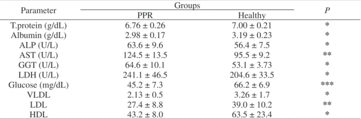
Table 2. Plasma biochemical parameters and total NEFA concentrations in kid goats naturally infected by PPR (n = 15) and in healthy controls (n = 15). Results are expressed as mean ± standard deviation.
Parameter PPR Groups Healthy P
T.protein (g/dL) 6.76 ± 0.26 7.00 ± 0.21 * Albumin (g/dL) 2.98 ± 0.17 3.19 ± 0.23 * ALP (U/L) 63.6 ± 9.6 56.4 ± 7.5 * AST (U/L) 124.5 ± 13.5 95.5 ± 9.2 ** GGT (U/L) 64.6 ± 10.1 53.1 ± 3.73 * LDH (U/L) 241.1 ± 46.5 204.6 ± 33.5 * Glucose (mg/dL) 45.2 ± 7.3 66.2 ± 6.9 *** VLDL 2.13 ± 0.5 3.26 ± 1.7 * LDL 27.4 ± 8.8 39.0 ± 10.2 ** HDL 43.2 ± 8.0 63.5 ± 23.4 *
DISCUSSION
In the Turkey, Eastern of Anatolia is one of the regions made intensive of livestock. The presence of PPRV infection in Turkey has been reported before different studies [19,41,44,52]. Although the clinical and postmortem findings may be sufficient for the diagnosis of PPR in the endemic areas, yet labara-tory confirmation is essential for definitive diagnosis because of the clinical similarity of PPR to rinderpest [34,36]. Immunocapture [35] and reverse transcrip-tion polymerase chain reactranscrip-tion (RT-PCR) followed by nucleotide sequencing [11,17,48] are the current diagnostic methods for all morbillivirus infections. Furthermore, due to simplicity, high sensitivity, and economy, several competitive enzyme-linked immu-nosorbent assays (c-ELISA) have been recognised as suitable systems for use for diagnosis and seroepide-miological surveillance [8,9,23,24,45]. We used both C-ELISA and RT-PCR in the diagnosis of suspected disease. We determined that the diagnostic value of necropsy materials such as lymph node, oro-nasal swap and blood were determined more valuable di-agnostic materials in the diagnosis of PPRV infection by RT-PCR.
The liver is not a primary target for the PPR virus and may be fatty degenerated [26,50]. Toplu et al. [50] and Yarım et al. [51] reported multifocal areas of coagulative necrosis and vacuolation of hepatocytes and abnormal liver function test in sheep naturally infected with PPR and significant increases in mean serum concentrations of AST and ALT. In this study, small multifocal necrotic foci and haemorrhagic areas were seen as macroscopically in the liver. Capsular fi-brosis, severe hidropic degeneration in the hepatocytes, multifocal necrosis foci, disorganisation in the hepatic cord, and some haemorrhagic areas were seen in the microscopic examination. Furthermore, proliferation of the bile ducts, and mononuclear cell infiltration were seen in the periportal perivascular areas. These histopatological findings were confirmed that the liver damage was formed during PPRV infection. Significant increases especially ALP, AST, and GGT, and signifi-cant decreases in total protein and albumin in the kids with PPR was consistent to the liver damage.
The liver plays a central role in the regulation of lipoprotein synthesis and degradation as well as in the storage of lipids in various lipids. Therefore, liver dysfunction can be associated with dys- or
paralipo-proteinaemia [47]. In this study, the decrease level of VLDL, LDL, and HDL in the kid goats with PPR were consistent findings with liver damage, and the cause of decrease could be inadequate synthesis of cholesterol that main structure of lipoproteins due to liver dysfunction.
The glucose level was lower in the PPR group than control. The cause of the low concentration in the infected kid goats could lose of their appetite.
The determination of lipid peroxidation status (MDA concentrations) is among the most widely used methods for determination of the oxidative stress. Increased malondialdehyde (MDA) concentration in plasma is a marker of lipid peroxidation [25,39]. Lipid peroxidation is a non-enzymatic chain reaction based on oxidation of mainly unsaturated fatty acids. It leads to the creation of lipid peroxides and other intermediates. These intermediates may influence the properties of cell membranes, and one of the most common of these intermediates is MDA [10,28]. In this study, plasma MDA concentrations were found to be increased in the kids with PPR compared to the control group, while decreases of GSHPx, and CAT activities were observed. Because of GSHPx and CAT are involved in the conversion of radicals into less effective metabolites, these changes coupled to the increase of MDA concentrations, suggest that an excessive ROS production occurred during PPR infection.
Vitamin E (in the form of α-tocopherol) is the
major lipid-soluble antioxidant of lipoproteins and biomembranes [27,31,49]. Vitamin C (ascorbic acid) functions as a potent water soluble chain-breaking antioxidant in the biological fluids, but it can not scavenge the radicals within the lipid region of the membranes [16]. The synergism between ascorbic acid and α-tocopherol in the inhibition of lipid peroxidation is well known. Vitamin C enhances the antioxidant
activity of vitamin E by recycling the α-tocopheroxyl
radical back to α-tocopherol and the depletion of the
α-tocopherol is markedly reduced [5,20,29,37]. In this study, the concentrations of serum vitamin E, and vitamin C, which are responsible for protecting the cells from damage caused by lipid peroxidation, were found significantly lower in the PPR group than in the control group. The decrease of plasma antioxidant vitamins could results to their over utilisation due to the disease related oxidative stress.
CONCLUSION
In conclusion, this study has highlighted the occurrence of an oxidative stress with important differ-ences in antioxidant status as reflected by assessment of some enzymatic and non-enzymatic antioxidants in kids infected by PPRV. There is no effective therapy for PPRV infection especially young goats, and due to oxidative stress might be an aggravating factor of the disease, it can be useful to add the antioxidant vitamins such as vitamin E and C, to the classical treatment procedures for get ride of the disease.
MANUFACTURERS
1IDVet Innovative Diagnostic kits. Grabels, France. 2Qiagen GmbH. Hilden, Germany.
3Techne TC-PLUS thermal cyclers. Staffordshire, UK. 4Siemens Healthcare Diagnostics Inc. Erlangen, Germany. 5Shimadzu Coorparation. Kyoto, Japan.
6SPSS Inc. Chicago, IL, USA.
Declaration of interest. The authors report no conflicts of
interest. The authors alone are responsible for the content and writing of the paper.
REFERENCES
1 Al-Majali A., Hussain M.N.O., Amarin N.M. & Majok A.A. 2008. Seroprevalance of, and risk factors for, peste des
petits ruminants in sheep and goats in Northern Jordan. Preventive Veterinary Medicine. 85(1-2): 1-8.
2 Anderson J., Mccayj A. & Butcher R.N. 1991. The use of monoclonal antibodies in competetive ELISA for the
detection of antibodies to rinderpest and petse des petits ruminants viruses. In: Jeggo M.H. (Ed). The sero-monitoring
of rinderpest throughout Africa-Phase one. In: The Proceedings of a final research Co-ordination meeting of the FAO/
IAEA/SIDA/OAU/IBAR/PARC Co-ordinated Research Programme (Ivory Coast. 1990). IAEA-TECDOC. 623: 43-53.
3 Banyard A.C., Parida S., Batten C., Oura C., Kwiatek O. & Libeau G. 2010. Global distribution of peste des petits
ruminants virus and prospects for improved diagnosis and control. Journal of General Virology. 91: 2885-2897.
4 Bao J., Li L., Wang Z., Barrett T., Suo L., Zhao W., Liu Y., Liu C. & Li J. 2008. Development of one-step
real-time RT-PCR assay for detection and quantitation of peste des petits ruminants virus. Journal of Virological Methods. 148(1-2): 232-236.
5 Bell A. 1995. Regulation of organic nutrient metabolism during transition from late pregnancy to early lactation. Journal
of Animal Science. 73: 2804-2819.
6 Brzezinska-Slebodzinska E., Miller J.K., Quigley J.D., Moore J.R. & Madsen F.C. 1994. Antioxidant status of
dairy cows supplemented prepartum with vitamin E and selenium. Journal of Dairy Science. 77: 3087-3095.
7 Castillo C., Hernandez J., Lopez-Alonso M., Miranda M. & Benedito J.L. 2003. Values of plasma lipid
hydrop-eroxides and total antioxidant status in healthy dairy cows: preliminary observations. Archives Animal Breeding. 46: 227-233.
8 Choi K.S., Nah J.J., Choi C.U., Ko Y.J., Sohn H.J., Libeau G., Kang S.Y. & Joo Y.S. 2003. Monoclonal
antibody-based competitive ELISA for simultaneous detection of rinderpest virus and peste des petits ruminants virus antibodies.
Veterinary Microbiology. 96: 1-16.
9 Choi K.S., Nah J.J., Ko Y.J., Kang S.Y. & Jo N.I. 2005. Rapid competitive enzyme-linked immunosorbent assay for
detection of antibodies to peste des petits ruminants virus. Clinical and Diagnostic Laboratory Immunology. 12(4): 542-547.
10 Comborti M. 1989. Three models of free radical-induced cell injury. Chemico-Biological Interactions. 72(1-2): 1-56. 11 Couacy-Hymann E., Roger F., Hurard C., Guillou J.P., Libeau G. & Diallo A. 2002. Rapid and sensitive detection
of peste des petits ruminants virusby a polymerase chain reaction assay. Journal of Virological Methods. 100: 17-25.
12 Couacy-Hymann E., Bodjo C., Danho T., Libeau G. & Diallo A. 2007. Evaluation of the virulence of some strains of
peste-des-petits-ruminantsvirus (PPRV) in experimentally infected West African dwarf goats. The Veterinary Journal. 173: 178-183.
13 Couacy-Hymann E., Bodjo S.C., Danho T., Kofi M.Y., Libeau G. & Diallo A. 2007. Early detection of viral
excre-tion from experimentally infected goats with peste-des-petits ruminants virus. Preventive Veterinary Medicine. 78: 85-88.
14 Davies K.J.A. 2000. Oxidative stress, antioxidant defences, and damage removal, repair and replacement systems.
15 Diop M., Sarr J. & Libeau G. 2005. Evaluation of novel diagnostic tools for peste des petits ruminants virus in
naturally infected goat herds. Epidemiology & Infection. 133: 711-717.
16 Eicher-Pruiett S.D., Morrill J.L., Blecha F., Higgins J.J., Anderson N.V. & Reddy P.G. 1992. Neutrophil and
lymphocyte response to supplementation with vitamin C and E in young calves. Journal of Dairy Science. 75: 1635-1642.
17 Forsyth M.A. & Barret T. 1995. Evaluation of polymerase chain reaction for the detection and characterisation of
rinderpest and peste des petits ruminants viruses for epidemiological studies. Virus Research. 39: 151-163.
18 Goth L. 1991. A simple method for determination of serum catalase activity and revision of reference range. Clinica
Chimica Acta. 196: 143-152.
19 Gulyaz V. & Ozkul A. 2005. Pathogenicity of a local peste des petits ruminants virus isolate in sheep in Turkey.
Tropi-cal Animal Health and Production. 37: 541-547.
20 Gutteridge J.M. 1993. Free radicals in disease processes: a compilation of cause and consequence. Free Radical
Research Communications. 19(3): 141-158.
21 Hallivell B. & Gutteridge J.M. 1985. The importance of free radicals and catalytic metal ions in human diseases.
Molecular Aspects of Medicine. 8(2): 189-193.
22 Herdt T.H. & Stowe H.D. 1991. Fat-soluble vitamin nutrition for dairy cattle. Veterinary Clinics of North America:
Food Animal Practice. 7: 391-415.
23 Hummel K.B., Erdman D.D., Heath J. & Bellini W.J. 1992. Baculovirus expression of the nucleoprotein gene of
measles virus and utility of the recombinant protein in diagnostic enzyme immunoassays. Journal of Clinical
Micro-biology. 30: 2874-2880.
24 Ismail T., Ahmad S., D’souza-Ault M., Bassiri M., Saliki J., Mebus C. & Yılma T. 1994. Cloning and expression
of the nucleocapsid gene of virulent Kabete O strain of rinderpest virus in baculovirus: use in differential diagnosis between vaccinated and infected animals. Virology. 198: 138-147.
25 Itze L. 1984. Ascorbic acid metabolism in ruminants. In: Wegger I., Tagwerker F.J. & Moustgaard J. (Eds). Workshop.
Ascorbic Acid in Domestic Animals. Copenhagen: Royal Danish Agr Soc., pp.120-130.
26 Khan H.A., Siddique M., Arshad M., Abubakar M., Akhtar M., Arshad M.J. & Ashraf M. 2009. Post-vaccination
antibodies profile against peste des petits ruminants (PPR) virüs in sheep and goats of Punjap, Pakistan. Tropical Animal
Health and Production. 41: 427-430.
27 Kleczkowski M., Klucinski W., Shaktur A. & Sikora J. 2005. Concentration of ascorbic acid in the blood of cows
affected with mastitis. Bulletin of the Veterinary Institute in Pulawy. 49: 203-207.
28 Kızıl O., Ozdemır H., Karahan M. & Kızıl M. 2007. Oxidative stres and alterations of antioxidant status in goats
naturally infected with Mycoplasma agalactia. Revue de Medecine Veterinaire. 158(6): 326-330.
29 Kohen R. & Nyska A. 2002. Oxidation of biological systems: oxidative stres phenomena, antioxidants, redox
reac-tions, and methods for their quantification. Toxicologic Pathology. 30: 620-650.
30 Kyaw A. 1978. A simple colorimetric method for ascorbic acid determination in blood plasma. Clinica Chimica Acta.
16: 151-157.
31 Lawrence R.A. & Burk R.F. 1976. Glutathione peroxidase activity in selenium-deficient rat liver. Biochemical and
Biophysical Research Communications. 71: 952-958.
32 Lefevre P.C. & Diallo A. 1990. Peste des petits ruminants. Revue Scientifique Et Technique. 9: 935-981.
33 Lefevre P.C., Diallo A., Schenkel F., Hussein S. & Staak G. 1991. Serological evidence of peste des petits ruminants
in Jordan. Veterinary Record. 128: 110-111.
34 Libeau G., Diallo A., Calvez D. & Lefevre P.C. 1992. A competitive ELISA using anti-N monoclonal antibodies for
specific detection of rinderpest in cattle and small ruminants. Veterinary Microbiology. 31: 147-160.
35 Libeau G., Diallo A., Colas F. & Guerre L. 1994. Rapid differential diagnosis of rinderpest and peste des petits
ruminants using an immunocapture ELISA. Veterinary Record. 134: 300-304.
36 Libeau G., Prehaud C., Lancelot R., Colas F., Guerre L., Bishop D.H. & Diallo A. 1995. Development of a
competi-tive ELISA for detecting antibodies to the petse des petits ruminants virus using a recombinant nucleoprotein. Research
in Veterinary Science. 58: 50-55.
37 Lowry O.H., Rosebrough N.J., Farr A.L. & Randall R.J. 1951. Protein measurement with folin phenol reagent. The
http://dx.doi.org/10.22456/1679-9216.81812
1527
38 Machlin L.J. & Bendich A. 1987. Free radical tissue damage: protective role of antioxidant nutrients. The FASEB
Journal. 1: 441-446.
39 Martinek R.G. 1964. Method for determination of vitamin E (total tocopherols) in serum. Clinical Chemistry. 10:
1078-1086.
40 Miller J.K., Brzezınska-Slebodzınska C. & Madsen F.C. 1993. Oxidative stress, antioxidants, and animal function.
Journal of Dairy Science. 76: 2812-2823.
41 Ozkul A., Akca Y., Alkan F., Barrett T., Karaoglu T., Dagalp S.B., Anderson J., Yesilbag K., Cokcaliskan C., Gencay A. & Burgu I. 2002. Prevalence, distribution, and host range of peste des petits ruminants virus, Turkey.
Emerging Infectious Diseases journal. 8: 708-712.
42 Placer A.Z., Linda L.C. & Johnson B. 1966. Estamination of product of lipid peroxidation (Malonly Dialdehyde) in
biochemical systems. Analytical Biochemistry. 16: 359-364.
43 Radostits O.M., Gay C.C., Hinchcliff K.W. & Constable P.D. 2007. Veterinary Medicine. A textbook of the diseases
of cattle, sheep, pigs, goats, and horses. 10th edn. London: Saunders Elsevier, 2065p.
44 Sahinduran S., Albay M.K., Sezer K., Ozmen O., Mamak N., Haligur M., Karakurum C. & Yıldız R. 2012.
Co-agulation profile, haematological and biochemical changes in kids naturally infected with peste des petits ruminants.
Tropical Animal Health and Production. 44: 453-457.
45 Saliki J.T., Libeau G., House J.A., Mebus C.A. & Dubovi E.J. 1993. Monoclonal antibody-based blocking
enzyme-linked immunosorbent assay for specific detection and titration of peste-des-petits-ruminants virus antibody in caprine and ovine sera. Journal of Clinical Microbiology. 31: 1075-1082.
46 Scott G.R. 1990. Peste des petits ruminants (goat plague) virus. In: Dinter Z. & Morein B. (Eds). Virus Infections of
Vertebrates. Amsterdam: Elsevier, pp. 355-361.
47 Seidel D. 1987. Lipoproteins in liver disease. Journal of Clinical Chemistry and Clinical Biochemistry. 25: 541-551. 48 Shaila M.S., Shamaki D., Forsyth M.A., Diallo A., Kitching R.P. & Barrett T. 1996. Geographic distribution
an-depidemiology of peste des petits ruminants virus. Virus Research. 43: 149-153.
49 Smith K.L., Todhunter D.A. & Schoenberger P.S. 1985. Environmental mastitis: cause, prevalence, prevention.
Journal of Dairy Science. 68: 1531-1553.
50 Toplu N. 2006. Characteristic and non-characteristic pathological findings in peste des petits ruminants (PPR) of sheep
in the Ege district of Turkey. Journal of Comparative Pathology. 13: 135-141.
51 Yarım G.F., Nisbet C., Yazıcı Z. & Gumusova S.O. 2006. Elevated serum total sialic acid concentrations in sheep
with peste des petits ruminants. Medycyna Weterynaryjna. 62: 1375-1377.
52 Yesilbag K., Yılmaz Z., Golcu E. & Ozkul A. 2005. Peste des petits ruminants outbreak in western Turkey. Veterinary