ROLE OF HISTONE VARIANT H3.3 IN TRANSCRIPTION AND
MITOTIC PROGRESSION
A DISSERTATION SUBMITTED TO
THE GRADUATE SCHOOL OF ENGINEERING AND SCIENCE OF BILKENT UNIVERSITY
IN PARTIAL FULFILLMENT OF THE REQUIREMENTS FOR THE DEGREE OF
DOCTOR OF PHILOSOPHY IN
MOLECULAR BIOLOGY AND GENETICS
By
Ayşegül Örs
April 2017
ROLE OF HISTONE VARIANT H3.3 IN TRANSCRIPTION AND MITOTIC PROGRESSION
By Ayşegül Örs April 2017
We certify that we have read this dissertation and that in our opinion it is fully adequate, in scope and in quality, as a thesis for the degree of Doctor of Philosophy in Molecular Biology and Genetics.
Işık Yuluğ (Advisor)
Mehmet Öztürk (Co-advisor)
İhsan Gürsel
Ayşe Elif Erson-Bensan
Uygar Halis Tazebay
Ali Osmay Güre Approved for the Graduate School of Engineering and Science
Ezhan Karaşan
ABSTRACT
ROLE OF HISTONE VARIANT H3.3 IN TRANSCRIPTION
AND MITOTIC PROGRESSION
Ayşegül Örs
Ph.D. in Molecular Biology and Genetics Advisor: Işık Yuluğ
Co-advisor: Mehmet Öztürk April 2017
Chromatin structure needs to be dynamic and flexible in order for the eukaryotic cellular processes to function correctly. Incorporation of histone variants into chromatin serves to increase epigenetic plasticity by conferring new structural and functional properties to chromatin. Histone variants are implicated in many cellular processes such as transcription or cell division and their deregulation is involved in tumorigenesis. H3.3 is an evolutionarily well conserved histone variant that differs by only a few amino-acids from its replication-dependent counterparts. With the aim of determining H3.3 function, novel knock-in/conditional knock-out mouse models were established and characterized. In these models, one of the two genes coding for H3.3, H3f3a or H3f3b has been modified to code for an N-terminal FLAG-FLAG-HA tagged H3.3A or H3.3B which can be depleted upon Cre expression. Nucleosome resolution genome-wide mapping FH-H3.3A and FH-H3.3B determined that H3.3A and H3.3B were similarly enriched at promoter regions and their enrichment levels positively correlated with high expression and gene body enrichment. They were also found enriched in telomeres and some repetitive DNA sequences. In a subset of these repetitive regions H3.3A and H3.3B showed differential enrichment properties. As double H3.3-KO mouse generation resulted lethal, mouse embryonic fibroblasts (MEFs) were isolated from FH-H3.3 mice and transformed. Using a combination of Cre recombinase mediated knock-out and RNA interference technology, a new cellular model was established where H3.3 expression was essentially depleted. Although H3.3 enrichment profiles were indicative of a role in active transcription, whole transcriptome analysis upon single H3.3 depletion in livers and an almost complete H3.3 depletion in MEFs yielded very few differentially regulated genes. Interestingly, H3.3
depleted MEFs showed a high increase in mitotic defects and abnormal nuclear structures. Thus, an important yet often understudied role for H3.3 in genomic maintenance during mitotic progression was highlighted.
Keywords: Histone variants, H3.3, H2A.Z, ChIP-Seq, RNA-Seq, liver, mouse model,
ÖZET
HİSTON VARYANTI H3.3’ÜN TRANSKRİPSİYONDA VE
MİTOZ BÖLÜNME İLERLEMESİNDEKİ ROLÜ
Ayşegül Örs
Moleküler Biyoloji ve Genetik, Doktora Tez danışmanı: Işık Yuluğ Eş tez danışmanı: Mehmet Öztürk
Nisan 2017
Ökaryotik hücresel süreçlerin doğru çalışabilmesi için kromatin yapısının dinamik ve esnek olması gerekir. Histon varyantlarının kromatine dahil edilmesi, kromatine yeni yapısal ve işlevsel özellikler kazandırarak, epigenetik esnekliği arttırır. Histon varyantları, transkripsiyon veya hücre bölünmesi gibi birçok hücresel fonksiyonların gerçekleştirilmesinde rol alır ve deregülasyonlarının kanserleşmede sürecinde etkisi vardır. H3.3 proteini, replikasyona-bağımlı eşdeğerlerinden sadece birkaç amino asit farklılık gösteren evrimsel olarak iyi korunmuş bir histon varyantıdır. H3.3 fonksiyonunu belirlemek amacıyla, yeni koşullu nakavt (knock-in/ conditional knock-out) fare modelleri oluşturulmuş ve tanımlanmıştır. Bu modellerde, H3.3’ü kodlayan iki genden biri, H3f3a veya H3f3b, N-terminal etiketli H3.3A veya H3.3B kodlamak üzere modifiye edilmiştir. Ayrıca cre rekombinaz ifadesiyle bu genlerin nakavt edilebilmektedir. Bu çalışmada, karaciğerde, nükleozom çözünürlüğünde, genom çapında FH-H3.3A ve FH-H3.3B yerleşme haritalaması gerçekleştirilmiştir. Bu analiz, H3.3A ve H3.3B'nin promotör bölgelerde benzer şekilde yoğunlaştıklarını ve yoğunluklarının, yüksek gen ekspresyonu ve gen boyunca yoğunlaşma ile pozitif korelasyonda olduğunu göstermiştir. H3.3’ün, ayrıca telomerlerde ve bazı tekrarlayan DNA dizilerinde yoğunlaştığı bulunmuştur. Bazı tekrarlayan bölgelerde H3.3A ve H3.3B, farklı şekilde yoğunlaşmıştır. İki H3.3 geninin de yok edildiği fare elde etme çalışmaları ölümcül sonuçlandığı için, FH-H3.3 farelerinden embriyonik fare fibroblastları (MEF) izole edilmiş ve ölümsüzleştirilmiştir. Cre aracılı nakavt ve RNA enterferans teknolojileri birlikte kullanılarak, H3.3 ifadesinin esas olarak susturulduğu yeni bir hücre modeli geliştirilmiştir. Genom boyunca yoğunlaşma profillerinin H3.3’ün aktif transkripsiyonda bir rolü olduğunu göstermesine rağmen, H3.3
nakavt modelleri üzerine yapılan transkriptom analizleri sonucu çok az sayıda gen etkilenmiştir. İlginç şekilde, H3.3 ifadesi susturulmuş MEF'lerde, mitotik defektler ve anormal nükleer yapılarında yüksek bir artış gözlenmiştir. Böylece, H3.3’ün, mitotik ilerleme sırasındaki genom bütünlüğünün korunmasında önemli ama çoğu zaman az üzerinde durulan bir rol üstlendiği vurgulanmıştır.
Anahtar kelimeler: Histon varyantları, H3.3, H2A.Z, ChIP-Seq, RNA-Seq, karaciğer, fare
Acknowledgements
Firstly, I would like to express my gratitude to my advisor Assoc. Prof. Dr. Işık Yuluğ for taking me on as her PhD student and offering her guidance during the last years of my PhD studies.
I am forever grateful to my co-advisor, Prof Dr. Mehmet Öztürk for his mentorship and for providing me with the opportunity to pursue my research in the Institute of Advanced Biosciences in Grenoble, France. His scientific guidance as well as his immeasurable professional and personal support allowed me to carry out my thesis in the best possible way.
I thank my supervisor Dr. Stefan Dimitrov for welcoming me in his research group at the Institute for Advanced Biosciences in Grenoble, France and providing me with his extensive guidance and the resources to develop my PhD project. He has always been available, supportive, generous and understanding throughout our collaboration and I consider it a privilege to have been part of his research group.
There are no words to describe the extent of my gratitude towards Dr. Kiran Padmanabhan. He has supervised my thesis and has been there with me through the good, the bad and the ugly. His resourceful knowledge and unique enthusiasm along with his everlasting support, guidance and patience allowed me to mature both scientifically and personally. I also thank him for his valuable input in the writing of my dissertation and for taking the time in his busy schedule to attend my defense.
I would like to express my deepest appreciations to the thesis committee members, Prof. Dr. İhsan Gürsel and Assoc. Prof. Dr. Ayşe Elif Erson-Bensan for their availability and valuable suggestions during meetings. Moreover, I would like to thank Prof Dr. Uygar Tazebay and Assoc. Prof. Dr. Ali Osmay Güre for accepting to read and evaluate my dissertation as members of the thesis jury.
A special thanks to our collaborators, Thomas Westerling, Razvan Chereji and especially Christophe Papin in analyzing the ChIP-Seq and RNA-Seq data presented in this study. I thank Bertrand Favier for his guidance and support with all animal models and experimentation as well as Patrick Vernet for his invaluable help in colony maintenance
and animal experiments. I extend my gratitude to the staff at the animal housing facilities in Plateforme de Haute Technologie Animale (PHTA) and Institute for Advanced Biosciences (IAB). I would also like to spare a line on this page to express my respect to all the mice used in this study and the valuable contribution of laboratory subjects to the advancement of scientific research everywhere.
I am most appreciative of the help and support of Mylène Pezet in the flow cytometry platform and of the staff in the microscopy platform in IAB.
The work presented in this thesis was conducted in part in the Dimitrov - Chromatin and Epigenetics group at the Institute of Advanced Biosciences (IAB) in Grenoble, France. This long collaboration was made possible thanks to funding from Agence National de la Recherche (ANR), the European Molecular Biology Organization (EMBO) short-term fellowship, Scientific and Technological Research Council of Turkey (TUBITAK) 2214/A doctoral research grant and French Ministry of Foreign Affairs scholarship.
During my thesis, I was lucky to be a part of two great laboratory families.
First, my gratitude goes to members of the former Ozturk group at Bilkent-MBG; Haluk Yüzügüllü, Özge Gürsoy Yüzügüllü, Şerif Şentürk, Çiğdem Özen, Dilek Çevik, Gökhan Yıldız, Mustafa Yılmaz, Emre Yurdusev, Hande Topel, Umur Keleş, Engin Demidizen, Derya Soner Cavga, Merve Deniz Abdüsselemoğlu and Yusuf İsmail Ertuna for the collaborative, supportive and positive work and learning environment they helped create during our time in Bilkent-MBG. I would like to sincerely thank Dilek Cevik and Pelin Telkoparan for their priceless friendship and endless support. I especially thank my colleague and house-mate Özlem Tufanlı as well as my other fellow PhD candidates, particularly Verda Bitirim, Gözde Güçlüler and Banu Bayyurt for their moral support and the fun times shared.
Second, a cordial thank you to past and present members of Dimitrov group at IAB; Damien Goutte-Gattat, Véronique Gerson, Geneviève Chevalier, Noémie Mandier, Thierry Gautier, Marc Block, Daniel Bouvard, Anne-Sophie Ribba and Emeline Fontaine with special mention of my fellow PhD candidates, Defne Dalkara, Lorrie Ramos, Yohan Roulland and Hiba Sabra. I particularly thank Sophie Barral for her initial mentorship in teaching me all chromatin based techniques used in the laboratory and for her valuable friendship. Working
in IAB also allowed me to meet amazing people and make great friends and I would like to express my thanks to Ayça Zeybek, Matteo Cattaneo, Hitoshi Shiota, Alexandar Kyumurkov and Mathieu Dangin for their friendship and encouragement over the years. Whether from a technical, scientific or personal point of view, having such a diverse working environment has trained me for the better and for the worse and was a real pleasure. I thank my parents, Hülya and Seyhun for all their love, support and patience throughout this thesis and my entire life. They have provided me with the best education possible, encouraged me to always push further and to keep all windows of opportunity open. I appreciate their sacrifices and I would not have been able to get to this stage without them. To my brother Ali Osman, a heartfelt thank you for his emotional, professional and “electronic” support and for being a role model for perfectionism and over-achievement throughout my life. I also wish to thank Onur Dallıağ, for his unfailing encouragement throughout these years.
Finally, I extend my gratitude to my other family members and friends in Turkey, in France and all over the world, who have cheered me on along the way and contributed to the successful completion of this thesis. I feel lucky to have a great family, amazing colleagues and lifelong friends.
“Gutta cavat lapidem non bis, sed saepe cadendo; sic homo fit sapiens non bis, sed saepe legendo”
Table of contents
Page ABSTRACT ... i ÖZET ... iii Acknowledgements ... vi Table of contents ... ixList of Figures ... xiii
List of Tables ... xv
Abbreviations ... xvi
Chapter 1. Introduction ... 1
1.1.Structural organization of chromatin ... 1
1.1.1. Histones ... 1
1.1.2. Nucleosome Core Particle and the Chromatosome ... 4
1.1.3. Higher-order organization of chromatin ... 5
1.2.Organization of chromatin function ... 8
1.2.1. Eukaryotic chromosome structure ... 8
1.2.2. The cell cycle and chromatin ... 8
1.2.3. Transcription and chromatin... 11
1.3.Epigenetic regulation ... 12
1.3.1. DNA methylation ... 14
1.3.2. Non-coding RNA... 14
1.3.3. Post-translational histone modifications... 15
1.3.4. Remodeling factors ... 18
1.3.5. Histone chaperones ... 19
1.3.6. Histone variants ... 20
1.4.H3.3 and the histone H3 family ... 25
1.4.1. Evolution of histone H3 variants ... 25
1.4.2. Genetic structure of H3 and H3.3 in mammals ... 25
1.4.3. The H3.3 containing nucleosome ... 29
1.4.4. H3 variant incorporation into chromatin ... 31
Page
1.5.1. Early studies on genome-scale H3.3 localization provide elements to
speculate on H3.3 function ... 32
1.5.2. Role of H3.3 in active transcription ... 33
1.5.3. H3.3 interaction with H2A.Z at promoters ... 33
1.5.4. Role of H3.3 in heterochromatin maintenance and genomic stability ... 34
1.5.5. H3.3 in development and sexual reproduction ... 36
1.5.6. H3.3 in tumorigenesis ... 36
1.6.Aim ... 39
Chapter 2. Materials and Methods ... 41
2.1.Materials ... 41
2.1.1. General laboratory chemicals and reagents ... 41
2.1.2. Cell culture chemicals and reagents ... 43
2.1.3. Primers ... 43
2.1.4. Lentiviral vectors and shRNA ... 47
2.1.5. Enzymes ... 47
2.1.6. Antibodies and beads ... 48
2.1.7. Equipment... 48
2.1.8. Software ... 50
2.2.Solutions and Media ... 51
2.2.1. Cell culture solutions and media ... 51
2.2.2. Genomic DNA Extraction and analysis ... 51
2.2.3. Western blot solutions and buffers ... 51
2.2.4. Nuclei isolation buffers ... 52
2.2.5. Chromatin preparation and immunoprecipitation buffers ... 54
2.3.Mouse models ... 55
2.4.Methods ... 55
2.4.1. General maintenance and handling of test subjects ... 55
2.4.2. Mouse embryonic fibroblast (MEF) isolation ... 55
2.4.3. General maintenance and handling of cell lines ... 56
2.4.4. Transformation of bacteria ... 57
Page
2.4.6. Genomic DNA isolation ... 57
2.4.7. Genotyping by PCR ... 57
2.4.8. Agarose Gel Electrophoresis ... 59
2.4.9. Lentivirus production for shRNA based knock-down in MEFs ... 59
2.4.10.Adenovirus infection for transient Cre expression in MEFs ... 59
2.4.11.RNA isolation ... 60
2.4.12.cDNA preparation ... 61
2.4.13.Quantitative PCR (qPCR) and analysis ... 61
2.4.14.Nuclei isolation from liver tissue ... 61
2.4.15.Nuclei isolation from cell lines... 62
2.4.16.Nuclei quantification and lysis ... 62
2.4.17.Whole cell protein extraction. ... 63
2.4.18.Western blot... 63
2.4.19.Native chromatin immunoprecipitation (N-ChIP) ... 64
2.4.20.Cross-linked chromatin immunoprecipitation (X-ChIP) ... 66
2.4.21.Immunofluorescent Staining and imaging... 67
2.4.22.Nuclear and mitotic defect evaluation ... 67
2.4.23.Flow cytometry cell cycle analysis with propidium iodide DNA staining ... 67
2.4.24.RNA and ChIP sequencing and bioinformatic analysis ... 67
2.4.25.Statistical analysis of qPCR data ... 68
Chapter 3. Results ... 69
3.1.Novel knock-in/ conditional knock-out mouse lines ... 69
3.1.1. Molecular description and validation of mouse models ... 69
3.1.2. WT, FH-H3.3A, FH-H3.3B, H3.3A-KO and H3.3B-KO mouse lines are phenotypically similar. ... 73
3.2.Genome-wide distribution of H3.3 at nucleosome resolution in the liver ... 75
3.2.1. Mono-nucleosome preparation from liver tissue ensures high-resolution profiling of H3.3 ... 75
3.2.2. H3.3 is highly enriched at TSS and its enrichment positively correlates with gene expression ... 77
Page
3.2.3. H3.3A and H3.3B have identical enrichment patterns at most genomic
sites except at some retroviral repeat elements ... 79
3.2.4. H2A.Z is co-localized with H3.3 around the TSS ... 81
3.3.Effect of H3.3 loss on transcriptome ... 83
3.3.1. Knock-out of a single H3.3 coding gene does not affect liver transcriptome ... 83
3.3.2. H3.3 depleted mouse embryonic fibroblasts ... 86
3.3.3. H3.3 depletion in MEFs has some, yet minimal effect on the transcriptome ... 91
3.4.H3.3 involvement in mitotic progression ... 95
3.4.1. H3.3 and H2A.Z are present at TSS of deregulated genes involved in cell cycle ... 95
3.4.2. H3.3 depletion does not affect H2A.Z enrichment at promoter regions 95
3.4.3. H3.3 depletion results in defective mitotic progression ... 98
Chapter 4. Discussion ... 100
Chapter 5. Perspectives ... 110
Complete H3.3 depletion in MEFs ... 110
Implication of the N-terminal tail of H3.3 and its phosphorylation in mitotic regulation ... 110
H3.3 implication in liver regeneration ... 111
References ... 112
Appendices ... 131
Appendix A – Flow cytometry analysis of the cell cycle by DNA content (PI incorporation) ... 131
Appendix B – Copyright Permissions ... 132
List of Figures
Figure Page
Figure 1-1. Secondary structure of histones. ... 2
Figure 1-2. Nucleosome assembly. ... 5
Figure 1-3. Levels of chromatin compaction in the eukaryotic nucleus. ... 6
Figure 1-4. The eukaryotic cell cycle. ... 9
Figure 1-5. Main mechanisms of epigenetic regulation. ... 13
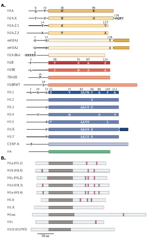
Figure 1-6. Human core and linker histone variants. ... 21
Figure 1-7. Genomic organization of H3 coding genes ... 25
Figure 1-8. Sequence alignment of processed transcripts of H3f3a and H3f3b... 28
Figure 1-9. Amino-acid sequence alignment of mammalian histone variants H3.3, H3.2 and H3.1. ... 29
Figure 1-10. Differential amino-acids between H3.3 and H3 variants in reference to the nucleosome structure. ... 30
Figure 1-11. H3.3 incorporation into chromatin. ... 31
Figure 1-12. Contribution of histone mutations and deregulations in their expression to tumorigenesis in humans. ... 37
Figure 3-1. Generation of H3.3A and H3.3B mouse models. ... 69
Figure 3-2. Genotype validation of FH-H3.3A and FH-H3.3B mice. ... 70
Figure 3-3. FH-H3.3A and FH-H3.3B expression in mouse livers. ... 71
Figure 3-4. H3.3 knock-out validation of WT, FH-H3.3A, H3.3A-KO, FH-H3.3B and H3.3B-KO mice by genotyping, RT-qPCR and Western blot. ... 72
Figure 3-5. Mono-nucleosome preparation for N-ChIP and ChIP validation. ... 76
Figure 3-6. ChIP-Seq data reveals enrichment of H3.3. ... 77
Figure 3-7. H3.3A and H3.3B are enriched around transcription start (TSS) and termination sites (TTS). ... 78
Figure 3-8. H3.3 is present at TSS and positively correlates with gene expression. ... 78
Figure 3-9. H3.3A and H3.3B show similar deposition patterns at most genomic regions. ... 79
Figure Page
Figure 3-11. Genome-wide enrichment pattern of FH-H3.3A, FH-H3.3B and H2A.Z
at TSS. ... 81
Figure 3-12. Normalized densities of H2A.Z, H3.3A and H3.3B within gene bodies expressed at different levels in mouse liver. ... 82
Figure 3-13. H3.3A, H3.3B and H2A.Z enrichment at TSS correlates with transcription levels. ... 84
Figure 3-14. Effect of single H3.3 loss on the transcriptome of adult mouse livers. ... 85
Figure 3-15. Generation of H3.3B-KO and H3.3B-KO / H3.3A-Kd MEFs. ... 86
Figure 3-16. Validation of WT, FH-H3.3A, H3.3A-KO, FH-H3.3B and H3.3B-KO MEFs by genotyping, RT-qPCR and Western blot. ... 87
Figure 3-17. H3.3 specific shRNA selection and validation of knock-down efficiency. ... 89
Figure 3-18. H3.3 expression and doubling times in H3.3B-KO / H3.3A-Kd MEF model. ... 90
Figure 3-19. Effect of H3.3 loss on MEF transcriptome... 91
Figure 3-20. Comparative analysis of the differentially expressed genes in H3.3 deficient embryos and in H3.3 depleted ESCs. ... 93
Figure 3-21. H3.3 and H2A.Z enrichment at transcription start sites (TSS) of mitotic genes showing differential mRNA expression. ... 97
Figure 3-22. Mitotic defects in H3.3 deficient MEFs. ... 99
Figure 4-1. Graphical abstract of thesis. ... 109
List of Tables
Table Page
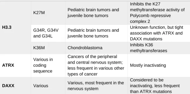
Table 1-1. Point mutations in H3.3 and its chaperones observed in human cancer ... 38
Table 2-1. Chemicals, reagents, enzymes and kits used for general laboratory purposes ... 41
Table 2-2. Chemicals, reagents, kits and media used in cell culture ... 43
Table 2-3. Primers used in genotyping mouse and cell lines from genomic DNA ... 44
Table 2-4. Primers used in RT-qPCR for gene expression ... 44
Table 2-5. Primers used in ChIP-qPCR ... 46
Table 2-6. Plasmids used for lentiviral shRNA transduction ... 47
Table 2-7. Enzymes and kits used in the study ... 47
Table 2-8. Antibodies used in the study ... 48
Table 2-9. Agarose resins and magnetic beads ... 48
Table 2-10. Instruments used in the study ... 48
Table 2-11. General laboratory equipment used in the study ... 49
Table 2-12. Software used in the study ... 50
Table 2-13. Cell lines and their relevant culture media ... 51
Table 2-14. Buffers used in genomic DNA extraction and analysis ... 51
Table 2-15. Buffers and solution used in western blot... 52
Table 2-16. Polyacrylamide Tris Gel preparation ... 52
Table 2-17. Buffer compositions used in nuclei isolation from liver tissue ... 53
Table 2-18. Buffer compositions used in nuclei isolation from cells ... 53
Table 2-19. Buffer compositions used in chromatin preparation and ChIP ... 54
Table 2-20. PCR reaction setup volumes for genotyping. ... 58
Table 2-21. Primer pairs used in genotyping of FH-H3.3A, FH-H3.3B and Actin-Cre mouse and cell lines ... 58
Table 2-22. PCR cycling conditions used in genotyping protocols ... 58
Table 3-1. Average litter size, weaned to born and female (F) to male (M) ratios in mouse models. ... 73
Abbreviations
Abbreviation Explanation
ACF ATP-utilizing
chromatin assembly and remodeling factor AEBSF 4-(2-Aminoethyl) benzenesulfonyl fluoride hydrochloride ALT Alternative Lengthening
of Telomeres
APS Ammonium PerSulfate ARE AU-Rich Element
ATRX
Alpha-Thalassemia/Mental Retardation Syndrome AUF1 AU-binding Factor 1
Bp Basepairs
CAF-1 Chromatin Assembly Factor-1
CDK Cyclin Dependent Kinase
Cds coding DNA sequence
CHD Chromodomain
Helicase DNA binding CpG Cytosine-Guanosine
motifs
DAXX Death Associated Protein
DMSO Dimethyl sulfoxide DNA DesoxyriboNucleic
Acid
DNMT DNA Methyl-Transferase
Dpf Days post fertilization DTT 1,4-dithiothreitol
Abbreviation Explanation
ECM ExtraCellular Matrix EDTA
EthyleneDiamineTetra-acetic Acid
EGTA Ethylene Glycol-bis (β-aminoethyl ether)-N,N,N',N'-Tetraacetic Acid
ERV Endogenous Retroviral element
ESC Embryonic Stem Cell FACS Fluorescence Assisted
Cell Sorting
FACT FAcilitates Chromatin Transcription
FBS Fetal Bovine Serum HAT Histone
Acetyl-Transferase
HDAC Histone DeACetylase HDM Histone DeMethylase HEPES 4-(2-hydroxyethyl)-1-piperazineethanesulfoni c acid
HJURP Holliday JUnction Recognition Protein HMT Histone Methyl
Transferase HP1 Heterochromatin
Protein 1
INO80 INOsitol requiring 80 ISWI Imitation Switch KCl Potassium Choride
kD Kilo Dalton
Abbreviation Explanation
KO Knock-out
LB Lysogeny/Luria Bertoni Broth
LiCl Lithium Chloride lncRNA Long non-coding RNA MEF Mouse Embryonic
Fibroblast
MNase Micrococcal Nuclease MOI Multiplicity of infection mRNA Messenger RNA MWCO Molecular Weight
Cut-Off
NaCl Sodium Chloride NaDOC Sodium Deoxycholate NAP1 Nucleosomes Assembly
Protein 1
NASP Nuclear Autoantigenic Sperm Protein
N-ChIP Native Chromatin ImmunoPrecipitation NCP Nucleosome Core
Particle
NEAA Non-Essential Amino-Acids NGS Next Generation Sequencing o/n Overnight OD Optical Density P/S Penicillin/ Streptomycin PBS Phosphate Buffered Saline
PCIA Phenol Chloroform Isoamyl Alcohol PCNA Proliferating Cell
Nuclear Antigen Abbreviation Explanation PCR Polymerase Chain Reaction PI Propopidium Iodide PLK Polo-Like Kinase Pol II RNA-polymerase II PTM Post-translational Modifications qPCR Quantitative PCR qs Quantum satis
(sufficient quantity for) RBP RNA Binding Protein RD Replication Dependent RNA RiboNucleic Acid RNAse A RiboNuclease A RT Room Temperature RT-qPCR Reverse
transcription-qPCR
SAC Spindle Attachment Checkpoint
SAM S-adenosyl methionine SDS Sodium dodecyl Sulfate
Seq Sequencing
SWI/SNF Switch/ Non-fermentable Sucrose tSCE Telomere sister
chromatin exchange UTR Untranslated region
WT WildType
X-ChIP Crosslinked Chromatin Immunoprecipitation
Chapter 1.
Introduction
In the eukaryotic organism, genomic information is organized into chromosomes. Each chromosome consists of a single, linear, double helix deoxyribonucleic acid (DNA) molecule tightly associated with highly conserved protein complexes. This organizational structure is called chromatin and more specifically corresponds to the complex of nuclear DNA and closely associated RNA and proteins.
The overall composition of chromatin is one third nucleic acids and two thirds proteins. Half of this protein fraction corresponds to very basic proteins called histones, the other half consists of various proteins called "non-histone proteins".
The nucleosome is the fundamental repeating unit of chromatin. It basically consists of DNA that’s wrapped in approximately 2 superhelical turns around an octameric core structure created by histone proteins 1.
Primarily, it was thought that chromatin’s only function was to compact DNA so that the large molecule would fit in the small volume of the eukaryotic nucleus. Histones were not subject to much interest since it was believed that the transcription machinery could easily override these small proteins as it was the case in bacteria 2. Over the last decades however, the increasing research in the field of epigenetics has seen histones transition from simple static building blocks to important dynamic actors in all physiological processes that involve interaction with DNA such as replication, transcription and maintenance of genomic integrity.
1.1. Structural organization of chromatin
1.1.1. Histones
Histones are the major protein constituents of chromatin. They are present in such large quantities that their mass is almost equal to that of DNA 3. Consequently, DNA quantification is commonly used as a means to determine histone mass when studying histones.
Histones are small and very basic proteins. They have approximately 100 amino-acids in their primary sequences (~15kD) with high lysine and arginine contents. There are five classes or histones: H1, H2A, H2B, H3 and H4. Based on their contribution to chromatin structure they can be divided into two groups: core histones consisting of H2A, H2B, H3, H4 that in pairs, form the core histone octamer and linker histone H1 that binds outwardly to the nucleosome core at entry and exit sites of linker DNA and contribute to the formation of the chromatosome. Histones are incorporated into chromatin through the action of specific histone chaperones 4,5.
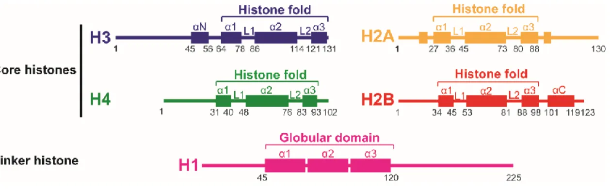
Figure 1-1. Secondary structure of histones. Core histones have a structure comprised of three regions, the
characteristic histone fold which consists of a large central alpha helix (α2) connected to two smaller alpha helices (α1, α3) on either side by two loops (L1, L2) and is flanked by the two N-terminal and C-terminal tails which remain mainly unstructured apart from some structures like the αN for histone H3. Linker histone H1 proteins have a central globular domain containing three helical regions flanked by variable N- and C-terminal domains. Representative figures constructed from 2,6
The histone fold motif is highly conserved and consists of a large central alpha helix connected by two loops to two smaller alpha-helices on either side. This central globular domain is flanked by a 15-45 amino-acid long N-terminal region and a much shorter C-terminal region of only a few residues (Figure 1-1) 7,8. While some parts of these extensions can be structured, they remain mostly flexible. Especially the N-terminal tails diverge notably between core histones and are subject to many post-translational modifications that play important roles in epigenetic signaling, as detailed in Section 1.3.3. The presence of the histone-fold motif allows for histones to form stable dimers via the “handshake” interaction which is the basis for the assembly of the histone octamer 8 (Figure 1-2).
Linker histones differ greatly from core histones and are far less conserved. They are enriched in lysine residues and lack the histone-fold motif. H1-like linker histones have a three-domain structure: a short unstructured aminoterminal domain of about 45 residues; a highly conserved globular core domain of about 75 residues; and a carboxy terminal domain of about 100 residues (Figure 1-1) 6,9.
Histones can also be classified into two distinct categories based on the time of their incorporation into chromatin during the cell cycle. These categories are replication dependent histones and replication independent histones. Replication-dependent (RD) histones are also known as “canonical”, “conventional”, “bulk” or “major” histones, and as suggested by their name, their expression and association with chromatin are coupled to DNA replication. They are loaded onto chromatin through the action of specialized proteins called histone chaperones. In metazoans, replication dependent histone transcripts present a very specific structure. They are coded by multicopy, intronless gene families organized in clusters with a stem-loop type structure to indicate transcription termination instead of the typical polyadenylation signal. This structure allows fast production of high levels of histone proteins needed for nucleosome assembly in the newly duplicated DNA during cell cycle progression. As an example, in humans, HIST1, a large histone cluster that contains 55 genes is located on chromosome 6 and HIST2 and HIST3, two smaller clusters containing 9 genes are located on chromosome 1. Despite some genomic rearrangements, gene number and organization of mouse histone gene clusters are strikingly similar their human counterparts. In mice, the large histone cluster Hist1 is located on chromosome 13 and contains 51 genes while the two smaller clusters, Hist2 and Hist3, are located on chromosomes 3 and 11 10. The expression of replication dependent histones is tightly regulated both at transcriptional and post-transcriptional levels and is restricted to the S-phase of the cell cycle to avoid histone toxicity 11–13.
In contrast, replication-independent histones, also called histone variants or replacement histones, are expressed at basal levels through-out the cell-cycle independently from DNA replication. Some histone variants have specific chaperones that load them on and evict them from chromatin. The genes that code for each histone variant are located outside the major histone clusters and present a much typical structure. They have at least one intron, a polyadenylation signal and often long 5’ and 3’ untranslated regions (UTR) 14. Histone variants can replace their corresponding conventional histones in a nucleosome and confer
new structural and functional properties. Histone variants and their roles in epigenetic regulation are further detailed in sections 1.3.6, 1.4 and 1.5.
1.1.2. Nucleosome Core Particle and the Chromatosome
The nucleosome core particle (NCP) was first discovered during nuclease digestion experiments of purified chromatin. When digested with nucleases for only a short period of time, protein-bound DNA is protected from digestion, whereas “free” DNA such as linker DNA, is accessible to nucleases and is degraded. High-resolution X-ray crystallization studies gave us detailed insight as to the atomic structure of the NCP 1,15.
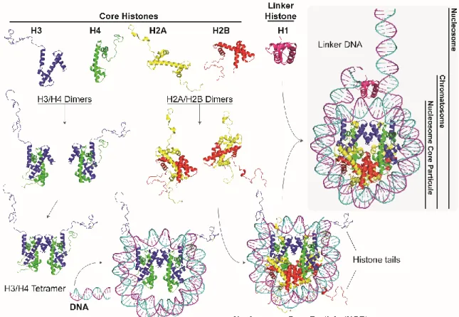
The histone octamer that constitutes the core of the NCP, consists of a pair of each of the core histones. For nucleosome assembly, first, H2A/H2B and H3/H4 dimers are formed. Then, two H3/H4 dimers associate via an interaction between the α2 and α3 helices of the H3 histones and form a tetramer. It is the H3/H4 tetramer that binds to DNA to form the intermediate core particle and the full nucleosome core particle assembly is completed with the association of two H2A/H2B dimers via the 2α and 3α helices of H2B and H4 (Figure 1-2) 1,16.
The handshake interactions between each pair of histones lead to the formation of β bridges between loops L1 and L2. These bridges form part of the DNA binding sites. The association between the histone octamer and the DNA fragment is mainly due to the insertion of the side chains of the arginine residues in the minor groove of the DNA double helix 3,8. While the structured histone regions are implicated in the majority of DNA-histone interactions, the less structured histone tails protrude from the nucleosome. Histone tails are more accessible to interact with neighboring nucleosomes or other factors. They are also target to many post-translational modifications (PTMs). The implication of PTMs in important cellular processes is detailed in section 1.3.3.
The chromatosome is the structure formed by the association of a H1 linker histone to the nucleosome core particle 17. Histone H1 binds to the NCP at the entry and exit sites of DNA 18. It associates over 20 base pairs (bp) of DNA at the entry and exit sites, greatly limiting their movements and thus locking the nucleosome in a closed position 19. The term nucleosome per se refers to the association of the NCP or chromatosome with one of its adjacent linker DNAs (Figure 1-2).
Figure 1-2. Nucleosome assembly. First, H3 (blue), H4 (green), H2A (yellow), H2B (red) dimerize through
“hand-shake” interactions to form H2A/H2B and H3/H4 dimers 8. Then, two H3/H4 dimers associate between each other to form a tetramer. It is the H3/H4 tetramer that binds to DNA (cyan, magenta) and the nucleosome core particle (NCP) assembly is completed with the association of two H2A/H2B dimers. Linker histone H1(hot pink) binds to the NCP forming the chromatosome 17,18. The nucleosome refers to the NCP associated to one of its adjacent linker DNA. N-terminal histone tails are usually accessible to various effectors. Image reconstructed from nucleosome core particle high resolution structure (PDB ID: 1KX5 15) and chromatosome structure (PDB ID: 4QLC 20) shown in cartoon representation using PyMol software 21.
1.1.3. Higher-order organization of chromatin
The main function of chromatin is to compact genetic material inside the nucleus all the while allowing access to different regions of DNA for the correct progression of physiological processes. In order to fulfill this function, it needs to remain highly dynamic and flexible. Through rearrangement of nucleosome arrays in various reproducible spatial conformations, chromatin achieves higher orders of organization 22.
The first order of compaction of chromatin is defined by the packaging of DNA with histones yielding a chromatin fiber of approximately 10-nm in diameter called the
nucleofilament (Figure 1-3). It is essentially a succession of nucleosomes separated by linker DNA and is familiarly called the “beads on a string” structure where “beads” refer to nucleosomes and “string” refers to DNA. This primary structure reduces DNA length by an approximate 6-fold and is generally permissive to transcription. However, under physiological conditions, nucleosomes are rarely found to stay in their “stretched-out” form. Instead, they are further folded and compacted at different levels to form “higher order structures” 2,23,24 (Figure 1-3).
Figure 1-3. Levels of chromatin compaction in the eukaryotic nucleus. DNA is wrapped around a histone
octamer to form the first order of compaction called the nucleofilament or the 10-nm fiber. Arrays of nucleofilaments fold further to form the 30-nm fiber. During interphase, this structure goes through different levels of compaction before reaching the highest level of compaction that is observed in the metaphase chromosome 2.
The secondary order of compaction for chromatin is the 30-nm chromatin fiber which is the result of a helical rearrangement of linear nucleosomes stabilized by linker proteins like H1 and HP1 (Figure 1-3). It allows a DNA compaction of almost 50 fold 25. Though still subject to controversy, two models have been put forward to explain the formation of this fiber. The first model, called the solenoid or the one-start helix model suggests that a single array of consecutive nucleosomes connected by linker histones, folds around an axis of symmetry to form the fiber with 6 to 8 nucleosomes per helical turn 25,26. The second model called the zig-zag or the two-start helix model, suggests that two nucleosome arrays are assembled in a zig-zag so that consecutive nucleosomes are found alternatively on each side of the fiber 27–29.
Unfortunately, both models have the disadvantage of being predominantly based on in vitro observations. Recent studies that aimed to elucidate which model is predominant in vivo, have suggested a coexistence of different structures where the formation of higher order structure would actually be determined by the environment of the nucleosomes 30,31. Chromatin architecture is sensitive to a large number of internal and external factors such as length of internucleosomal DNA, presence of histone variants or post-translational modifications, ionic conditions or binding of chromatin architecture proteins 32,33.
To date, the in vivo structure of the 30-nm fiber has not been determined and there is even doubt to its actual existence 34,35. Although higher levels of organization do exist, with the most evident example being the metaphasic chromosome, the precise structure and the sequential hierarchy of such organization is subject to intensive research.
The ability of chromatin to be arranged and rearranged in different orders of structure following intranuclear signals demonstrates its’ dynamic nature. Organisms have evolved different mechanisms to introduce variation into chromatin. Total or partial reorganization of chromatin allows for new functional properties, that allow in particular, transcription, replication or repair of DNA.
1.2. Organization of chromatin function
1.2.1. Eukaryotic chromosome structure
For genetic information to be accurately and effectively passed on during subsequent cell divisions, a chromosome must be able to replicate and the doubled material be equally divided between daughter cells, all the while maintaining a damage free DNA. Three specialized elements in the chromosome control these processes 2. First, the presence of many replication origins on eukaryotic chromosomes allows for rapid duplication of the genetic material during S phase of the cell cycle. The duplicated DNA molecules, i.e. sister chromatids, are maintained in proximity by cohesins and stay attached at their centromeres. During cell division, centromeres serve as a docking point for kinetochore assembly which will allow the sister chromatids to segregate into two daughter cells 36,37. Finally, telomeres, specialized repetitive sequences, ensure correct replication and protection of the ends of the linear DNA molecule 36,38,39. The architectural structure of the chromosomes can be best observed during metaphase of the cell cycle when DNA has duplicated and chromatin is at its most condensed state (Figure 1-3).
In the eukaryotic nucleus, nucleosomes are not arranged in a regular, homogenous manner. Instead, chromatin shows different levels of compaction dependent on cell-cycle stage and nuclear processes such as replication, transcription and repair. Furthermore, the nucleus is organized into function-specific sub-nuclear domains even though these domains are not architecturally fixed nor delimited by membranes. This compartmentalization links chromatin structure to genomic function under the control of physiologic signaling 40.
1.2.2. The cell cycle and chromatin
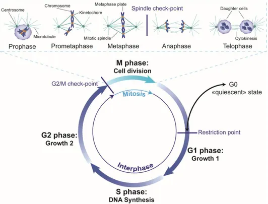
The cell cycle enables the duplication of genetic information and its division into two identical daughter cells. These two events determine the phases of the cell cycle. DNA replication takes place during the S phase which stands for synthesis and chromosome segregation and cell division takes place during the M phase which stands for mitosis. Between these two phases are the gap or growth phases, G1 that follows the M-phase and G2 that follows the S-phase. These gap phases not only serve to prepare the cell for growth but also allow for checking that conditions are favorable both internally and externally for proper cell cycle progression. As a matter of fact, if conditions are not suitable, cells may
the cell cycle or die. Interphase corresponds to the G1, S and G2 phases. There are three known checkpoints: the G1 checkpoint also called the restriction point because cells pass this point are committed to DNA replication and can no longer extend their stay in G1 or enter G0, the G2/M checkpoint which checks for DNA damage before cell division and the Metaphase checkpoint also called the spindle checkpoint which checks for correct alignment of chromosomes on the metaphase plate (Figure 1-4).
Figure 1-4. The eukaryotic cell cycle. The cell cycle consists of an S phase, when DNA replication takes
place, and a mitosis phase, when cell division takes place, separated by the two growth or gap phases G1 and G2 during which the cell prepares for the ensuing phases and checks that conditions are favorable for cell cycle progression. If conditions are not favorable, during G1, the cell may enter a quiescent state called G0 until conditions become favorable or the cell dies. There are 3 known checkpoints: the restriction point at the end of G1 when the cell commits to DNA replication, the G2/M checkpoint which checks for DNA damage before cell division and the spindle checkpoint at the end of metaphase which checks for correct attachment of sister chromatids at their kinetochores and their alignment on the metaphase plate. Interphase corresponds to G1, S and G2 phases. An expanded view of mitosis is presented at the top of the figure. In prophase, chromatin condenses and the centrosomes migrate to opposite poles of the cell emanating microtubules in all directions. In prometaphase, the nuclear membrane is disintegrated and microtubules attach to chromosomes at their kinetochores which migrate toward the center. During metaphase, chromosomes are aligned on the metaphase plate and the cell pursues onto anaphase only if all chromosomes are correctly aligned and attached to the mitotic spindle. In anaphase, the sister chromatids separate, finally in telophase the DNA decondenses, the nuclear envelope reforms and the two daughter cells form by cytokinesis 2. Figure adapted from Pines, 2011 41 with permission (Appendix B).
Mitosis is divided into five phases. During the first phase, prophase, chromatin condenses to form individual chromosomes. The two centrosomes each migrate to opposite poles of the cell and emanate microtubules in all directions and the nuclear membrane starts being absorbed by the endoplasmic reticulum. During prometaphase, the nuclear envelope is disintegrated and fully condensed chromosomes migrate towards the center of the cell and align at the spindle equator also called the metaphase plate. The mitotic spindle consists of the set of microtubules linking the chromosomes at their centromere to the centrosomes. In metaphase, chromosomes remain on the metaphase plate and the cells “checks” for correct orientation and attachment of each chromosome to the mitotic spindle. If cells pass the “mitotic checkpoint”, mitosis continues on to anaphase where sister chromatids are separated towards each pole of the spindle which starts to elongate as the daughter cells separate. During telophase, the chromosomes are released from the spindle microtubules as they begin to de-condense and the nuclear envelope begins to form around the daughter cells. Cytokinesis occurs almost simultaneously and leads to the separation of the cytoplasm of the two daughter cells. It consists of the formation of a contractile ring in the middle of the cell that gradually tightens to leave a small very dense structure called the residual body between the cells. The disappearance of this structure marks the separation of the daughter cells and the end of mitosis 2 (Figure 1-4).
During mitosis, chromatin is highly condensed with a varying level of compaction depending on the specific phase of mitosis. The highest level of compaction is achieved in metaphase and chromosomes can be easily visualized in their characteristic X-shape. The organization of chromatin in the mitotic chromosomes depends on the arrangements of the fibers into an axial skeleton composed of structural maintenance of chromosome (SMC) proteins 42,43, in particular, condensins I and II 44. Mitosis, and therefore the structure of the mitotic chromosome, is associated with an almost complete absence of transcription. Intriguingly, this absence of transcription does not come directly from the high level of compaction of DNA. As a matter of fact, the level of compaction does not necessarily entail a total loss of the accessibility of DNA to various transcription factors 45. It would seem that it is rather the fact that the structure of chromatin is relatively "frozen" during mitosis leading to the fact that RNA polymerase can no longer advance along the DNA strand 46. As a result of the transcriptional arrest at this stage, all effectors and enzymes needed for mitotic progression should be readily available in the cell.
Mitosis is a tightly regulated process. Mistakes in distribution of sister chromatids could lead to the loss of significant genetic information and cause irreparable damage to the cells. This regulation is carried out by mainly two mechanisms. The first one consists of the degradation of effectors by proteolysis after they have completed their function and the second mechanism uses signaling through phosphorylation by different types of kinases
47,48, such as the members of the cyclin dependent kinase (CDK), Aurora 49 and Polo-like
kinase (PLK) families 50.
At interphase, the mitotic chromosome structure does not persist. Chromosomes adapt a more relaxed conformation and yet remain organized into nuclear domains called chromosome territories 51. A gene’s expression can be highly altered depending on its nuclear sub-location. Two main areas can be defined based on chromatin condensation levels. Euchromatin has a loose chromatin structure, is rich in actively transcribed genes and replicates in early S-phase. Heterochromatin on the other hand is highly condensed, gene poor, enriched in repetitive elements and it replicates at the end of S phase. Heterochromatin can be further divided into constitutive and facultative heterochromatin. Constitutive heterochromatin is always compact and is characteristic of centric, pericentric and telomeric regions harboring repetitive DNA elements and imprinted genes which are heritably silenced throughout the whole organism. On the other hand, facultative heterochromatin is formed in gene rich regions but remains transcriptionally inactive. However, it can become de-condensed, hence reactivated, in certain contexts that can be temporal (developmental, cell cycle specific), spatial (changes in nuclear localization due to external signals) or hereditary (mono-allelic gene expression) 52. Inside the nucleus, heterochromatic regions of a chromosome are often found at the periphery, closely associated to lamina, whereas gene-rich regions are directed towards the center 51. Furthermore, constitutive and facultative heterochromatin are characterized by distinct epigenetic marks such as specific histone post-translational modifications and associated proteins 33,53.
1.2.3. Transcription and chromatin
Transcription is an extremely complex process involving many levels of control. Its regulation is essential for development and cellular homeostasis as it allows the differential expression of genes according to cell type and cell cycle stage. Deregulation of transcription
severely impacts cellular functions and is implicated in the development of many pathological conditions. Although transcription is one of the most studied areas in science, many mechanisms still remain to be elucidated.
Like the cell cycle, transcription is a cyclic process. Briefly, during initiation, activators bind specific DNA sequences upstream of the promoter leading to the binding of effectors such as transcription factors which position RNA polymerase II (Pol II) and constitute the pre-initiation complex. RNA synthesis is initiated when a transcription factor melts a short DNA sequence and places it in the Pol II cleft. In higher eukaryotes, Pol II is paused at promoter-proximal regions and the carboxy-terminal domain (CTD) of RNA Pol II needs to be phosphorylated for elongation to proceed. Effective elongation is proceeded by termination which consists of the release of mRNA from Pol II and consequently release of Pol II form DNA.
As stated in the previous section, at interphase, euchromatin and heterochromatin define areas of differential levels of transcription based on condensation levels of chromatin. However, even though euchromatin is less compact than heterochromatin, it still has a dense nucleosome structure. Nucleosomes constitute obstacles to transcription and need to be displaced or replaced by chromatin remodeling to allow access to genetic information 54. Chromatin structure can block initiation of transcription and delay its elongation 55,56. Therefore, uncovering mechanisms that control nucleosome interaction with DNA and other proteins at sites of transcription is an important step in understanding the regulation of transcription.
1.3. Epigenetic regulation
The notion of epigenetics, in its modern sense, is based on the work of biologist Conrad H. Waddington (1942). He initially defined epigenetics as “the branch of biology which studies the causal interactions between genes and their products, which bring the phenotype into being” 57. Historically, any event that could not be clarified by genetic phenomena would be classified into “epigenetics”. Today, the term “epigenetics” is used to refer to heritable changes in gene expression that are independent from direct modifications to the DNA sequence 23,58. It’s a collection of marks affixed to the genome that orchestrate the reorganization of chromatin into the functional domains described above, allowing a
marks persist even after the external stimuli that lead to their deposition are no longer relevant. They are stable and play a role in cellular memory, thus being responsible for long term gene expression profiles. Nonetheless, epigenetic marks can be modified or reversed depending on the environment 23,58.
The main mechanisms of epigenetic regulation of gene expression in mammals are DNA methylation, interactions with non-coding RNAs, remodeling of local chromatin structures by energetically displacing nucleosomes and/or altering histone-DNA interactions, reversible post-translational modifications on histone tails and incorporation of histone variants 59–61 (Figure 1-5). These mechanisms are briefly described below with an emphasis on histone variants.
Figure 1-5. Main mechanisms of epigenetic regulation. Several epigenetic mechanisms are employed
individually or in combination by eukaryotic cells to introduce variation into chromatin and allow for the proper progression of cellular processes 59–61. The main mechanisms of epigenetic regulation of gene expression in mammals are 1- covalent modifications to DNA such as cytosine methylation (5mc) and hydroxymethylcytosine (5hmC) (depicted as yellow stars), 2- interactions with non-coding RNAs (depicted as purple strands coating chromatin), 3- remodeling of local chromatin structures by energetically displacing nucleosomes and/or altering histone-DNA interactions using chromatin remodellers, 4- reversible post-translational modifications on histone tails such as acetylation (green triangle), phosphorylation (purple circle) and methylation (orange hexagon) and 5- incorporation of histone variants depicted by pink and green quarter circles.
1.3.1. DNA methylation
DNA methylation is a post-replicative modification that, in mammals consists of the binding of a methyl (CH3) group almost exclusively to the carbon 5 of the pyrimidine ring of Cytosine residues in CpG dinucleotides 62. This covalent addition of a methyl group to a cytosine is catalyzed by DNA methyltransferases (DNMTs). Some DNMTs bind new methyl groups (de novo methylation) to DNA while others function to maintain existing methyl marks through mitosis, thus contributing to epigenetic memory (methylation maintenance) 63. Regions with high density of CpG dinucleotides are called CpG islands and are usually located on gene promoters and/or the first exons of mammalian genes 64. In combination with other epigenetic marks like histone PTMs and RNA, DNA methylation and its binding proteins are associated with heterochromatin formation thus to silencing of transcription 65–68.
DNA methylation is involved in the establishment and maintenance through cell division of repetitive and centromeric DNA silencing, X chromosome inactivation in females and mammalian imprinting.
1.3.2. Non-coding RNA
Even though only a small fraction of the genome is actually translated, a large number of transcripts are implicated in important cellular functions. In recent years, increasing evidence showed that microRNAs (miRNA), Piwi-interacting RNAs (piRNA), endogenous silencing RNAs (siRNA) and long non-coding RNAs (lncRNA) play important roles in regulatory mechanisms at both transcriptional and post-transcriptional levels 69–71.
Non-coding RNAs do not code for any functional proteins by definition and their expression can be strongly tissue-specific 72. Through various mechanisms not completely understood for most, non-coding RNAs are able to regulate gene expression. For example, their association with specific proteins involved in gene regulation like transcription or remodeling factors, could serve as a trap that prevents those factors to act on their relevant targets thus impeding their function. On the other hand, association with lncRNAs can also serve to guide proteins to their specific targets.
Finally, lncRNA can also serve as docking platform that would centralize various effector proteins to the same genomic site 73. Non-coding RNAs are known to associate with many
factors involved in chromatin reorganization and methylation such as DNMTs and heterochromatin protein (HP1). One of the most well-known examples of non-coding RNA based epigenetic regulation is the XIST RNA involved in the silencing of the supernumerary female X chromosome in mammals 74. Although, the detailed mechanisms by which non-coding RNAs act remain to be elucidated, expanding research has provided insight to their involvement in dynamic regulation of chromatin and epigenetic memory, including genomic imprinting, DNA methylation and transcription regulation 68,69,73,75.
1.3.3. Post-translational histone modifications
Histones are targets to a large number of post-translational modifications (PTMs). These modifications include the addition of small chemical groups such as acetyl, methyl and phosphate groups as well as the addition of bigger globular proteins such as ubiquitin and SUMO 76–78. A majority of these modifications are found on the easily accessible histone termini, but globular domains situated at the core of the nucleosome can also be affected. Some of these modifications are specifically associated with cellular processes such as transcription, DNA repair or chromatin condensation.
Post-translational histone modifications, together with their “writers”, “erasers” and “readers” form the basis of the “histone code” hypothesis. This hypothesis suggests that specific PTMs, alone or in combination with others, serve as signals that regulate cellular processes 79.
In the majority of cases, PTMs act indirectly as docking sites for modification recognizing effector proteins that can in turn recruit other proteins or change or maintain chromatin structure 76. Some PTMs can also act directly on nucleosome structure by altering the charge of a residue, thereby changing histone-DNA or inter-histone affinities 80 or facilitating its remodeling 81.
The most studied post-translational histone modifying enzyme families are histone acetyltransferases (HAT) / deacetylases (HDAC) for acetylations and histones methyltransferases (HMT) / demethylases (HDM) for methylations 82–84. Acetylation and methylation along with phosphorylation and ubiquitination, are the most researched histone modifications. The action mechanisms of some principle modifications are briefly detailed in the following paragraphs.
Acetylation of histones
Acetylation occurs with the addition of an acetyl molecule on the amine group of lysine residues and is mainly present on the N-terminus of H3 and H4 histones. It is catalyzed by histone acetyl transferases (HATs) through transfer of an acetyl group from acetyl-CoA molecule to the ε-amino terminal group of lysine residues, while the removal of acetyl groups is ensured by histone deacetylases (HDACs) that recognize acetylated lysines via their bromodomains 84.
Acetylation serves to neutralize the charge of the residue, thus by loosening the interaction of H3-H4 with DNA, facilitates access to DNA 85. Henceforth, acetylation is associated with active transcription and hypoacetylation is a mark of transcriptionally inactive chromatin 86. However, studies based on mutation of lysine residues revealed that the observed effect on transcription is not dependent on the specific position of lysine residues but is rather due to the modification of the general charge of the N-terminal tail 87,88. Active promoters and euchromatic regions are enriched with histone acetylations. One of the best studied PTMs is the acetylation of H4K16 found at promoters and actively transcribed genes. By neutralizing the charge of the lysine residue, H4K16ac alters the interaction of H4 with the neighboring nucleosome and prevents formation of higher order chromatin structure. Notably, this modification needs to be removed from chromatin during the G2/M phases of the cell cycle to allow for proper chromosome condensation 89,90. Methylation of histones
Histone methylation mainly occurs on lysine and arginine residues prominently on histones H3 and H4. However, recently, other residues such as glutamine, aspartic acid and proline were determined to be targets for methylation. Lysine residues can be mono, di or trimethylated whereas arginine residues can be mono- or dimethylated. Histone methyltransferases (HMT) are responsible for the transfer of a methyl group from the S-adenosyl methionine (SAM). Most HMTs contain a highly-conserved SET domain that catalyzes this transfer. Even though histone methylation was initially thought to be permanent, the identification of histone demethylases indicated that this type of modification could be reversed 76,77.
Contrary to acetylation, methylations do not alter the charge of the residues they target and show no consensus in their function. Each enzyme is generally specific to a residue and the function of a methylation is dependent on the position of the target residue as well as the number of bound methyl groups. In particular, methylations of H3K4, H3K36 and H3K79 are associated with active transcription whereas the di- and trimethylation of H3K9 and H3K27 are rather linked to heterochromatin and repression of transcription 82,84.
The presence of these marks allows recruitment of proteins necessary for the formation of heterochromatin. H3K9me2 or H3K9me3 allows the recruitment of HP1 91,92 whereas H3K27me2 and H3K9me3 are associated with the recruitment of Polycomb proteins 93. Interestingly however, the mono-methylation of H3K9 or H3K27 is associated with active transcription. H4K20 is another example for different functions dependent on the number of attached methyl groups. While H4K20me1 presence in promoters is associated with active transcription, H4K20me2 is associated with DNA damage response and H3K20me3 with repression of transcription 94,95.
Phosphorylation of histones
Histone phosphorylation is catalyzed by specific kinases that transfer a phosphate group from ATP to the hydroxyl group of mainly serine, tyrosine or threonine residues, thus introducing a negative charge to the histone. This process is reversible through hydrolysis by phosphatases 96,97.
In general, phosphorylation is an important element in cellular signaling as it can rapidly regulate protein function and localization. Until recently, phosphorylation of histones seemed to attract lesser attention in comparison to acetylation or methylation. However, recent research has elucidated prominent functions for this modification 96. One of the most studied examples of histone phosphorylation is the phosphorylation of the serine residue at position 139 of the histone H2A variant H2A.X. This phosphorylation is an important initiator event in DNA damage response and repair 98.
Phosphorylation also plays an important role during mitosis as H3S10ph and H3S28ph are necessary for chromosome condensation at the beginning of mitosis 99–101. In particular, H3S10 phosphorylation by Aurora B kinase allows the recruitment of Hst2p, a histone deacetylase, which removes an acetyl group from the H4 lysine 16. The N-terminal end of H4 can then interact with the same end of the neighboring nucleosome and allow
condensation of chromatin 101,102. The H3S10ph mark is also antagonistic to the H3K9me3 mark which results in the dissociation of HP1 and probably serves in the proper condensation of the mitotic chromosome 103. H3S10ph is also linked to regulation of transcription as it is coupled to the acetylation of nearby residues at K14 and K9 104,105. Likewise, H3S28 phosphorylation interferes with H3K27me3 and interaction with the PRC2 repressive complex, inducing a methyl-acetylation switch 106. Similarly, the presence of H3T3ph during mitosis weakens the association of TFIID to H3K4me3 that serves to repress transcription during mitosis 107.
In addition to the three marks briefly described above, histones can be targets to various chemical groups and peptides such as ADP-ribosylation, deamination, propionylation, butyrylation, citrullination and crotonylation and other marks are being discovered constantly 77,78. Notably, with 21 potential modification sites, the N-terminal tail of histone H3 (first 40 residues) is the most heavily modified tail of all four core histones 77.
In general, by acting individually or in combination, PTMs offer a means to alter the interaction of nucleosomes with DNA, other nucleosomes and external factors (transcription factors, repair proteins, etc.) with direct implications on chromatin structure and function.
1.3.4. Remodeling factors
Variation in chromatin can also be introduced by non-covalent modification of chromatin thanks to chromatin remodeling factors. Through ATP-dependent mechanisms, chromatin remodeling complexes regulate chromatin accessibility through displacement, removal or reassembly of nucleosomes on DNA. Their effect can be global, throughout the nucleus, or localized to specific genomic sites 108–110. The removal of a nucleosome may be followed by its replacement by another nucleosome containing a histone variant.
There are four families of chromatin remodelers defined by the structure of their ATPase domains. All four families share a high affinity for nucleosomes, histone modifications, DNAse dependent ATPase domains involved in DNA/histone interactions and domains or proteins that interact with transcription factors 111.
The SWI / SNF family (SWitch / Non-Fermentable Sucrose) is defined by the presence of an N-terminal domain SANT and at least one bromo-domain in the C-terminal part. The
first members of this family were first discovered in yeast 112,113. Later they were described to be involved in all mechanisms requiring nucleosome sliding and removal such as transcription, development, DNA repair, replication. However they haven’t been described in chromatin assembly 114.
The INO80 (INOsitol requiring 80) family members have a split ATPase domain with a "spacer" in the middle of its sequence. This structure allows interaction with the Rvb1 or Rvb2 proteins and adds helicase activity to the complex. Members of the INO80 family are involved in diverse functions such as activation of transcription, DNA repair and histone dimer repositioning 115. More specifically, they are responsible for the regulation of the incorporation of H2A.Z into chromatin 116. They are also involved in repair mechanisms, particularly in the repair of double strand breaks 117,118.
The CHD (Chromodomain-Helicase-DNA binding) family is defined by the presence of at least one chromodomain, usually at the N-terminal position. Chromodomains specifically recognize methylated histones. Members of the CHD family are mainly involved in the regulation of gene expression through sliding and removal of nucleosomes 119,120 and have important roles in development 121. NURD (NUcleosome Remodelling and Deacetylase) is a particular member of this family, in addition to its ATP-ase activity, it has a constant HDAC (Histone deacetylase) subunit 122 which is associated with repression of transcription.
The ISWI (Imitation SWItch) family members are characterized by the presence of two SANT domains in the C-terminal region, but differ from the SWI / SNF family members by the absence of a bromodomain. This family is also involved in regulation of transcription through nucleosome sliding and reassembly 123. ACF (ATP-utilizing chromatin assembly and remodeling factor) is a member of this family and regulates the nucleosome spacing together with NAP1 chaperone. ACF also enables the association of new histone octamers on DNA after replication 111.
1.3.5. Histone chaperones
Histone chaperone proteins ensure the storage, deposition and removal of histones into and from chromatin 124–126. They interact with the hydrophobic surfaces of histones and prevent them from interacting with nucleic acids or any charged cellular constituents other than the