https://doi.org/10.1007/s11010-020-03964-8
The role of labile Zn
2+
and Zn
2+
–transporters in the pathophysiology
of mitochondria dysfunction in cardiomyocytes
Belma Turan
1,2· Erkan Tuncay
2Received: 27 August 2020 / Accepted: 23 October 2020
© Springer Science+Business Media, LLC, part of Springer Nature 2020
Abstract
An important energy supplier of cardiomyocytes is mitochondria, similar to other mammalian cells. Studies have
demon-strated that any defect in the normal processes controlled by mitochondria can lead to abnormal ROS production, thereby
high oxidative stress as well as lack of ATP. Taken into consideration, the relationship between mitochondrial dysfunction
and overproduction of ROS as well as the relation between increased ROS and high-level release of intracellular labile Zn
2+,
those bring into consideration the importance of the events related with those stimuli in cardiomyocytes responsible from
cellular Zn
2+-homeostasis and responsible Zn
2+-transporters associated with the Zn
2+-homeostasis and Zn
2+-signaling.
Zn
2+-signaling, controlled by cellular Zn
2+-homeostatic mechanisms, is regulated with intracellular labile Zn
2+levels,
which are controlled, especially, with the two Zn
2+-transporter families; ZIPs and ZnTs. Our experimental studies in
mam-malian cardiomyocytes and human heart tissue showed that Zn
2+-transporters localizes to mitochondria besides sarco(endo)
plasmic reticulum and Golgi under physiological condition. The protein levels as well as functions of those transporters
can re-distribute under pathological conditions, therefore, they can interplay among organelles in cardiomyocytes to adjust
a proper intracellular labile Zn
2+level. In the present review, we aimed to summarize the already known Zn
2+-transporters
localize to mitochondria and function to stabilize not only the cellular Zn
2+level but also cellular oxidative stress status. In
conclusion, one can propose that a detailed understanding of cellular Zn
2+-homeostasis and Zn
2+-signaling through
mito-chondria may emphasize the importance of new mitomito-chondria-targeting agents for prevention and/or therapy of cardiovascular
dysfunction in humans.
Keywords
Zinc · Heart · Hyperglycemia · Hyperinsulinemia · Aging · Mitochondria · Zinc-transporters
Introduction
Mitochondria, similar to most mammalian cells, occupy the
large part of a cardiomyocyte and play vital roles in alive
cells. Under physiological conditions, mitochondria mainly
function to provide the required energy to the beating heart
via producing ATP through oxidative phosphorylation [
1
–
7
].
Therefore, those abundant mitochondria maintain the energy
need of cells, as a perfect ATP source, to support
contrac-tion, metabolism, and ion homeostasis in cardiomyocytes.
Since cell metabolic activity besides energy is derived from
mitochondria under physiological conditions, therefore,
mitochondrial dysfunction is considered to be a therapeutic
target for pathological conditions including cardiac
dysfunc-tion [
8
]. Any abnormalities in mitochondrial fission–fusion
dynamics (i.e. altered expression of mitochondrial proteins)
and bioenergetics can lead to cardiovascular diseases [
9
,
10
].
In other words, mitochondrial dysfunction, including
struc-tural and metabolic alterations, contributes to heart diseases
besides others.
Studies pointed out that oxidative stress is the main
molecular mediators of heart diseases in patients and
experi-mental animals while these mediators regulate both the
deg-radation and remodeling processes in the heart [
7
,
11
]. In
that regard, it has been shown that not only reactive oxygen
species (ROS) but also reactive nitrogen species (RNS) play
important in the development of cellular abnormalities such
as defective Ca
2+-handling (causing cardiac arrhythmia) as
* Belma Turan
[email protected]; [email protected]
1 Department of Biophysics, Faculty of Medicine, Lokman
Hekim University, Ankara, Turkey
2 Department of Biophysics, Faculty of Medicine, Ankara
well as inducing hypertrophic signaling, apoptosis, and
necrosis [
12
–
15
]. Often, these alterations are caused by
genetic mutations in mitochondrial DNA [
16
]. In line with
that statement, now, it is also well known that mitochondrial
dysfunction and associated ROS over-generation lead mainly
to extensive oxidative stress and less ATP production, which
in turn causes the activation of mitochondrial-driven cell
death via the opening of mPTP [
8
,
17
,
18
].
We, previously, have shown that Zn
2+is releasing into
the cytosol during the cardiac excitation-contraction cycle
in a manner of both Ca
2+and redox-dependent and can
trig-ger ROS production via inducing changes in metal-binding
properties of metallothioneins [
19
,
20
]. Furthermore, over
ROS production can induce a high level of intracellular Zn
2+releases under pathological stimuli such as hyperglycemia
and/or exposure directly to oxidants [
21
–
25
]. Indeed, we
demonstrated that disturbances in cellular Zn
2+levels in
cardiomyocytes could contribute and/or exacerbate heart
dysfunction observed under chronic hyperglycemic
condi-tions [
18
,
26
–
28
].
It has been also shown that a significant increase in
intra-cellular free Zn
2+could induce marked increases in
mito-chondrial matrix/cristae area and matrix volume together
with increased lysosome numbers in mammalian
cardiomyo-cytes. Also, there were notable clustering and vacuolated
mitochondrion markedly disrupted and damaged myofibrils
and electron-dense small granules with some implications of
fission-fusion defects in the mitochondria in those cells [
18
,
26
]. In terms of functional changes in those Zn
2+exposed
cardiomyocytes, there was marked depolarization in
mito-chondrial membrane potential as well as a high level of
ROS production [
28
,
29
]. Those findings are highlighting
the close association between cellular free Zn
2+level,
oxi-dative stress, and mitochondrial function in cardiomyocytes
under not only pathological stimuli but also for their
physi-ological function.
Therefore, a better understanding of this cellular
cross-talk might help to develop new ways to prevent and/or treat
heart diseases. Under the light of this hypothesis, here,
we aimed to document and discuss the current data in this
subject.
Labile Zn
2+plays an important role
in the regulation of cardiac cell function
Both experimental and clinical studies demonstrate that
impairment of Zn
2+-homeostasis leads to alterations in the
body which leads to induce a variety of health problems
[
30
–
32
]. Among them, zinc-deficiency can affect human
health, including cardiovascular function among others
[
33
–
35
]. However, there are some controversies related
to the labile Zn
2+role in mammalian cells, particularly in
cardiomyocytes, such as its opposing effects. The recent
and early studies indicate that Zn
2+is a co-factor for
sev-eral enzymes in the antioxidant defense system, thereby,
protects cells against oxidative damage [
31
,
36
–
41
]. Also,
Zn
2+acts in the stabilization of membranes inhibit the
enzyme nicotinamide adenine dinucleotide phosphate
oxidase (NADPH-Oxidase), a pro-oxidant enzyme, and
induces metallothionein synthesis [
42
]. However, studies
also emphasized that elevated intracellular labile Zn
2+is
toxic for cardiomyocytes similar to those of other cells,
through essentially its action on the modulation of protein
gene expression and mitochondrial and SER functions [
26
,
28
,
29
,
43
–
45
].
Correspondingly, it is reported that an optimal ratio of
labile Zn
2+level to labile Ca
2+level in cytosol and
mito-chondria can be preserved to combat oxidative stress by
the protection of cardiomyocyte-injury by different stimuli
including high Zn
2+through a well-controlled mitochondrial
function [
46
–
49
]. Of note, it has been previously shown that
the total intracellular labile Zn
2+level in ventricular
cardio-myocytes is less than 1-nM in both rat and rabbit ventricular
cardiomyocytes under physiological conditions [
45
,
50
,
51
].
Under pathological conditions, including hyperglycemia,
hyperinsulinemia, and aging as well as acute oxidant
expo-sures, its level can increase either over twofold or 30-fold
[
19
,
20
,
25
,
29
,
45
,
48
,
50
]. Together, it should be
empha-sized that there are important cellular toxicity of high
intra-cellular labile Zn
2+in cardiomyocytes and this type of
toxic-ity can in turn lead to the Ca
2+dyshomeostasis, impairment
in excitation-contraction coupling as well as mitochondrial
dysfunction. These alterations will result from important
elevation in the production of ROS and/or RNS, apoptosis,
and cell death in cells including cardiomyocytes [
19
,
26
,
28
,
39
,
45
,
52
–
56
]. Although the exact molecular mechanisms of
high intracellular labile Zn
2+toxicity in cells, its interactions
with cysteinyl thiols of proteins thereby its participation in
the redox reactions seems to be at most its molecular effect
in ventricular cardiomyocytes [
21
,
26
]. Furthermore, in our
previous studies performed in heart preparations, we have
shown that all these toxic changes and damages via high
intracellular labile Zn
2+in tissue and cell levels were at most
associated with increases in not only ROS but also RNS
levels. Correspondingly, the light and electron microscopy
examinations of cardiomyocytes incubated exposed to high
Zn
2+demonstrated clear hypertrophy in cardiomyocytes,
and increased numbers of lysosomes and lipid droplets in
the interstitial area, besides markedly disrupted and
dam-aged myofibrils [
18
,
26
]. Therefore, it seems that
intracel-lular high Zn
2+toxicity is closely associated with increased
oxidative stress, while increased oxidative stress can induce
further increase in intracellular labile Zn
2+through Zn
2+release from subcellular stores [
28
,
45
,
57
]. Altogether, one
can propose that increased intracellular Zn
2+is leading to
the induction of deleterious changes to stimulate different
cardiac dysfunction [
25
,
28
,
57
,
58
].
Two faces of zinc in biological systems: Zinc
and oxidative stress
Zinc is not only a co-factor for many enzymes involved in
the physiological role of the antioxidant defense system but
also protects cells against oxidative damage through
sta-bilizing the homeostasis of several intracellular pathways.
Among its activities, it plays an important role in
restor-ing impaired energetic metabolism via the stabilization of
membranes, ionic homeostasis as well as it mediates the
phosphorylation and oxidation of several proteins, kinases,
and enzymes [
25
,
59
,
60
]. Studies also have shown that it
plays an important role in the conversion of two
superox-ide radicals to hydrogen peroxsuperox-ide and molecular oxygen,
reducing the toxicity of ROS [
61
]. However, we and others
demonstrated its toxic effect that an increase in intracellular
labile Zn
2+level can elevate in cardiomyocytes by ROS/RNS
through in a process dependent on Zn
2+release from
intra-cellular stores [
31
,
45
,
53
,
62
]. Correspondingly, through
the contribution of elevated ROS/RNS to the damage and
dysfunction in cardiomyocytes, one can interpret why there
is a close relationship between increased intracellular labile
Zn
2+level and deleterious changes in several signaling
path-ways in the heart [
18
,
21
,
25
,
26
,
28
,
45
,
53
,
62
].
Similar to the intracellular Ca
2+-homeostasis, the
intra-cellular Zn
2+-homeostasis is dynamically maintained by a
variety of proteins, kinases, and enzymes as well as sharing
the same intracellular stores which are distributed in distinct
cellular compartments of cardiomyocytes [
9
,
19
,
47
,
57
,
63
].
Those actors responsible for the homeostasis, are very
sensi-tive to increased oxidasensi-tive stress in cell levels.
Although Zn
2+itself is not a direct redox-active element,
it plays an important and complex interplay in many cells
including cardiomyocytes [
45
]. It has been shown
modula-tion of intracellular labile Zn
2+level in cells by the redox
state (i.e. increased ROS) [
64
]. Together with that property,
it increases the antioxidant capacity of the cells as well
as it can lead to the release of toxic ROS [
21
,
65
],
well-acceptable evidence of its two faces properties. Therefore,
it has both properties in the antioxidant network and
redox-regulated signaling in cells [
66
]. It has been demonstrated
that labile Zn
2+-coordination environments with cysteine
ligands oxidizing the sulfur-ligands together with reducing
with concomitant release and binding of labile Zn
2+[
45
,
53
,
65
,
67
]. Moreover, early studies have been demonstrated that
high intracellular labile Zn
2+elevates ROS in living cells
by activating the enzyme nicotinamide adenine dinucleotide
phosphate (NADPH) oxidase [
67
,
68
]. Besides, in another
study, it has been shown that labile Zn
2+can protect cells
against oxidative damage through acting on the stabilization
of membranes and inhibiting NADPH-oxidase, which is a
pro-oxidant enzyme and induces metallothionein synthesis
[
69
–
71
]. Besides, other studies mentioned that it can act
as an antioxidant by affecting the expression of
glutamate-cysteine ligase to neutralize free radicals directly or
indi-rectly [
72
–
74
]. Under hyperglycemic conditions, such as
dia-betes, studies demonstrated zinc-associated improvements in
insulin sensitivity and glycemic control through reduction
of the synthesis of ROS, thereby inhibiting the activation of
oxidative stress pathways [
75
]. Those studies emphasized a
zinc-favorous action on glucose transport into the cells [
76
,
77
]. Together, hyperglycemic cardiomyocytes had high basal
labile Zn
2+, being associated with increased levels of not
only increased ROS but also increased RNS in those
cardio-myocytes [
28
,
78
]. Furthermore, we have demonstrated that
an antioxidant application could provide a balanced oxidant/
antioxidant level in the heart due to the prevention of the
altered cellular redox state, though directly normalization of
macromolecular complex responsible for both intracellular
Ca
2+- and Zn
2+-homeostasis in hyperglycemic
cardiomyo-cytes from the diabetic rats [
25
]. Studies emphasized how it
is important to maintain an adequate concentration of zinc
in the cell compartments for the essentiality of the proper
functioning of the antioxidant defense system. Moreover,
oxidative stress appears to be capable of altering the
expres-sion of proteins responsible for the Zn
2+-homeostasis [
79
].
The ion Zn
2+can act as a pro-oxidant when its
concentra-tion is either deficient or in excess and becomes
pro-inflam-matory and pro-apoptotic, whereas it has an important role
in the antioxidant defense system through regulation of
glu-tathione peroxidase and in the expression of metallothionein,
as well as it is a co-factor for superoxide dismutase.
Interest-ingly, it has been also shown that a low zinc concentration
could induce an important level of oxidative stress which
further leads to cell death and promotes the production of
ROS [
80
,
81
]. It is noteworthy that, zinc as a multifunctional
micronutrient, intracellular labile Zn
2+in biological systems
has two faces, particularly under pathophysiological
condi-tions, at most, depends on its level.
Labile Zn
2+‑mediated alterations
in cardiomyocytes through its
phosphorylation and oxidation actions
of intracellular proteins
Several in vivo and in vitro studies strongly indicate that
systemic and cellular Zn
2+-homeostasis are important
pro-cesses in mammalian life and are controlled with different
regulatory proteins. Intracellular labile Zn
2+in
cardiomyo-cytes has multiple functions to provide cardioprotection in
the preventions of different pathological conditions in the
heart. Although zinc is important against oxidative stress
and cytoprotection processes in the heart, its role in
induc-tion together with regulainduc-tion of proteins remains largely not
known yet. Correspondingly, we have shown that
hyper-glycemic cardiomyocytes from experimental diabetic rats
have higher resting intracellular labile Zn
2+level, linking
increased both ROS and RNS levels in those
cardiomyo-cytes [
25
,
28
,
57
]. In further observations, we determined a
marked decrease in the activity of protein phosphatase 1 and
2A, a significant increase in the phosphorylation levels of
extracellular signal-regulated kinase1/2, RyR2, and
acces-sory protein of RyR2 macromolecular complex, FKBP12.6,
as well as protein kinase A (PKA) and calcium calmodulin
kinase II (CaMKII). To confirm the high intracellular labile
Zn
2+induced changes in those proteins and kinases, we
performed in vitro studies with rat ventricular
cardiomyo-cytes incubated with either a zinc-ionophore of 1-hydroxy
pyridine-2-thione or ZnCl
2. Then we determined first the
phosphorylation levels of RyR2 and FKBP12.6 and then the
phosphorylation levels of PKA and CaMKII together with
activation in transcription factors such as NFκB and GSK
and other endogenous actors such as Akt [
25
,
26
]. There
were marked increases in the phosphorylation levels of those
proteins and kinases in those incubated cells. In early
stud-ies, we have also demonstrated that either high labile Zn
2+or
increased oxidative stress could induced markedly increased
levels of oxidation in protein thiols [
21
,
45
,
66
,
82
]. Further
studies supported our above results. They have shown that
high intracellular labile Zn
2+inhibits the activity of adenylyl
cyclases, the hormone, and forskolin stimulation of cAMP
synthesis in N18TG2 cells [
83
]. It also caused inhibition of
substrate phosphorylation by CaMKII such as to produce
a concentration-dependent inhibition of phospholamban
phosphorylation in the presence of Ca
2+and calmodulin
[
84
]. Those above observations, under in vivo and in vitro
high Zn
2+conditions, further supported the hypothesis that
a Zn
2+-disbalance could affect different signaling pathways
resulting in several cellulars in different signaling networks.
Among them, the critical roles of intracellular high labile
Zn
2+in the redox signaling pathway together with its role in
maintaining the normal structure and physiology of cellular
actors should be one of the main reasons besides others [
53
,
85
–
90
]. Supporting to those data, early studies mentioned
that Zn
2+has multiple functional effects on kinases
includ-ing PKC and cAMP-dependent protein kinase [
91
].
Overall, one can propose that intracellular high labile
Zn
2+in cardiomyocytes under pathological conditions,
seems to be closely associated with alterations in several
cellular proteins, responsible for higher levels of
phospho-rylation and oxidation of the actors of this machinery as well
as a high level of ROS and RNS. Therefore, it can be
sum-marized that an intracellular labile Zn
2+level is modulated
by the redox state of the cells (being associated with the
levels of both ROS and RNS [
92
]. Indeed, zinc-coordination
environments with cysteine ligands have a property in which
the sulfur-ligands can be oxidized and then reduced with
concomitant release and binding of labile Zn
2+while it is
about 30% buffering capacity emanates from sulfur donors
(thiols), serving as redox buffer capacity [
92
,
93
]. However,
all the above effects strongly are depending on its level in
cells. Zn
2+can increase the antioxidant capacity of the cells
beside it can lead to the release of toxic ROS [
19
,
28
,
45
].
So far, the cellular toxicity of excess labile Zn
2+in
cardio-myocytes can induce a dyshomeostasis in intracellular labile
Ca
2+, and thereby, an impairment in excitation-contraction
coupling, as well as high-level production of ROS and/or
RNS and loss of signaling quiescence leading to apoptosis
in cells and cell death [
19
,
39
,
45
,
53
,
54
,
94
,
95
].
Zn
2+‑transporters mediate the control
of cellular Zn
2+among intracellular
compartments of cardiomyocytes
Together, our studies and literature data performed in
mam-malian tissues as well as human heart tissues provide strong
evidence for two faces of zinc as a supplement or toxic
through intracellular labile Zn
2+in the function of organs
under physiological and pathological conditions,
includ-ing diabetes, metabolic syndrome or obesity [
18
,
26
,
28
,
54
,
96
–
100
]. Correspondingly, studies have shown how
low levels of zinc have adverse effects on physiological
and metabolic functions (particularly linked to obesity) in
humans as well as its high levels are detrimental to organs
including the heart [
18
,
19
,
28
,
47
,
54
,
96
]. Today, it is well
documented that cellular homeostasis of labile Zn
2+is
regu-lated and controlled efficiently with two families of specific
Zn
2+-transporters. One family named SLC39A family has 14
members and functions to carry labile Zn
2+into the cytosol
in cells (ZIPs) whereas the second family is the SLC30A
family which has 10 members and carries labile Zn
2+out
off cytosol (ZnTs). Alterations in their expression and/or
localization can lead to intracellular labile Zn
2+homeosta-sis which can underline several pathophysiological stimuli
further leading to cellular damages [
48
,
57
,
95
,
101
,
102
].
Also, there is a close correlation between alterations in
intra-cellular labile Zn
2+level and progression of many diseases
including heart diseases, therefore, alterations in expression
and/or function of any Zn
2+-transporters can be one of the
reasons for the development of diseases in mammalians.
This event is a strong clue why those transporters are
play-ing important roles in a human health situation.
ZIPs are expressed in different cell types in mammalians
which regulate intracellular free Zn
2+and have crucial roles
in physiology and pathophysiology. It is shown that ZIP1
[
103
–
108
], ZIP2 [
107
–
110
], ZIP3 [
107
–
110
], ZIP7 [
57
,
79
,
111
–
116
] and ZIP8 [
79
,
105
,
115
,
117
–
119
] are identified in
widespread mammary tissues and cells. Besides, ZIP4
pro-tein is found in skin, chondrocytes, odontoblasts, fibroblast,
pancreas, gastrointestinal tract, kidney, and hippocampal
neurons [
120
–
123
], ZIP5 is found in the pancreas, kidney,
liver, stomach, intestine, and hepatocytes [
120
–
123
], ZIP6 is
fouınd in several cancer tissues, neuroblastoma cells, T
lym-phocytes, peripheral blood mononuclear cells [
124
–
130
],
while ZIP9 is fouınd in the prostate, HeLa cells [131, 132].
ZIP10 has been shown in testis, kidney, breast, pancreatic
α-cells [
118
,
119
,
133
–
136
], whereas ZIP11 is found in testis
and digestive system, glands [
110
,
137
,
138
]. Further studies
have shown that ZIP12 is found in the brain, lung, testis, and
retina, neurons, endothelial, smooth muscle, and
intersti-tial cells [
110
,
139
,
140
], while ZIP13 is found in bone, fat
and adipose tissue, and also in hepatocytes [
115
,
141
–
143
].
The last member of the ZIPs family, ZIP14 has been shown
in bone and adipose tissue [
79
,
115
,
135
,
144
–
147
]. The
expressions of ZIP7, ZIP8, and ZIP14 have also been shown
in hepatocytes and heart, as well [
29
,
148
].
In mammalian tissues and cells, it has been identified
10 ZnTs in that member, which are responsible for Zn
2+efflux from the cytosol in cells. ZnTs are expressing in
dif-ferent types of tissues and cells including the brain, liver,
gut, fat, heart, intestine, stomach, prostate, retina, pancreas,
testis, muscle, and many types of cells including secretory
cells and pancreatic β-cells. Studies demonstrated that ZnT1
presents in peripheral blood mononuclear cells [
104
–
107
,
130
,
149
,
150
], whereas ZnT2 is found in the mammary
gland, prostate, retina, pancreas, small intestine, and kidney
[
103
–
107
,
110
], ZnT3 is found in prostate glands [
106
,
107
,
109
,
110
,
151
], while ZnT4 is found in various tissues such
as skin, chondrocytes, odontoblasts and fibroblast, pancreas,
gastrointestinal tract, kidney, and hippocampal neurons
[
120
–
123
,
141
], ZnT5 is found in bone and heart [
79
,
105
,
123
,
152
,
153
]. ZnT6 is generally found in cancer tissues,
and neuroblastoma cells, T lymphocytes, peripheral blood
mononuclear cells [
124
–
126
,
128
–
130
]. ZnT7 is found in
different main organ tissues such as the brain, liver, gut, fat,
heart, intestine, stomach, prostate, retina, pancreas, testis,
muscle, and many types of cells including secretory cells,
pancreatic β-cells [
29
,
48
,
57
,
111
,
112
,
116
,
154
–
159
].
ZnT8 is found in the pancreas, thyroid, heart, testis, and
several cell types including cardiomyocytes, islet cells,
pan-creatic cells, endocrine cells, adrenal glands, insulin
gran-ules, pancreas, thyroid, adrenal gland [
48
,
57
,
159
–
170
]. The
last two members of that family, ZnT9 is found in prostate,
brain, muscle, kidney, HeLa cells [
131
,
171
,
172
], while
ZnT10 is found in testis, kidney, breast, pancreatic α-cells,
red blood cells, brain, liver, erythroid, and kidney [
118
,
119
,
133
–
135
,
173
,
174
].
Labile Zn
2+is not only an essential structural constituent
of many intracellular actors but also it has a central role in
excitation-contraction coupling in cardiomyocytes.
There-fore, any change in its physiological range could initiate
induction of deleterious changes directly and/or indirectly
in the heart [
19
,
45
,
53
]. In those considerations
particu-larly in recent years, there are some research and review
articles mentioned why Zn
2+-transporters are important
for several organ proper functions in mammalians through
being responsible for the re-distribution of subcellular labile
Zn
2+levels at cell levels. For instance, in the last 5 years,
it is published over 200 articles focused on the impact
of Zn
2+-transporters in health and disease [
47
,
48
,
102
,
175
–
188
].
The already shown roles of already known several
Zn
2+-transporters (for sure not all) are summarized in
Tables
1
and
2
with their references. The phenotypes of those
Zn
2+-transporters knockout mice and variants have been also
characterized in mammalian tissues and cells [
117
,
189
–
191
]
and the results of early studies on Zn
2+-transporters are
under consideration particularly during the last 20 years
[
106
,
110
,
177
,
179
–
181
,
183
,
192
–
198
].
Structure and function of mitochondria
in cardiomyocytes under pathophysiological
conditions via high intracellular labile Zn
2+Mitochondria in the mammalian heart are the major sources
of the high-energy compound, ATP, which have multiple
activities, and one of the vital organelles in eukaryotes
including cardiac cells, as well [
2
,
6
,
218
]. Mitochondria
are classified as either subsarcolemmal or interfibrillar in
cardiomyocytes. There are two aqueous spaces such as the
intermembrane space and the matrix of two lipid bilayer
membranes, while the outer membrane has a role as the
boundary between the cytoplasm and mitochondria.
Impor-tantly, that part contains multiple receptors and transporters
to perform communication between mitochondria and other
organelles, such as Sarco(endo)plasmic reticulum, SER, as
well as cytoplasm [
171
,
219
–
221
]. The morphology of
car-diac mitochondria, as well as their physiology, is available
to support the cell viability under different pathological
situ-ations, such as diabetes or aging [
25
,
27
,
29
].
Correspond-ingly, studies emphasize a close apposition between SER
and mitochondria representing a key platform responsible
for the regulation of different fundamental cellular pathways
under physiological conditions, including redox-regulation
of the cells [
222
]. Studies imply that any alteration in the
SER-mitochondria axis can cause an onset and progression
of several diseases, including cardiovascular disorders [
29
,
48
,
223
,
224
].
Mitochondria play a central role in the heart
homeo-stasis in mammalians. In general, electron microscopy of
analysis of cardiac mitochondria showed that they have an
elliptical shape with either lamelliform or tubular numerous
transverse cristae. They have also numerous sharp
angula-tions, mall dense granules which are deposits of divalent
cations present in the mitochondrial matrix [
225
]. The Zn
2+is required in the matrix of the mitochondria for the function
of proteins and special ion transporters within mitochondrial
compartments [
226
–
232
]. Labile Zn
2+is detected in the
mitochondria of mammalian neuronal cells [
231
], which is
compartmentalized into the mitochondrial membrane [
231
]
associated with release from that compartment further
lead-ing to cell death [
229
].
It can be stated that labile Zn
2+can be detected in
the mitochondria of mammalian cardiac cells using
Zn
2+-responsive fluorophores [
47
,
50
,
230
] [. Although the
mitochondrial labile Zn
2+is low compared to either cytosol
or SER in cardiomyocytes under physiological conditions,
it can increase over normal values under pathological
condi-tions, including hyperglycemia [
47
]. Even early studies
men-tioned the toxic effects of elevated intracellular labile Zn
2+for mammalian cells through its action on the modulation
of gene expression and mitochondrial function [
43
,
45
,
233
,
234
]. Furthermore, it has been pointed out the importance of
an optimal range for the ratio of intracellular Zn
2+to Ca
2+in
both cytosol and mitochondria to protect cardiomyocytes via
controlling oxidative stress through regulation of
mitochon-drial function with Zn
2+[
46
,
235
]. Additional studies have
also shown a close association between elevated cytosolic
labile Zn
2+and impairment of mitochondrial respiration
under pathological stimuli in mammalian cells [
235
,
236
].
Some studies indicate that there is a close relation
between mitochondrial Zn
2+and mitochondrial membrane
potential in either neurons or cardiomyocytes [
28
,
47
,
228
,
230
]. It is an interesting process that any disruption of
mitochondrial membrane potential results in the release of
Zn
2+to the cytosol whereas high labile Zn
2+can induce
serious disruption of mitochondrial membrane potential in
those cells. This release of mitochondrial labile Zn
2+can be
a contributing cause of cellular damage and/or death during
pathological stimuli [
28
,
229
]. Interestingly, Dineley and
co-workers [
237
] have shown a loss of membrane potential
and elevation of ROS in rat brain mitochondria by high Zn
2+.
One of the impacts of combined effects of labile Zn
2+and
Ca
2+is on the openings of mitochondrial permeability
tran-sition pore and increased the production of ROS, which are
also closely associated with the induction of ER stress and
apoptosis [
238
,
239
]. Likely, the mitochondrial membrane
potential is known to be not only an important driving force
for ATP production during oxidative phosphorylation, but
also for the mitophagy, and for the transport of proteins and
ions such as Ca
2+and Zn
2+in cells including
cardiomyo-cytes [
10
,
18
,
29
,
48
,
240
].
Zinc is generally as Zn
2+in biological macromolecules
of mammalian cells [
31
,
36
,
38
,
39
], however, it can be very
toxic to most living cells when they expose to it beyond its
normal physiological levels [
28
,
45
,
241
]. Being one of the
most affected organelles, mitochondria in cardiomyocytes
have detectable labile Zn
2+besides labile Ca
2+[
27
,
29
,
48
].
Although mitochondrial labile Zn
2+level is low compared
to the cytosol and SER in cardiomyocytes it can get very
high under pathological conditions, such as
hyperglyce-mia and hyperinsulinehyperglyce-mia as well as aging [
10
,
47
,
48
,
50
].
Exposure to high Zn
2+and/or increases in intracellular labile
Zn
2+via different signaling stimuli can increase the
mito-chondrial labile Zn
2+level while it, in turn, induces serious
increases in ROS production and decreases in ATP level of
cardiomyocytes [
18
,
47
,
48
]. More importantly, we, here and
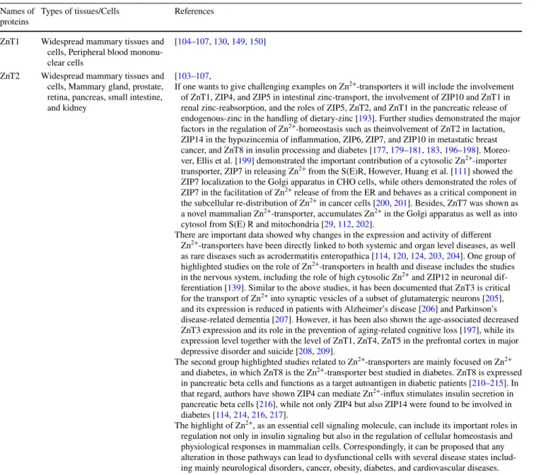
Table 1 Distribution of Zn2+-transporters in mammalian tissues/cells responsible of Zn2+-influx into cytosol (ZIPs)Names of proteins Types of tissues/Cells References
ZIP1 Widespread mammary tissues and cells [103–108]
ZIP2 Widespread mammary tissues and cells [103–105, 107, 108]
ZIP3 Widespread mammary tissues and cells, prostate glands [107–110]
ZIP4 Skin, chondrocytes, odontoblasts and fibroblast, pancreas, gastrointestinal tract, kidney, and
hip-pocampal neurons [120–123]
ZIP5 Pancreas, kidney, liver, stomach, and intestine, hepatocytes [79, 105, 153, 264] ZIP6 several cancer tissues, neuroblastoma cells, T lymphocytes, peripheral blood mononuclear cells [124–130] ZIP7 Widespread mammary tissues and cells, hepatocytes, cardiomyocytes [57, 79, 111–116] ZIP8 Widespread mammary tissues and hepatocytes, red blood cells, [79, 105, 115, 117–119]
ZIP9 Prostate, HeLa cells [131, 132]
ZIP10 Testis, kidney, breast, pancreatic α cells, red blood cells, brain, liver, erythroid, and kidney [118, 119, 133–136]
ZIP11 Testis and digestive system, glands [110, 137, 138]
ZIP12 Brain, lung, testis, and retina, neurons, endothelial, smooth muscle, and interstitial cells [110, 139, 140]
ZIP13 Bone, fat tissue, adipose tissue, hepatocytes [115, 141–143]
previously, have shown that exposure to high Zn
2+induced
marked increases in mitochondrial matrix/cristae area and
matrix volume together with an increased lysosome in
car-diomyocytes [
26
,
179
]. Together, the notable clustering and
vacuolated mitochondrion markedly disrupted and damaged
myofibrils, and electron-dense small granules were observed
in Zn
2+-exposed cardiomyocytes [
26
]. Those changes were
also including notable increases in mitochondrial matrix/
cristae area and matrix volume, together with some signs
indicating fission-fusion defects in the mitochondria, in
a manner of its concentration-dependent [
26
]. High Zn
2+exposure also caused a marked depolarization in
mitochon-drial membrane potential, as well [
28
,
29
,
48
]. Additional
studies have also shown a close association between
intra-cellular high labile Zn
2+and impairment of mitochondrial
respiration in a variety of pathological conditions in
mam-malian cells [
235
,
236
]. One can state that if intracellular
labile Zn
2+gets over its physiological level, it can stimulate
one or more deleterious changes, such as marked
altera-tions in mitochondrion morphology and function as well as
marked changes in the phosphorylation/oxidation levels of
cytosolic signaling proteins [
47
,
48
]. Moreover, it has been
demonstrated that both extra-and intracellular high-level
Zn
2+modulates L-type Ca
2+-channel properties, as well
as its regulation by β-adrenergic agonists independently of
altering the cellular redox status but associated with cellular
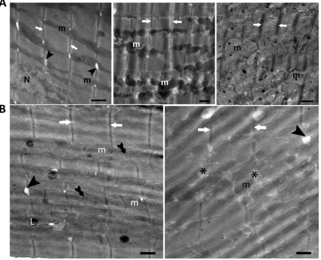
Table 2 Distribution of Zn2+-transporters in mammalian tissues/cells responsible for Zn2+-efflux of cytosol (ZnTs)Names of
proteins Types of tissues/Cells References ZnT1 Widespread mammary tissues and
cells, Peripheral blood mononu-clear cells
[104–107, 130, 149, 150] ZnT2 Widespread mammary tissues and
cells, Mammary gland, prostate, retina, pancreas, small intestine, and kidney
[103–107,
If one wants to give challenging examples on Zn2+-transporters it will include the involvement
of ZnT1, ZIP4, and ZIP5 in intestinal zinc-transport, the involvement of ZIP10 and ZnT1 in renal zinc-reabsorption, and the roles of ZIP5, ZnT2, and ZnT1 in the pancreatic release of endogenous-zinc in the handling of dietary-zinc [193]. Further studies demonstrated the major factors in the regulation of Zn2+-homeostasis such as theinvolvement of ZnT2 in lactation,
ZIP14 in the hypozincemia of inflammation, ZIP6, ZIP7, and ZIP10 in metastatic breast cancer, and ZnT8 in insulin processing and diabetes [177, 179–181, 183, 196–198]. Moreo-ver, Ellis et al. [199] demonstrated the important contribution of a cytosolic Zn2+-importer
transporter, ZIP7 in releasing Zn2+ from the S(E)R, However, Huang et al. [111] showed the
ZIP7 localization to the Golgi apparatus in CHO cells, while others demonstrated the roles of ZIP7 in the facilitation of Zn2+ release of from the ER and behaves as a critical component in
the subcellular re-distribution of Zn2+ in cancer cells [200, 201]. Besides, ZnT7 was shown as
a novel mammalian Zn2+-transporter, accumulates Zn2+ in the Golgi apparatus as well as into
cytosol from S(E) R and mitochondria [29, 112, 202].
There are important data showed why changes in the expression and activity of different Zn2+-transporters have been directly linked to both systemic and organ level diseases, as well
as rare diseases such as acrodermatitis enteropathica [114, 120, 124, 203, 204]. One group of highlighted studies on the role of Zn2+-transporters in health and disease includes the studies
in the nervous system, including the role of high cytosolic Zn2+ and ZIP12 in neuronal
dif-ferentiation [139]. Similar to the above studies, it has been documented that ZnT3 is critical for the transport of Zn2+ into synaptic vesicles of a subset of glutamatergic neurons [205],
and its expression is reduced in patients with Alzheimer’s disease [206] and Parkinson’s disease-related dementia [207]. However, it has been also shown the age-associated decreased ZnT3 expression and its role in the prevention of aging-related cognitive loss [197], while its expression level together with the level of ZnT1, ZnT4, ZnT5 in the prefrontal cortex in major depressive disorder and suicide [208, 209].
The second group highlighted studies related to Zn2+-transporters are mainly focused on Zn2+
and diabetes, in which ZnT8 is the Zn2+-transporter best studied in diabetes. ZnT8 is expressed
in pancreatic beta cells and functions as a target autoantigen in diabetic patients [210–215]. In that regard, authors have shown ZIP4 can mediate Zn2+-influx stimulates insulin secretion in
pancreatic beta cells [216], while not only ZIP4 but also ZIP14 were found to be involved in diabetes [114, 214, 216, 217].
The highlight of Zn2+, as an essential cell signaling molecule, can include its important roles in
regulation not only in insulin signaling but also in the regulation of cellular homeostasis and physiological responses in mammalian cells. Correspondingly, it can be proposed that any alteration in those pathways can lead to dysfunctional cells with several disease states includ-ing mainly neurological disorders, cancer, obesity, diabetes, and cardiovascular diseases.
ATP level [
93
]. However, in an early study by Traynelis et al.
demonstrated contradictory data demonstrating the
inhibi-tion of both L-type and T-type Ca
2+currents with high Zn
2+in neuronal cells [
242
]. Correspondingly, others had
dem-onstrated a more sensitivity of K
+-channels to high Zn
2+than those of Na
+-channels in neural cells [
243
], whereas a
recent data has been shown activation of the M-type
(includ-ing Kv7 channels) K
+-channels by high intracellular labile
Zn
2+[
244
].
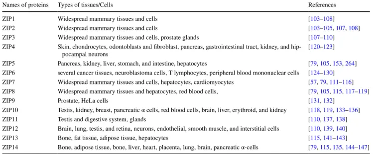
Here, we incubated ventricular cardiomyocytes with
different zinc-compounds and using light and electron
microscopy analysis, the heart tissue, and cardiomyocytes.
The electron microscopy analysis showed that incubation
of cardiomyocytes with a Zn
2+-ionophore, Zn
2+-pyrithione
(ZnPT; 0.01-μM for 1-h) induced elongation in mitochondria
leading to a significant increase in a sarcomere length, and
clear irregular cristae appearance of mitochondrion located
between myofibrils, together with electron-dense matrix
(Fig.
1A
, left). A tenfold increase in ZnPT concentration
induced marked changes in the shapes of the mitochondria
such as fragmentation, rounding, and swollen (Fig.
1A
,
middle). In incubation of the cells with the highest ZnPT
concentration (1-μM), the mitochondria appeared more
elec-tron-lucent while the loss of the matrix density (Fig.
1A
,
right). When cardiomyocytes incubated with 10-μM ZnPO
4(1-h), there was more disorganized mitochondrial cristae,
and electron-lucent matrix, and partitioned mitochondria in
the cells (Fig.
1B
, left). The cardiomyocytes incubated with
0.1 μM ZnCl
2(1-h), clustered mitochondria, slight
intrami-tochondrial edema, and enlargement of T-tubules and highly
localized lysosomes were observed (Fig.
1B
, right). In this
regard, it has been demonstrated concentration-dependent
Zn
2+inhibition of mitochondrial complex I [
236
], as well as
Zn
2+entry into mitochondria via uniporter inducing
mito-chondrial dysfunction, at most, via ROS production and
con-tributing to mitochondrial Ca
2+deregulation [
245
].
As a consequence mentioned above paragraphs, the
impaired mitochondrial function through exposure to high
Zn
2+and/or increase intracellular labile Zn
2+might lead to
several cardiovascular diseases. Therefore, one can
empha-size the importance of a well-controlled intracellular labile
Zn
2+through the mitochondria as a novel therapeutic
tar-get for cardiac complications under pathological conditions
including
oxida tive stres s
. Indeed, studies pointed out that
cardiac mitochondria, similar to SER, also play an
impor-tant role in regulating not only Ca
2+-homeostasis but also
Ca
2+-homeostasis via acting as a sponge to buffer both ions
in cardiomyocytes [
19
,
21
,
25
,
29
,
45
,
48
]. So far, it has
been shown that both elevated labile ion levels such as Zn
2+and Ca
2+in the cytosol are deleterious in cardiomyocytes,
and therefore their well-controlled levels in the cytosol are
necessary to maintain a physiologic function of the heart.
Supporting the last statement, we, recently, have shown that
mitochondria played an important role to maintain cytosolic
labile Zn
2+level though uptake high Zn
2+from cytosol
increased due to high-level release from SER in
hypergly-cemic or hypertrophic ventricular cardiomyocytes [
29
,
48
].
Therefore, one can interpret that mitochondria contribute to
cellular Zn
2+-muffling between cellular compartments under
pathological conditions via affecting S(E)R-mitochondria
coupling [
246
–
250
].
Distribution of Zn
2+‑transporters
in mitochondria of cardiomyocytes
Similar to others, there are several Zn
2+-signaling pathways
to control the intracellular Zn2+ homeostasis in
cardiomyo-cytes. Of note, the intracellular Zn
2+-signaling can easily
interfere with the Ca
2+-signaling in cardiomyocytes, under
both physiological and pathological conditions [
19
–
21
,
25
,
45
,
58
]. A piece of widespread information on cellular
regu-lation of cytosolic Zn
2+-signaling through Zn
2+-transporters,
Zn
2+-binding molecules, −fingers, and Zn
2+-sensors in
sev-eral tissues and cell types are very well documented [
96
,
111
,
190
,
193
,
199
,
200
,
251
–
255
], the distribution and
function of those carries in subcellular organelles are not
well clarified in cardiomyocytes yet.
Recently we and others have demonstrated that
Zn
2+-transporters induced developmental and
physiologi-cal defects in mammalians including cardiomyopathy in the
heart [
27
,
29
,
57
]. Following demonstrating the distribution
of labile in the cytosol, SER, and mitochondria of
cardiomyo-cytes using eCALWY probes [
50
] and the important roles of
both ZIP7 and ZnT7 to mediate ER stress in hyperglycemic
cardiomyocytes [
57
], we first demonstrated the subcellular
localizations of ZIP8, ZIP14 and ZnT8 in cardiomyocytes
besides ZIP7 and ZnT7 in cardiomyocytes [
148
]. By using
the Huygens program for co-localization values of those
trans-porters, we calculated Pearson’s coefficients (PC) for
ZIP8-SER and ZIP8-sarcolemma as 44 ± 3% and 60 ± 2%,
respec-tively. The PC values of ZIP14 were 50 ± 8% and 42 ± 3%
for SER and sarcolemma, while those PC values of ZnT8
were 66 ± 3% and 80 ± 2% for SER and sarcolemma [
148
].
Those PCs strongly supported the high-level localization
of those three Zn
2+-transporters on sarcolemma ventricular
cardiomyocytes. In the same study, authors demonstrated that
the expression levels of ZIP14 and ZnT8 were significantly
high in the human heart with serious failure, whereas ZIP8
level was significantly low than those of controls, through,
at most, increased oxidative and ER stress. Correspondingly,
we have shown that the expression levels of ZIP7, ZnT7, and
ZIP14 were decreased with no change in ZIP8 of high
carbo-hydrate diet-induced metabolic syndrome rat cardiomyocytes
[
102
]. Furthermore, in our other study, there were significant
increases in the expression levels of ZIP7, ZIP14, and ZnT8
along with decreases in the ZIP8 and ZnT7 levels in the heart
tissue from transverse aortic constriction model induced
hypertrophic young rats [
159
,
202
].
Recently, authors also studied the role and localization
of Zn
2+–transporters on mitochondria in aged ventricular
cardiomyocytes. Together with high ROS level in those
cells, the examination of the distribution of cellular labile
Zn
2+among suborganelles, such as S(E)R and
mitochon-dria parallel to cytosolic labile Zn
2+showed that the
cyto-solic was markedly high, at most, due to increased ZIP7
level with decreased ZnT7 level [
48
]. In that study, it was
for the first time demonstrated that labile Zn
2+level in
isolated mitochondria was significantly high while it was
decreased in isolated SER, supporting the hypothesis of
re-distribution of Zn
2+–transporters under the
pathologi-cal condition to buffer the intracellular labile Zn
2+level.
Fig. 1 The electron microscopy analysis of left ventricular cardio-myocytes incubated with a Zn2+-ionophore, Zn2+-pyrithione, ZnPT
(0.01-μM, 0.1-μM, or 1-μM for 1-h) (A; left, middle, right, respec-tively), with 10-μM ZnPO4 (1-h; B, left), or with 0.1 μM ZnCl2 (1-h;
B, right). Shorten symbols; m: mitochondria, arrow: Z-line, L: lyso-some, N: nucleus, tailed arrow: partitioned mitochondrion, arrow-head: T-tubule, asterisk: intramitochondrial edema. Magnification: ×12,930 and bars: 500 nm
Supporting the re-distribution of labile Zn
2+among
cyto-sol and organelles through Zn
2+–transporters, the
West-ern-blotting data demonstrated that the levels of ZnT7
and ZnT8 were increased in isolated mitochondria with
no changes in ZIP7 and ZIP8 levels [
48
]. Those changes
have positive responses to the mitochondria-targeting
anti-oxidant (MitoTEMPO) treatment of those cells, as well.
Moreover, another transporter, the ZIP14 protein level
was significantly low in isolated mitochondria from aged
ventricular cardiomyocytes with a positive response to
an application of the mitochondria targeting antioxidant
[
256
].
Correspondingly, early studies pointed out a relatively
low expressing levels of Zn
2+–transporters such as ZIP7
and ZnT7 in mammalian heart tissues [
111
,
112
,
235
]. An
interesting study by Seo et al. focused on showing the
local-ization of ZnT2 in mammary epithelial cells (HC11) and
they found that ZnT2 localized to the inner mitochondrial
membrane and acts as an auxiliary Zn
2+importer into
mito-chondria [
257
]. In a recent study, authors also have shown
the localization of ZIP1 on mitochondria and responsible
for Zn
2+–entry into mitochondria in HeLa cells [
258
].
Although limited data are demonstrating the importance of
mitochondrial labile Zn
2+and the mitochondrial
localiza-tion of Zn
2+-transporters, our and earlier studies
empha-sized the role of excess labile Zn
2+likeness to Ca
2+, in the
injury of cells, including cardiomyocytes, through excess
ROS production alone and/or together with mitochondrial
dysfunction [
26
,
28
,
234
,
259
–
261
]. However, there are
controversies about how high Zn
2+can affect mitochondria
function: Excess Zn
2+could induced increases have been
reported to induce mitochondrial Zn
2+uptake, resulting in
a longer loss of mitochondrial membrane potential in
cul-tured neurons, besides prolonged duration of ROS
produc-tion [
44
], whereas other reports demonstrated that high-level
Zn
2+did not acutely depolarize mitochondria [
262
,
263
].
Besides, a high Zn
2+could induce a clear depolarization in
mitochondrial membrane potential parallel to high ROS
pro-duction ventricular cardiomyocytes while high intracellular
Zn
2+including hyperglycemic ventricular cardiomyocytes
presented high ROS production as well as a clear
depolar-ized mitochondrial membrane potential [
28
,
29
,
57
]. All the
above studies are calling an important question whether or
not high labile Zn
2+is an effective inhibitor of mitochondrial
function under any pathological stimuli, therefore, this event
is providing an important interest to a clarification of that
question.
The already known documents showing re-distribution
of some Zn
2+-transporters localized to the mitochondria in
mammalian ventricular cardiomyocytes under pathological
conditions are summarized in Table
3
.
Conclusions
Considering the already shown data, it is acceptable to
men-tion the intracellular labile Zn
2+as a critical signaling
mole-cule in normal cell physiology as well as in
pathophysiologi-cal conditions, such as aging, diabetes, insulin resistance,
or heart failure in mammalians. As mentioned previously,
cellular Zn
2+-homeostasis is tightly controlled by different
regulatory signaling pathways including Zn
2+-transporters
alone and/or the pathways associated with Zn
2+-transporters.
In another insight, coordinated regulation of Zn
2+uptake,
efflux, distribution, and storage in cardiomyocytes is a
very important issue for a proper heart function in humans.
Although experimental data clearly show the multiple
bio-logic functions of intracellular labile Zn
2+there are yet
some controversies among them, and, therefore, none of
them are more clear than the others to provide
cardiopro-tection in pathological cardiac tissue. Overall, here, we
tried to document the prevalence of important relationships
between intracellular labile Zn
2+and Zn
2+-transporters,
particularly localized to mitochondria, under physiological
as well as under any pathological stimuli such as
hypergly-cemia, hyperinsulinemia, cardiomyopathy, heart failure, or
aging (Fig.
2
). Therefore, we first emphasized the
possibil-ity of an association between intracellular labile Zn
2+and
Zn
2+-transporters in mitochondria as therapeutic targets in
heart dysfunction. Second, we proposed the importance of
possible new therapeutic agents particularly targeting
mito-chondrial Zn
2+-transporters, potentiality control that
rela-tionship in cardiac cells.
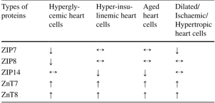
Table 3 The re-distribution of some Zn2+-transporters localized to
the mitochondria in mammalian ventricular cardiomyocytes under pathological conditions
Here, the symbols ↑, ↓, and ↔ are representing increased, decreased and unchanged protein expression levels in associated pathological conditions (re-organized from references, 29, 48, 57, 82, 148, 159, 179, 256). All measurements are performed in isolated ventricular rat cardiomyocytes. All changes are statistically significant compared to those of control cardiomyocytes (p < 0.05)
Types of
proteins Hypergly-cemic heart cells Hyper-insu-linemic heart cells Aged heart cells Dilated/ Ischaemic/ Hypertropic heart cells ZIP7 ↓ ↔ ↔ ↓ ZIP8 ↓ ↔ ↔ ↔ ZIP14 ↔ ↓ ↓ ↔ ZnT7 ↑ ↑ ↑ ↑ ZnT8 ↑ ↑ ↑ ↑
Acknowledgments Thanks to Dr. D. Billur for her electron microscopy analysis. This work was supported by grants (No. SGAB-216S979) from The Scientific and Technological Research Council of Turkey.
Compliance with ethical standards
Conflict of interest The authors declare no conflicts of interest.
References
1. Ernster L, Schatz G (1981) Mitochondria: a historical review. J Cell Biol 91:227s–255s. https ://doi.org/10.1083/jcb.91.3.227s 2. Yang D, Oyaizu Y, Oyaizu H, Olsen GJ, Woese CR (1985)
Mito-chondrial origins. Proc Natl Acad Sci U S A 82:4443–4447. https ://doi.org/10.1073/pnas.82.13.4443
3. Gray MW, Burger G, Cedergren R, Golding GB, Lemieux C, Sankoff D, Turmel M, Lang BF (1999) A genomics approach to mitochondrial evolution. Biol Bull 196:400–403. https ://doi. org/10.2307/15429 80
4. Gustafsson AB, Gottlieb RA (2008) Recycle or die: the role of autophagy in cardioprotection. J Mol Cell Cardiol 44:654–661. https ://doi.org/10.1016/j.yjmcc .2008.01.010
5. Hoppel CL, Tandler B, Fujioka H, Riva A (2009) Dynamic organization of mitochondria in human heart and in myocar-dial disease. Int J Biochem Cell Biol 41:1949–1956. https ://doi. org/10.1016/j.bioce l.2009.05.004
6. Friedman JR, Nunnari J (2014) Mitochondrial form and function. Nature 505:335–343. https ://doi.org/10.1038/natur e1298 5 7. Zhao Q, Sun Q, Zhou L, Liu K, Jiao K (2019) Complex
regula-tion of mitochondrial funcregula-tion during cardiac development. J Am Heart Assoc 8:e012731. https ://doi.org/10.1161/jaha.119.01273 1
8. Bonora M, Wieckowsk MR, Chinopoulos C, Kepp O, Kroemer G, Galluzzi L, Pinton P (2015) Molecular mechanisms of cell death: central implication of ATP synthase in mitochondrial per-meability transition. Oncogene 34:1608. https ://doi.org/10.1038/ onc.2014.462
9. Olgar Y, Tuncay E, Billur D, Durak A, Ozdemir S, Turan B (2020) Ticagrelor reverses the mitochondrial dysfunction through preventing accumulated autophagosomes-dependent apoptosis and ER stress in insulin-resistant H9c2 myocytes. Mol Cell Biochem 469:97–107. https ://doi.org/10.1007/s1101 0-020-03731 -9
10. Olgar Y, Tuncay E, Degirmenci S, Billur D, Dhingra R, Kir-shenbaum L, Turan B (2020) Ageing-associated increase in SGLT2 disrupts mitochondrial/sarcoplasmic reticulum Ca(2+) homeostasis and promotes cardiac dysfunction. J Cell Mol Med 24:8567–8578. https ://doi.org/10.1111/jcmm.15483
11. Münzel T, Camici GG, Maack C, Bonetti NR, Fuster V, Kovacic JC (2017) Impact of oxidative stress on the heart and vasculature: part 2 of a 3-part series. J Am Coll Cardiol 70:212–229. https ://doi.org/10.1016/j.jacc.2017.05.035 12. Münzel T, Gori T, Bruno RM, Taddei S (2010) Is oxidative
stress a therapeutic target in cardiovascular disease? Eur Heart J 31:2741–2748. https ://doi.org/10.1093/eurhe artj/ehq39 6 13. Afanas’ev I (2011) ROS and RNS signaling in heart disorders:
could antioxidant treatment be successful? Oxidative Med Cell Longev 2011:293769. https ://doi.org/10.1155/2011/29376 9 14. Afanas’ev I (2011) Reactive oxygen species signaling in
can-cer: comparison with aging. Aging Dis 2:219–230
15. Griendling KK, Touyz RM, Zweier JL, Dikalov S, Chilian W, Chen YR, Harrison DG, Bhatnagar A (2016) Measurement of reactive oxygen species, reactive nitrogen species, and redox-dependent signaling in the cardiovascular system: a scientific statement from the American Heart Association. Circ Res 119:e39–e75. https ://doi.org/10.1161/res.00000 00000 00011 0 16. Bray AW, Ballinger SW (2017) Mitochondrial DNA muta-tions and cardiovascular disease. Curr Opin Cardiol. https :// doi.org/10.1097/hco.00000 00000 00038 3
17. Peoples JN, Saraf A, Ghazal N, Pham TT, Kwong JQ (2019) Mitochondrial dysfunction and oxidative stress in heart dis-ease. Exp Mol Med 51:1–13. https ://doi.org/10.1038/s1227 6-019-0355-7
18. Degirmenci S, Olgar Y, Durak A, Tuncay E, Turan B (2018) Cytosolic increased labile Zn(2+) contributes to arrhythmo-genic action potentials in left ventricular cardiomyocytes through protein thiol oxidation and cellular ATP depletion. J Fig. 2 A summarized representation to demonstrate the
re-distribu-tion of intracellular labile Zn2+ levels in the cytosol ([Zn2+] i),
mito-chondria ([Zn2+]
Mit), and Sarco(endo)plasmic reticulum ([Zn2+]SER)
as well as the Zn2+–transporters in left ventricular cardiomyocytes
under any pathological stimuli (hyperglycemia, hyperinsulinemia, cardiomyopathy, heart failure, aging, etc.) (B) comparison to that of physiological condition (A). The presentation is summarized from our already published articles [29, 48, 57, 82, 148, 159, 179, 256]