Original Paper
Med Princ PractDo Statins Affect Thyroid Volume and Nodule
Size in Patients with Hyperlipidemia in a
Region with Mild-to-Moderate Iodine Deficiency?
A Prospective Study
Canan Demir
aCuneyd Anil
aYusuf Bozkus
aUmut Mousa
aAltug Kut
bAsli Nar
aNeslihan B. Tutuncu
aaDepartment of Endocrinology and Metabolism, Baskent University School of Medicine, Ankara, Turkey; bDepartment Family Medicine, Baskent University School of Medicine, Ankara, Turkey
Received: June 4, 2017 Accepted: January 7, 2018 Published online: January 23, 2018
Cuneyd Anil © 2018 The Author(s)
Significance of the Study
• This study compared the thyroid function and morphology of hyperlipidemic patients who received atorvastatin or rosuvastatin with those of a control group of non-statin-treated hyperlipidemic pa-tients. There was an association between rosuvastatin treatment and reduction in thyroid volume and maximum nodule diameter. This could be a new treatment alternative for benign and malignant pro-liferative thyroid diseases.
DOI: 10.1159/000486748
Keywords
Hyperlipidemia · HMG-CoA reductase inhibitor · Statins · Atorvastatin · Rosuvastatin · Thyroid volume · Thyroid nodule
Abstract
Objective: The objective of this study was to assess the
anti-proliferative pleiotropic effects of statins on thyroid func-tion, volume, and nodularity. Subjects and Methods: One hundred and six hyperlipidemic patients were included in this prospective study. The 69 patients in the statin groups received atorvastatin (16 received 10 mg and 18 received 20 mg) or rosuvastatin (20 received 10 mg and 15 received 20 mg). The 37 patients in the control group, assessed as not requiring drugs, made only lifestyle changes. Upon
admis-sion and after 6 months, all patients were evaluated by ultra-sonography as well as for lipid variables (total cholesterol, high- and low-density lipoprotein cholesterol, and triglycer-ides) and thyroid function and structure. Results: After 6 months, no differences in thyroid function, thyroid volume, the number of thyroid nodules, or nodule size were ob-served in the statin and control groups. In a subgroup analy-sis, total thyroid volume had decreased more in patients re-ceiving 20 mg of rosuvastatin than that in the control group (p < 0.05). Maximum nodule size had decreased more in those receiving 10 mg of rosuvastatin (p < 0.05).
Conclu-sions: Our results suggest an association between
rosuvas-tatin treatment and smaller thyroid volume and maximum nodule diameter; this could be attributable to the antiprolif-erative effects of statin therapy on the thyroid.
© 2018 The Author(s) Published by S. Karger AG, Basel
Introduction
Statins inhibit 3-hydroxy-3-methylglutaryl-coenzyme A (HMG-CoA) reductase enzyme, the rate-limiting step in cholesterol synthesis, and thus prevent cholesterol syn-thesis through mevalonate [1]. The effects of statins apart from lipid reduction are known as pleiotropic effects and are believed to be attributable to the inhibition of meval-onate and isoprenoid compound synthesis via the inhibi-tion of HMG-CoA reductase [2, 3]. The mevalonate path-way provides the bioactive molecules essential for numer-ous cellular processes. The by-products of the mevalonate pathway, farnesyl pyrophosphate and geranylgeranyl py-rophosphate, enable G proteins to be transported from within cells to the cell membranes and activated by post-translational modification.
The resultant activated proteins of the Ras super-fam-ily participate in controlling processes such as cell prolif-eration and differentiation, and the regulation of the actin cytoskeleton, membrane traffic, and nuclear transport. Many studies have shown that the mechanism responsi-ble for statin pleiotropy is the inhibition of the Ras super-family guanosine triphosphate (GTP)ases, which act as regulators in guanosine diphosphate/GTP transforma-tion, and whose membrane localizations and functions depend on posttranslational protein isoprenylation [4– 6]. When the pleiotropic effects of statins were first rec-ognized, various studies investigated their antitumor, an-tiproliferative, and apoptotic effects [7–10].
Abnormal epithelial cell proliferation, which is com-mon in thyroid tissue, may present as nodular or non-nodular thyroid growth or neoplasia [11]. However, little is known about the pleiotropic effects of statins on thy-roid cells. A possible thythy-roid-statin association was first reported in 1999, when Vitale et al. [12] showed in vitro that statins induce apoptosis in “multiplying thyroid cells” independently of p53, through protein synthesis and a mechanism based on caspase enzymes. Rat thyroid cells with Ras-transformation were found to be more sen-sitive to HMG-CoA reductase inhibitors than normal cells. Ras mutation activation and other oncogenes are common in thyroid tumors; ras mutations may also be present in nodular hyperplasia of the thyroid [13, 14]. In a 2006 study performed on rats, Laezza et al. [15] showed that HMG-CoA reductase inhibitors inhibit propylthio-uracil-induced goiter with p21 ras-MAPK (mitogen-acti-vated protein kinase) pathway modulation. Thus, statins may offer a new treatment alternative for benign and ma-lignant proliferative thyroid diseases. Only 2 retrospec-tive and no prospecretrospec-tive clinical studies on the pleiotropic
effects of statins on the thyroid have been published in English [16, 17]. In this study, the objective was to pro-spectively compare the change in thyroid function and morphology of hyperlipidemic patients receiving atorv-astatin or rosuvatorv-astatin with those of a control group of non-statin-treated hyperlipidemic patients in a region with mild-to-moderate iodine deficiency. Thyroid vol-ume and morphology are affected by iodine intake, with the incidence of goiter and nodules being higher in io-dine-deficient regions [18, 19].
Subjects and Methods
Subjects
This prospective, controlled study took place from August 2012 to March 2014 at the Department of Endocrinology and Metabo-lism, Baskent University School of Medicine, Ankara, Turkey. The study was approved by the local institutional review board and written informed consent was obtained from all participants.
One hundred and seventeen patients who presented to our hos-pital and were diagnosed with hypercholesterolemia were en-rolled. The National Cholesterol Education Program Adult Treat-ment Panel (NCEP ATP) III guidelines [20] on drug treatTreat-ment indications were used to plan the hyperlipidemia treatment. After assessment for the risk of coronary heart disease, 80 patients with indications for drug treatment and no exclusion criteria according to NCEP ATP III guidelines (despite a 4-week first-stage diet) were assigned to various “statin” groups. Exclusion criteria included previous statin treatment, a known current or previous thyroid disease, current levothyroxine replacement therapy, current or previous use of antithyroid drugs, radioactive iodine therapy, and current or previous use of drugs that may affect thyroid functions. Eleven patients were excluded from the drug intervention groups
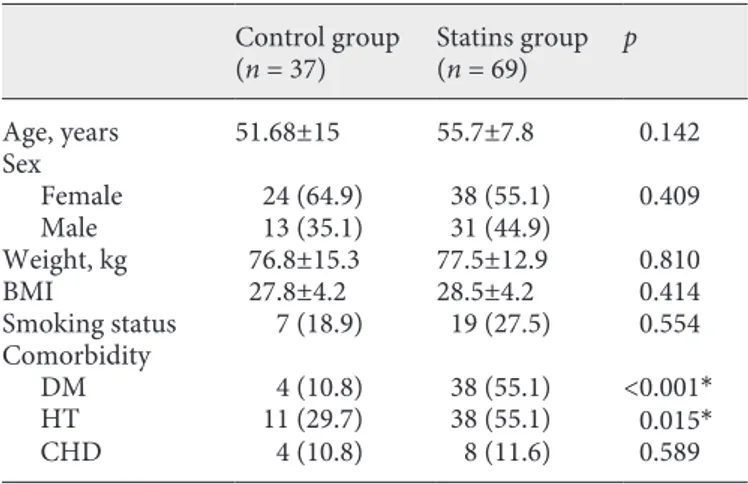
Table 1. Relevant characteristics of the study population
Control group (n = 37) Statins group(n = 69) p Age, years 51.68±15 55.7±7.8 0.142 Sex Female 24 (64.9) 38 (55.1) 0.409 Male 13 (35.1) 31 (44.9) Weight, kg 76.8±15.3 77.5±12.9 0.810 BMI 27.8±4.2 28.5±4.2 0.414 Smoking status 7 (18.9) 19 (27.5) 0.554 Comorbidity DM 4 (10.8) 38 (55.1) <0.001* HT 11 (29.7) 38 (55.1) 0.015* CHD 4 (10.8) 8 (11.6) 0.589
Values are expressed as means ± SD or n (%). DM, diabetes mellitus; HT, hypertension; CHD, coronary heart disease.
because of lack of compliance. The control group comprised 37 patients who were not given statins. The study was ultimately com-pleted with 106 patients (37 controls and 69 in the statin group). Of these 106, 62 were female (58.5%) and 44 were male (41.5%). Their mean age was 53.22 ± 10.26 years (range 25–81 years). Forty-two patients (39.6%) had type 2 diabetes (DM), 49 (46.2%) had hypertension (HT), and 12 (11.3%) had coronary heart disease. Their body mass index was 28.26 ± 4.29 and their rate of smoking was 24.5%. All patients were residing in Ankara, a region with mild-to-moderate iodine deficiency (Table 1) [21].
The 37 (34.9%) patients in the control group were prescribed lifestyle and dietary changes for their hyperlipidemia. Of the 69 patients receiving drug treatment, 20 received rosuvastatin 10 mg (19.8%),15 received rosuvastatin 20 mg (14.9%),16 received ator-vastatin 10 mg (15.8%), and 18 received atorator-vastatin 20 mg (17.8%). The mean rosuvastatin and atorvastatin dose was 14.3 and 21.7 mg, respectively.
Methods
Patients assessed as requiring statin treatment were assigned by a simple random sampling method to receive either atorvastatin (10–20 mg daily) or rosuvastatin (10–20 mg daily). The study groups were as follows: group 1 (assessed as not requiring statin treatment), were advised to make therapeutic lifestyle changes (n = 37), group 2 received rosuvastatin (n = 35: 20 received 10 mg [R10] and 15 received 20 mg [R20]), and group 3 received atorv-astatin (n = 34: 16 received 10 mg [A10] and 18 received 20 mg [A20]). Patients receiving statins were also advised about thera-peutic lifestyle changes [20].
The patients in the control group were scheduled for follow-up at 0 and 6 months and those in the statin groups at 0, 1, and 6 months. Serum concentrations of low-density lipoprotein choles-terol (LDL-c), high-density lipoprotein cholescholes-terol (HDL-c), tri-glycerides (TG), aspartate aminotransferase, alanine aminotrans-ferase, gamma glutamyl transaminotrans-ferase, creatine kinase, and thyrotro-pin-stimulating hormone (TSH) were measured at admission and at 6 months. Concentrations of hepatic enzymes and creatine ki-nase were measured in the first month of treatment in patients receiving statins. Total cholesterol, HDL-c, and TG concentrations were measured using enzymatic assay (Boehringer, Mannheim, Germany). LDL-c was calculated using the Friedewald formula: LDL-c = total cholesterol − (HDL-c + TG/5). Thyroid function was evaluated by measuring the relevant variables by immunoche-moluminescence assays with an automated analyzer (Immulite 2000; Diagnostic Products, Los Angeles, CA, USA).
Thyroid ultrasonography was performed on all patients at ad-mission and at 6 months by the same researcher (C.D.) using a 10-MHz linear probe (Logic 5 Pro; GE Medical Systems, Madison, WI, USA). The size of the thyroid gland and each nodule identified was measured in 3 dimensions. The volume of the thyroid gland was calculated by using the following ellipsoid formula:
Volume (mL) = Depth (cm) × Width (cm) × Length (cm) × 0.479 [22].
Statistical Analysis
Statistical analyses were performed by using SPSS for Windows v21.0. Mean values in independent groups were compared by us-ing an independent-samples t test and means of nonparametric data with the Mann-Whitney U test. Rates of groups were com-pared by using the χ2 test. Mean values in dependent groups were
compared by using a paired-samples t test and means of nonpara-metric data of dependent groups with the Wilcoxon test. p < 0.05 was considered statistically significant.
Results
The statin treatment groups did not differ significant-ly from each other or the control group with respect to sex, age, weight, BMI, smoking status, or the presence of coronary heart disease. As expected, a statistically signifi-cant greater proportion of patients had DM and HT in the drug treatment groups than in the control group.
In addition to statin treatment, the routine use of 3 other groups of drugs for longer than 6 months was also assessed: (a) drugs for DM (oral antidiabetic drugs in-cluding metformin, sulfonylurea, glinide, acarbose, and gliptins as well as insulin and combination therapies); (b) drugs for HT (angiotensin-converting enzyme inhibitors, angiotensin receptor blockers, β blockers, calcium-chan-nel blockers, and diuretics, or combinations thereof); and (c) acetylsalicylic acid, vitamin D, and omega-3. While all these drugs were used significantly less frequently in the control group than in the drug treatment groups, the drug subgroups did not differ in this respect.
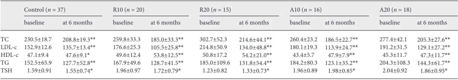
Table 2. Mean lipid concentrations before and after intervention in all groups
Control (n = 37) R10 (n = 20) R20 (n = 15) A10 (n = 16) A20 (n = 18)
baseline at 6 months baseline at 6 months baseline at 6 months baseline at 6 months baseline at 6 months TC 230.5±18.7 208.8±19.3** 259.8±33.3 185.0±33.3** 302.7±52.3 214.6±44.1** 260.4±23.2 186.5±22.7** 277.4±42.1 205.3±27.6** LDL-c 152.9±12.6 135.7±13.4** 176.6±25.3 105.5±25.8** 214.8±50.9 134.0±48.8** 180.1±19.3 113.9±24.7** 191.2±31.5 129.1±27.2** HDL-c 47.1±9.4 47.6±9.1* 49.6±12.4 53.8±12.5** 50.8±17.2 54.2±21.0** 43.4±5.7 47.9±7.9** 45.3±11.7 47.3±11.7** TG 152.5±65.9 127.7±52.8** 167.9±49.6 128.7±41.5** 185.0±109.6 131.8±54.4** 184.2±80.3 123.1±35.2** 204.3±108.3 144.3±61.7** TSH 1.59±0.91 1.55±0.74* 1.96±0.97 1.72±0.79* 1.23±0.82 1.33±0.73* 1.96±0.89 1.98±0.85* 2.04±0.92 1.86±0.95*
Values are expressed as means ± SD. R10, rosuvastatin 10 mg; R20, rosuvastatin 20 mg; A10, atorvastatin 10 mg; A20, atorvastatin 20 mg; TC, total cholesterol; LDL-c, low-density lipoprotein cholesterol; HDL-c, high density lipoprotein cholesterol; TG, triglycerides; TSH, thyrotropin-stimulating hormone. * p < 0.05; ** p < 0.05.
Following treatment, there were statistically signifi-cant decreases from the levels at baseline of total choles-terol, LDL-c, and TG, in both the control and treatment groups (p < 0.01). An apparent increase from baseline lev-els in HDL concentrations was not statistically significant in the control group at the end of the study; however, these increases were statistically significant in the drug treatment groups (p < 0.05). No statistically significant changes were observed between pre- and posttreatment TSH concentrations in any group (Table 2).
Upon entry to the study, the mean total thyroid vol-ume of all 106 patients was 12.46 ± 5.21 mL, and at 6 months it was 12.39 ± 5.11 mL (p > 0.05), a nonsignificant change. Although there were no statistically significant changes in total thyroid volume at 6 months in groups 1, 2 and 3, a significant reduction was seen in the R20 sub-group of sub-group 2 after treatment (p = 0.034; Table 3).
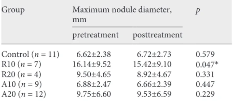
Nodules were detected in 43 (40.56 %) patients; 21 had single nodules and 22 had multiple nodules. The mean
maximum nodule diameter upon entry was 6.62 ± 2.38 mm in the control group and 10.31 ± 2.93 mm in the drug treatment groups. There was no change in the number of nodules at 6 months.
The posttreatment maximum nodule diameter was 6.72 ± 2.73 mm (a mean increase of 0.10 mm) in the con-trol group and 9.98 ± 6.76 (a mean decrease of 0.33 mm) in the drug group; these changes were not statistically sig-nificant. However, compared with the values at admis-sion, a statistically significant decrease in maximum nod-ule diameter was observed in the R10 group (n = 18) at 6 months. Seven of these 18 patients had nodules with a mean diameter of 16.14 ± 9.5 mm at admission. Although the number of nodules did not change after treatment, the mean nodule diameter decreased to 15.42 ± 9.1 mm (p = 0.047; Table 4).
Discussion
This study found no statistically significant differences in TSH concentrations, thyroid volume, the number of nodules, or maximum nodule diameters between patients who did and did not receive statin treatment.
At six months of treatment, there was no difference in the patients receiving the 2 types of statins. However, a comparison of the subgroups with the corresponding val-ues in the control group revealed that total thyroid vol-ume was significantly smaller in the R20 group (p < 0.05), and maximum nodule diameter significantly smaller in the R10 group (p < 0.05). The lack of reduction in the maximum nodule diameter in the R20 group could have been due to the smaller number of patients with nodules in this group (R10 n = 7 vs. R20 n = 4) or to the relatively short duration of the study or both. The effects on thyroid volume after 6 months of treatment with 20 mg rosuvas-tatin are in agreement with data obtained from retrospec-tive patient surveys and in vitro and in vivo animal stud-ies, and they may be related to the antiproliferative and apoptotic effects of prenylation inhibition on the meval-onate pathway [11, 12, 16, 17]. The number of nodules did not change with treatment.
First discovered in 1976 and in continuous use since 1981, statins have pleiotropic effects that have been known and studied extensively for 15 years [23]. Most of these effects are associated with the inhibition of the mev-alonate pathway [3, 4].
Vitale et al. [12] showed in vitro that statins induce apoptosis by multiplying thyroid cells independently of p53 through protein synthesis and a mechanism based on
Table 3. Thyroid volume before and after intervention
Group Thyroid volume, mL p
pretreatment posttreatment Control (n = 37) 12.4±4.4 12.4±4.46 0.97 R10 (n = 20) 13.2±8.7 13.1±8.5 0.16 R20 (n = 15) 12.0±4.3 11.5±4.0 0.03* A10 (n = 16) 11.7±4.1 11.5±4.2 0.18 A20 (n = 18) 12.4±3.4 12.2±3.3 0.07
Values are expressed as means ± SD. R10, rosuvastatin 10 mg; R20, rosuvastatin 20 mg; A10, atorvastatin 10 mg; A20, atorvastatin 20 mg. * p < 0.05.
Table 4. Mean maximum nodule diameter before and after
inter-vention
Group Maximum nodule diameter,
mm p pretreatment posttreatment Control (n = 11) 6.62±2.38 6.72±2.73 0.579 R10 (n = 7) 16.14±9.52 15.42±9.10 0.047* R20 (n = 4) 9.50±4.65 8.92±4.67 0.331 A10 (n = 9) 6.88±2.47 6.66±2.39 0.447 A20 (n = 12) 9.75±6.60 9.53±6.59 0.229
Values are expressed as means ± SD. R10, rosuvastatin 10 mg; R20, rosuvastatin 20 mg; A10, atorvastatin 10 mg; A20, atorvastatin 20 mg. * p < 0.05.
caspase enzymes. Apoptosis, a process of the self-destruc-tion of cells, may be physiological or pathological. An apoptotic pathway may be initiated by various cytokines, growth factors, hormone deficiencies, or drugs, via trig-gering the apoptotic system threshold. Various genes are associated with the regulation of apoptosis. The p53 gene controls the RNA transcription of pro- and antiapoptotic genes and plays a key role in cell death [13, 24].
Lovastatin, an HMG-CoA reductase inhibitor, has been shown to induce apoptosis in malignant glial tumors and prostate stromal tumors [25, 26]. Lovastatin inhibits the reduction of HMG-CoA reductase to mevalonate, the precursor of isopentenyl pyrophosphate. It also prevents the formation of mevalonate, geranyl pyrophosphate, farnesyl pyrophosphate, and all-trans-geranylgeranyl py-rophosphate, thus preventing these molecules from pro-liferating with farnesyl and geranylgeranyl transferase, and also preventing the transfer of the Ras super-family (Rho, Raf, Rac, and Rap), which plays a role in major cel-lular functions like cell adhesion or motility, to small GTP-binding proteins. It was observed that in cases of prenyl modification deficiency, small GTP-binding pro-teins fail to form a complex with target propro-teins, and this disruption in their functions induces apoptosis.
To investigate the apoptotic effects of HMG-CoA re-ductase inhibition on thyroid cell cultures, Vitale et al. [12] studied the effects of lovastatin on normal and neo-plastic thyroid cells, and observed cellular rounding in 24 h and DNA fragmentation and apoptosis in 48 h, in both normal and neoplastic cadaver thyroid cell series. When mevalonate and the protein synthesis inhibitor, cyclohex-imide, were added, all effects of lovastatin were blocked in a dose-dependent manner. Their finding that lovas-tatin is a potent inducer of apoptosis in multiplying thy-roid cell cultures supports the contention that prenyl-ation inhibitors may have therapeutic potential, not only in prostate cancers and prostatic hyperplasia, but also in the proliferative processes of the thyroid.
In another study, rat thyroid cells with Ras-transfor-mation were found to be more sensitive to HMG-CoA reductase inhibitors than normal cells. Ras mutation ac-tivation and other oncogenes are common in thyroid tu-mors, and ras mutations may also be present in the nodu-lar hyperplasia of the thyroid. This suggests that statins have a potential use for treatment of benign and malig-nant diseases of the thyroid [12, 14, 24].
Zhong et al. [24] studied the effects of lovastatin on apoptosis and the induction of differentiation in ana-plastic thyroid carcinoma (ATC) cells refractory to con-ventional treatments. As a result, a high dose (50 μM) of
lovastatin was found to induce apoptosis, while smaller doses (25 μM) were seen to trigger cytomorphological differentiation. An increase in the expression of thyro-globulin was also detected in the ATC cells. These results supported the use of HMG-CoA reductase inhibitors for redifferentiation in the treatment of ATC. In a more re-cent study [27], this group demonstrated that lovastatin at lower concentrations exerted an antiproliferation ef-fect on human ATC cells through increases of p27 medi-ated by the reduction of Rho geranylgeranylation. These results suggested that lovastatin increases the levels of p27 protein through actions on RhoA/ROCK signaling and ubiquitination pathways, and inhibited proliferation of ARO cells by increasing the level of p27 protein. They concluded that lovastatin exerts both antiproliferative and differentiation-inducing effects on human ATC cells, probably rendering them more sensitive to radioactive io-dine therapy.
A preclinical study indicated that epidermal growth factor (EGF) is involved in the proliferation and migra-tion of differentiated thyroid cancer [28]. Overexpression of EGF or EGF receptor (EGFR) was observed in most of the thyroid cancer cells including the ATC cells, and was associated with a poor prognosis in patients with meta-static thyroid cancer. EGFR was therefore identified as a novel therapeutic target for treating patients with ATC [29, 30].
Several studies have demonstrated that lovastatin and simvastatin can inhibit cell adhesion, migration, and in-vasion [31, 32]. Recently, it has been shown that EGF can induce Cyr61 expression through the activation of the EGFR/ERK/CREB signaling pathway, and that EGF-in-duced Cyr61 is involved in the regulation of cell migra-tion in ATC cells [33]. Combined treatment with lovas-tatin and troglitazone markedly inhibited cell migration by inhibiting EGF-induced Cyr61 expression in ATC cells. The authors reported that these results may provide a useful future strategy in the treatment of ATC [34].
In a 2006 study conducted on rats, Laezza et al. [15] reported that HMG-CoA reductase inhibitors inhibited propylthiouracyl-induced goiter by p21 ras-MAPK path-way modulation. Even though different molecular and genetic processes have been proposed, results show that lovastatin is an effective inhibitor in goitrogenesis with p21 ras farnesylation inhibition. The authors concluded that mevalonate pathway inhibition may create antipro-liferative effects in both benign and malignant thyroid tissue.
The first clinical study on thyroid volume and nodule prevalence in dyslipidemia patients on statins was
con-ducted in 2008 by Cappelli et al. [16]. They retrospective-ly assessed 137 patients who had been using statins for at least 5 years, and found a reduction in thyroid volume, and the prevalence, number, and volume of nodules with HMG-CoA reductase inhibitor treatment. Nodule preva-lence among patients who had been receiving various statin treatments in the previous 5 years was 36.3% where-as the rate in a nontreatment control group wwhere-as 67.9% (p < 0.001). Thyroid and nodule volumes were also sig-nificantly smaller in the group that had received statins for a long time.
In another retrospective study on 70 DM subjects who were on statins for long periods and lived in a region with iodine deficiency, no difference in thyroid size, or nodule prevalence, number, and volume was found between the drug and control groups (n = 98). However, when pa-tients were grouped into age brackets, thyroid nodules were less prevalent in patients aged 60–65 years (n = 12) than in control patients in the same age bracket (n = 27) (33 vs. 70%, p = 0.04). These findings are thought to be associated with the possible antiproliferative effects of statins, an association believed to be weak in regions with iodine deficiency [17].
In vitro and in vivo studies on breast cancer and mela-noma suggest that only hydrophobic statins have useful pleiotropic effects [35, 36]. However, Cappelli et al. [16], in a retrospective study on 137 patients on statins (104 on simvastatin [hydrophobic], 29 on atorvastatin [hydro-philic], 1 on pravastatin, and 1 on rosuvastatin), did not detect a relationship between the number and volume of thyroid nodules and molecule type. While the duration of statin use is reportedly a stronger predictor than the daily dose for a reduction in the risk of developing prostate cancer, Shannon et al. [37] concluded that nodules are less prevalent among patients taking a greater mean daily dose, independent of duration.
In our study, hydrophilic rosuvastatin (20 mg) had a significant effect on total thyroid volume. The maximum reduction in nodule diameter was observed in the R10 group. The lack of a reduction in maximum nodule diam-eter in patients in the R20 group may be attributable ei-ther to the small number of patients with nodules (n = 4) in this group, or to the absence of a relationship between the pleiotropic effects of statins on the thyroid gland and the dosage.
Very few published studies have focused on the rela-tionship between the pleiotropic effects of statins and common thyroid diseases. Existing studies are in vitro studies, animal studies, case observations, or retrospec-tive studies. We obtained results in 2 of our study groups
that suggest that the antiproliferative effects of statins can result in smaller thyroid volumes and nodule diameters. Statins may thus have beneficial therapeutic effects in thyroid diseases. Our study was limited by its short dura-tion and small case numbers.
Future monitoring studies with larger groups of pa-tients and of longer duration are needed to fully under-stand the molecular mechanisms of the action of prenyl-ation inhibitors on antiproliferative and apoptotic path-ways, and to determine the appropriate and usable molecules and daily dosages. Our findings support the possible use of HMG-CoA reductase inhibitors in the treatment of benign and malignant diseases of the thy-roid.
Conclusion
We compared the effects of 2 statins on thyroid func-tion, volume, and nodule prevalence in a prospective, controlled setting. Posttreatment, we found a significant decrease in total thyroid volume in the R20 group and maximum nodule diameter in the R10 group compared with the changes in the control group. Rosuvastatin may exert antiproliferative effects on cells by inhibiting the mevalonate synthesis pathway and decreasing isoprenoid compound formation.
Acknowledgement
This study was supported by the Research Fund of the Baskent University (project No. KA12/02).
Disclosure Statement
The authors declare no conflicts of interest.
References 1 Takemoto M, Liao JK: Pleiotropic effects of 3-hydroxy-3-methylglutaryl coenzyme A red inhibitors. Arterioscler Thromb Vasc Biol 2001;21:1712–1719.
2 Liao JK, Laufs U: Pleiotropic effects of statins. Annu Rev Pharmacol Toxicol 2005;45:89– 118.
3 Vaughan CJ, Murphy MB, Buckley BM: Statins do more than just lower cholesterol. Lancet 1996;348:1079–1082.
4 Buhaescu I, Izzedine H: Mevalonate pathway: a review of clinical and therapeutical implica-tions. Clin Biochem 2007;40:575–584.
5 Goldstein JL, Brown MS: Regulation of the mevalonate pathway. Nature 1990;343:425– 430.
6 Bonetti PO, Lerman LO, Napoli C, et al: Statin effects beyond lipid lowering – are they clini-cally relevant? Eur Heart J 2003;24:225–248. 7 Dulak J, Józkowicz A: Anti-angiogenic and
anti-inflammatory effects of statins: relevance to anti-cancer therapy. Curr Cancer Drug Targets 2005;5:579–594.
8 Vallianou NG, Kostantinou A, Kougias M, et al: Statins and cancer. Anticancer Agents Med Chem 2014;14:706–712.
9 Alexandre L, Clark AB, Bhutta HY, et al: Statin use is associated with reduced risk of histologic subtypes of esophageal cancer: a nested case-control analysis. Gastroenterolo-gy 2014;146:661–668.
10 Farina HG, Bublik D, Alonso DF, et al: Lovas-tatin alters cytoskeleton and inhibits experi-mental metastasis of mammary carcinoma cells. Clin Exp Metastasis 2002;19:551–559. 11 Bifulco M: Therapeutic potential of statins in
thyroid proliferative disease. Nat Clin Pract Endocrinol Metab 2008;4:242–243.
12 Vitale M, Di Matola T, Rossi G, et al: Prenyl-transferase inhibitors induce apoptosis in proliferating thyroid cells through a p53-in-dependent, CrmA-sensitive, and caspase-3- like protease-dependent mechanism. Endo-crinology 1999;140:698–704.
13 Demierre MF, Higgins PD, Gruber SB, et al: Statins and cancer prevention. Nat Rev Can-cer 2005;5:930–945.
14 Hofman A, John P, Schaffarich MP, et al: Statins as a new therapeutic approach in de-differentiated thyroid cancer? Nuklear Med-izin 2006;45:28–30.
15 Laezza C, Mazziotti G, Fiorentino L, et al: HMG-CoA reductase inhibitors inhibit rat propylthiouracil-induced goiter by modulat-ing the ras-MAPK pathway. J Mol Med 2006; 84:967–973.
16 Cappelli C, Castellano M, Pirola I, et al: Re-duced thyroid volume and nodularity in dys-lipidaemic patients on statin treatment. Clin Endocrinol 2008;68:16–21.
17 Chon MG, Suk JH, Oh KH, et al: Influence of long-term statin use in type 2 diabetic patients on thyroid nodularity in iodine-sufficient area. Exp Clin Endocrinol Diabetes 2011;119: 497–501.
18 Aghini-Lombardi F, Antonageli L, Martino E, et al: The spectrum of thyroid disorders in an iodine-deficient community: the Pescopaga-no survey. J Clin EndocriPescopaga-nol Metab 1999;84: 561–566.
19 Andersen-Ranberg K, Jetine B, Hoier-Mad-sen M, et al: Thyroid function, morphology and prevalence of thyroid disease in a popula-tion based study of Danish centenarians. J Am Geriatr Soc 1998;47:1238–1243.
20 Executive summary of the third report of the National Cholesterol Education Program (NCEP) Expert Panel on Detection, Evalua-tion, and Treatment of High Blood Choles-terol in Adults (Adult Treatment Panel III). JAMA 2001;285:2486–2497.
21 Erdoğan MF, Ağbaht K, Altınsu T, et al: Cur-rent iodine status in Turkey. J Endocrinol In-vest 2009;32:617–622.
22 Brunn J, Block U, Ruf G, et al: Volumetric analysis of thyroid lobes by real-time ultra-sound. Dtsch Med Wochenschr 1981;106: 1338–1340.
23 Endo A, Kuroda M, Tanzawa K: Competitive inhibition of 3-hydroxy-3-methylglutaryl co-enzyme A reductase by 236A and ML-236B fungal metabolites, having hypocholes-terolemic activity. FEBS Lett 1976;72:323– 326.
24 Zhong W, Wang C, Chang T, Lee W: Lovas-tatin induces apoptosis of anaplastic thyroid cancer cells via inhibitor of protein geranyl-geranylation and de novo protein synthesis. Endocrinology 2003;144:3852–3859. 25 Jones KD, Couldwell WT, Hinton DR, et al:
Lovastatin induces growth inhibition and apoptosis in human malignant glioma cells. Biochem Biophy Res Commun 1994;205: 1681–1687.
26 Padayatty SJ, Marcelli M, Shao TC, et al: Lov-astatin induced apoptosis in prostate stromal cells. J Clin Endocrinol Metab 1997;82:1434– 1439.
27 Zhong WB, Hsu SP, Ho PY, et al: Lovastatin inhibits proliferation of anaplastic thyroid cancer cells regulation of p27 by interfering with the Rho/ROCK-mediated pathway. Bio-chem Pharmacol 2011;82:1663–1672.
28 Hoelting T, Siperstein AE, Clark OH, et al: Epidermal growth factor enhances prolifera-tion, migraprolifera-tion, and invasion of follicular and papillary thyroid cancer in vitro and in vivo. Clin Endocrinol Metab 1994;79:401–408. 29 van der Laan BF, Freeman JL, Asa SL:
Expres-sion of growth factors and growth factor re-ceptors in normal and tumorous human thy-roid tissues. Thythy-roid 1995;5:67–73. 30 Ensinger C, Spizzo G, Moser P, et al:
Epider-mal growth factor receptor as a novel thera-peutic target in anaplastic thyroid carcino-mas. Ann NY Acad Sci 2004;1030:69–77. 31 Zhong WB, Liang YC, Wang CY, et al:
Lovas-tatin suppresses invasiveness of anaplastic thyroid cancer cells by inhibiting Rho gera-nylgeranylation and RhoA/ROCK signaling. Endocr Relat Cancer 2005;12:615–629. 32 Xiao Y, Liang L, Pan Y, et al: Inhibitory effects
of simvastatin on migration and invasion of rheumatoid fibroblast-like synoviocytes by preventing geranylgeranylation of RhoA. Rheumatol Int 2013;33:389–399.
33 Chin LH, Hsu SP, Zhong WB, et al: Involve-ment of cysteine-rich protein 61 in the epider-mal growth factor-induced migration of hu-man anaplastic thyroid cancer cells. Mol Car-cinog 2016;55:622–632.
34 Chin LH, Hsu SP, Zhong WB, et al: Combined treatment with troglitazone and lovastatin in-hibited epidermal growth factor-induced mi-gration through the downregulation of cyste-ine-rich protein 61 in human anaplastic thy-roid cancer cells. PLoS One 2015;10:e0118. 35 Farina HG, Bublik DR, Alonso DF, et al:
Lov-astatin alters cytoskeleton and inhibits exper-imental metastasis of mammary carcinoma cells. Clin Exp Metastasis 2002;19:551–559. 36 He L, Mo H, Hadisusilo S, et al: Isoprenoids
suppress the growth of murine B16 melano-mas in vitro and in vivo. J Nutr 1997;127:668– 674.
37 Shannon J, Tewoderos S, Garzotto M, et al: Statins and prostate cancer risk: a case-con-trol study. Am J Epidemiol 2005;162:318– 325.