栽藻造押8629原愿圆圆源缘员苑圆 8629-82210956 耘皂葬蚤造押ijopress岳员远猿援糟燥皂
corneal biomechanical analysis of unilateral
keratoconus
窑Clinical Research窑
1Department of Ophthalmology, Bulent Ecevit University, Kozlu 67600, Zonguldak, Turkey
2Department of Ophthalmology, Gazi University, Ankara 06500, Turkey
3Department of Ophthalmology, Medipol University, Istanbul 34214, Turkey
4Ulucanlar Eye Research and Education Hospital, Ankara 06340, Turkey
5Kudret Eye Hospital, Ankara 06700, Turkey
Correspondence to: Orhan Ayar. Department of Ophthalmology, Bulent Ecevit University, Kozlu 67600, Zonguldak, Turkey. [email protected]
Received: 2014-04-25 Accepted: 2015-03-17
Abstract
·
AIM: To evaluate and compare corneal biomechanical findings measured by ocular response analyzer, topographic and pachymetric findings in patients with unilateral keratoconus patients and healthy controls.·
METHODS: This is an observational, case -control study. Patients with keratoconus in one eye and forme fruste keratoconus in the fellow eye were compared with sex and age matched with controls healthy subjects. All subjects were evaluated with rotating scheimpflug imaging system. The receiver -operating -characteristic curves were analyzed to evaluate the sensitivity and specificity of the parameters.·
RESULTS: Twenty-seven patients with keratoconus in one eye and forme fruste keratoconus in the fellow eye were compared with 40 eyes of 40 normal subjects. Corneal hysteresis (CH) was 8.0 依1.7 mm Hg in keratoconus group, 8.3 依1.6 mm Hg in forme fruste keratoconus group, and 9.8 依1.6 mm Hg in control groups ( =0.54 between keratoconus and forme fruste keratoconus groups, <0.01 between control group and other groups). Corneal resistance factor (CRF) was 7.1 依2.2 mm Hg in keratoconus group, 7.8 依1.2 mm Hg in forme fruste keratoconus group and 9.9依1.5 mm Hg in control group ( <0.001 between control group and other groups). Using receiver-operating-characteristic analysis, the area under curve values of the parameters to distinguish forme fruste keratoconus from control subjects were: CH(0.768), CRF (0.866). Best cut-off points were 9.3 mm Hg and 8.8 mm Hg for CH and CRF respectively.
·
CONCLUSION: Ocular response analyzer parameters (CH and CRF) are found to be significantly lower in forme fruste keratoconus patients compared to normal control subjects.·
KEYWORDS: forme fruste keratoconus; keratoconus; ocular response analyzerDOI:10.3980/j.issn.2222-3959.2015.06.11
Ayar O, Ozmen MC, Muftuoglu O, Akdemir MO, Koc M, Ozulken K. corneal biomechanical analysis of unilateral keratoconus.
2015;8(6):1141-1145
INTRODUCTION
K
eratoconus is an ectatic disorder characterized with corneal thinning, protrusion and conical corneal shape. Although it is a bilateral disease with strong genetic component, it may be significantly asymmetric with no or very subtle changes that is called as ''unilateral keratoconus''[1-3]. It is believed that the disease will likely occur in the fellow eyes of patients with unilateral keratoconus if observed for a sufficient follow up time [4-9]. "Forme fruste keratoconus (FFKCN)" or "subclinical keratoconus" terms are used for eyes that show subtle topographic changes below the threshold of keratoconus suspicion, thus rendering topographic parameters useless in differentiating FFKCN from healthy eyes [1,7,8]. However, studies showed that keratoconic corneas and even FFKCN are biomechanically compromised compared to normal corneas, as measured with the ocular response analyzer (ORA)[9-12].Long-term results found that laser keratomileusis (LASIK) is safe and effective for the treatment of refractive errors[13,14]. However, post-LASIK corneal ectasia remains one of the rare but potentially sight threatening complication of excimer laser refractive surgery [15,16]. Corneal refractive surgery should not be attempted on FFKCN patients or on apparently normal fellow eyes of unilateral keratoconus patients because of high risk of developing corneal ectasia after surgery[3,5,9].
Despite preoperative screening with corneal topography, tomography, or recently developed new indices, post-LASIK ectasia is still possible, even in the absence of FFKCN[17-19].
Therefore assessment of corneal biomechanical data aside from the topographical or pachymetric data might help identify those eyes that have a high risk of developing FFKCN.
In this study we compared corneal biomechanical parameters of unilateral keratoconus patients with FFKCN in the fellow eyes and healthy control subjects to determine the ability of biomechanical parameters in differentiating the very early stages of keratoconus that may help to reduce the risk of ectasia after corneal refractive surgery.
SUBJECTS AND METHODS
This retrospective case control study adhered to the tenets of the Declaration of Helsinki. Patients examined at Kudret Eye Hospital from October 2009 to September 2012 were retrospectively enrolled. Along with detailed ocular examination, keratoconus was defined with clinical examination with one or more of the following clinical signs of keratoconus: Munson's sign, Rizutti's sign, slit lamp examination ( stromal thinning, Fleischer ring, Vogt striae), and topographic findings [ asymmetric bow tie pattern with or without skewed radial axis (SRAX), central elevation on the posterior surface of cornea, inferior steepening of the anterior surface of the cornea, abnormal Belin/Ambrossi佼 Enhanced Ectasia criteria and percentage thickness increase (PTI)].
Eyes were divided into 3 groups. Patients diagnosed with keratoconus in one eye (group 1) with no slit lamp or topography findings of keratoconus (FFKCN) in the fellow eyes (group 2) were enrolled retrospectively in the study group. Control group (group 3) was selected from data of consecutive candidates for excimer laser refractive surgery [<6.0 diopters (D) of sphere, <3.0 D of cylinder]. Only one eye of each consecutively numbered case in control group were evaluated (right eye for odd numbers, left eye for even numbers). Patients who had previous corneal surgery, ocular trauma, corneal scarring and any concurrent ocular disease that may potentially affect the outcomes were excluded. The patients were asked to discontinue contact lens wear 72h before the examination. Each patient received a detailed assessment for keratoconus or refractive surgery including medical history assessment, refraction, complete ocular examination, topography, Pentacam (Oculus Optikger覿te GmbH, Wetzlar, Germany) tomography, pachymetry and ORA measurements.
All of the measurements were taken by an experienced technician. While taking scheimpflug tomography measurement the patient was seated comfortably and properly positioned. The eyelids had to be open without any help. After a few blinking the patients were asked to open and look through the fixation target. Then scheimpflug camera captures the images and only the best-quality images were included in the study.
Similarly, ORA measurements were taken whilst the patient was seated comfortably and properly positioned at the forehead strap. After good alignment of the eye and probe, a good quality measurement with well-defined readings was included in the study.
ORA device records bidirectional applanation pressure measurements using adjusted air pressure and a reflected infrared signal. One of the measurements is recorded while the air pressure is moving the cornea inwards passing the applanation point, and the other as the cornea returns to its normal shape. Two different pressures are obtained from this inward and outward movement of cornea. The difference between these two pressures is corneal hysteresis (CH). CH is thought to be a result of viscous damping of cornea. Corneal resistance factor (CRF) is considered to be an indicator of the overall resistance of the cornea[11,12,20-22]. Steep, flat and average central keratometry, corneal astigmatism, corneal thickness at the apex (CTap) and at the thinnest point (CTth), maps including tangential, sagittal and Belin-Ambrossi佼 criteria were obtained in each measurement. The inferior-superior asymmetry (I-S) of sagittal and tangential maps were calculated by subtracting the mean corneal curvature of superior 3 data points (60 degrees, 90 degrees, 120 degrees) from the mean value of inferior 3 data points (240 degrees, 270 degrees, 300 degrees). The steepest point of sagittal and tangential maps, the maximum value of posterior elevation map of the back corneal surface, the maximum value of difference map taken from Belin-Ambrossi佼 enhanced ectasia display of Pentacam device, corneal volume was recorded. Ten variables (CH, CRF, peak1, peak2, P1, Pm, P2, TP1, Tpm, TP2) of ORA measurements were evaluated.
The data was normally distributed ( >0.05, analyzed with Shapiro-Wilk test). Parametric, independent samples test was used to compare each parameter between groups. Receiver operating characteristic (ROC) curves were used to determine the predictive accuracy of the test parameters as described by the area under the curve (AUC) and to calculate sensitivity and specificity of these parameters. value less than 0.05 was considered statistically significant.
We certify that all applicable institutional and governmental regulations concerning the ethical use of human volunteers were followed during this research.
RESULTS
Mean patient age was 26.5依7.2y (range 17-37y) in unilateral keratoconus patients and 28.6依5.1y (range 19-41y) in the control group ( >0.05).There were 16 males and 11 females in unilateral keratoconus group and 20 males and 20 females were in the control group ( >0.05). CH and CRF values of male (9.06 and 9.26 respectively) and female (9.17 and 8.63 respectively) subjects are similar ( >0.05).The demographic, mean keratometry, topographic, pachymetry and
栽藻造押8629原愿圆圆源缘员苑圆 8629-82210956 耘皂葬蚤造押ijopress岳员远猿援糟燥皂
biomechanical data for eyes with clinical keratoconus (group 1), FFKCN eyes (group 2), and control group (group 3) are given in Table 1.
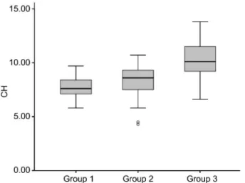
The mean K readings were statistically significantly higher in group 1 compared to group 2 ( <0.001) and group 3 ( < 0.001). Mean CH and CRF values were significantly lower in group 1 than group 3 ( <0.001) whereas there was not a significantly difference in groups 1 and 2 ( >0.05). Mean CH and CRF values of group 2 were also found to be significantly lower than those of group 3 ( <0.001 for both) (Figures 1, 2). Corneal thickness at the apex values were significantly different in group 1 and group 3 ( <0.001) and group 2 and group 3 ( <0.01). Corneal thickness at the thinnest point was significantly different in all groups ( <0.05).
IS-S, IS-T, the steepest point of the sagittal and tangential curvature map, back difference of Belin-Ambrossi佼 map, the maximum point of posterior elevation map values were all statistically significantly higher in group 1 compared with groups 2 and 3 ( <0.001) and also higher in group 2 compared with group 3 ( <0.01). Steep, flat and mean keratometry values were not statistically different in groups 2 and 3 ( >0.05).
The results of ROC curve analysis (area under curve, standard error, significance level, best cut-off point, and sensitivity and specificity of best cut-off points for each parameter tested) for keratoconus group versus control group are given in Table 2, and for FFKCN group versus control group are given in Table 3. Using ROC analysis, the area Table 1 Demographic, mean keratometry, pachymetry and biomechanical (CH and CRF) data for keratoconic
eyes (group 1), FFKCN eyes (group 2) and control group (group 3)
Parameters Group 1 Group 2 Group 3 P Age (a) 26.5±7.2 26.5±7.2 28.6±5.1 >0.05 Km (D) 46.4±2.5 43.9±1.3 43.5±1.2 Group 1 vs 2: <0.001 Group 1 vs 3: <0.001 Group 2 vs 3: =0.177 Ks (D) 48.2±3.0 44.5±1.4 44.1±1.3 Group 1 vs 2: <0.001 Group 1 vs 3: <0.001 Group 2 vs 3: =0.196 Kf (D) 44.7±2.3 43.4±1.3 42.9±1.3 Group 1 vs 2: =0.008 Group 1 vs 3: <0.001 Group 2 vs 3: =0.188 IS-S (D) 5.30±3.80 0.68±0.90 0.01±0.43 Group 1 vs 2: <0.001 Group 1 vs 3: <0.001 Group 2 vs 3: <0.001 IS-T (D) 5.50±3.30 0.83±0.90 0.14±0.37 Group 1 vs 2: <0.001 Group 1 vs 3: <0.001 Group 2 vs 3: <0.01 Ssteepest K (D) 51.2±3.6 45.3±1.3 44.3±1.3 Group 1 vs 2: <0.001 Group 1 vs 3: <0.001
Group 2 vs 3: <0.01 Tsteepest K (D) 52.1±4.4 45.6±1.6 44.4±1.3
Group 1 vs 2: <0.001 Group 1 vs 3: <0.001 Group 2 vs 3: <0.01 BDE (µm) 37.0±15.8 15.7±5.6 4.8±3.1 Group 1 vs 2: <0.001 Group 1 vs 3: <0.001
Group 2 vs 3: <0.001 CTap (μm) 480.9±30.9 495.6±35.1 548.4±28.9 Group 1 vs 3: <0.001 Group 1 vs 2: >0.05 Group 2 vs 3: <0.01 CTth (μm) 467.2±30.7 489.6±35.7 545.1±29.3 Group 1 vs 2: =0.021 Group 1 vs 3: <0.001 Group 2 vs 3: <0.01 P1 (mm Hg) 29.9±7.4 30.7±4.9 34.3±5.6 Group 1 vs 3: =0.012 Group 1 vs 2: >0.05 Group 2 vs 3: =0.016 P2 (mm Hg) 20.4±4.2 21.3±6.2 21.4±6.9 Group 1 vs 2: >0.05 Group 1 vs 3: >0.05 Group 2 vs 3: >0.05 CH (mm Hg) 8.0±1.7 8.3±1.6 9.8±1.6 Group 1 vs 2: =0.542 Group 1 vs 3: <0.001 Group 2 vs 3: <0.001 CRF (mm Hg) 7.1±2.2 7.8±1.2 9.9±1.5 Group 1 vs 2: =0.161 Group 1 vs 3: <0.001 Group 2 vs 3: <0.001 Km: Mean keratometry; Ks: Steep keratometry; Kf: Flat keratometry; IS-S: Inferior-superior difference on sagittal map; IS-T: Inferior-superior difference on tangential map; Ssteepest K: Steepest keratometry on sagittal map; Tsteepest K: Steepest keratometry on tangential map; BDE: Back difference elevation on Belin-Ambrossió enhanced ectasia display; CTap: Corneal thickness at the apex; CTth: Corneal thickness of the thinnest point; P1: First applanation pressure; P2: Second applanation pressure; CH: Corneal hysteresis; CRF: Corneal resistance factor. Parametric, independent samples t test was used to compare each parameter between groups.
s
x
±
under curve values of the parameters to distinguish FFKCN from control subjects were: CH (0.768), CRF (0.866). Best cut-off points were 9.3 mm Hg and 8.8 mm Hg for CH and CRF respectively.
DISCUSSION
Keratoconus is a bilateral, progressive and asymmetric corneal dystrophy approximately seen 1 in 2000 individuals in general population [23]. Holland [5] reported that, if observed for a sufficient period of time, most patients with unilateral keratoconus develops signs of disease in the fellow
eye. Since it is an asymmetrical bilateral disease, fellow eye of a unilateral keratoconus case which shows no clinical or topographical keratoconus sign is expected to have some degree of biomechanical instability which is defined as "forme fruste keratoconus"[12].
Undiagnosed FFKCN is the main cause of iatrogenic corneal ectasia after corneal refractive surgical procedures[3,10,12].The threshold values of keratoconus is not well-described so certain diagnosis is difficult. Also keratoectasia may develop after excimer laser procedures even with the absence of the assumed risk factors such as low residual stromal thickness (<250滋m), abnormal preoperative topographic findings and indices[3,13,14]. This suggests us iatrogenic keratoectasia may be due to additional weakening of biomechanically instable cornea by LASIK.
There are two main causes of corneal biomechanical alterations: treatment related (corneal refractive surgery, postoperative edema and cross-linking), and disease related (keratoconus, Fuch's endothelial dystrophy and diabetes mellitus)[5]. Alterations caused by surgical procedures and the ability to detect early changes in FFKCN make this measurement popular especially in refractive surgery. It is known that keratoconic eyes are more elastic and less rigid than normal. Previously several studies have reported lower CH and CRF values in FFKCN and clinical keratoconus[9-12,24]. Fontes [24] reported lower CH and CRF values in the fellow eyes of unilateral keratoconus patients but it was statistically not significant. Authors explained this insignificance with small sample size and projected that an ideal sample of 20 patients with unilateral keratoconus would provide a better understanding and more conclusive results. In the present study we aimed to show the ability of ORA measurements in diagnosing FFKCN in unilateral keratoconus patients and determine the difference of biomechanical properties in FFKCN and normal healthy eyes. Similar results in CH and CRF in keratoconus and FFKCN patients showed us that groups have similar biomechanical properties. CH and CRF both were significantly lower in the topographically normal fellow eye of the keratoconic eye when compared with the normal healthy eyes ( <0.01). Selected cut-off points for CH and
Table 2 ROC curve analysis parameters to distinguish keratoconus group (group 1) from control subjects (group 3)
Parameters AUC SE Sig. Best cut-off Sensitivity Specificity
CH (mm Hg) 0.851 0.051 <0.001 8.6 0.825 0.815
CRF (mm Hg) 0.922 0.042 <0.001 8.3 0.900 0.926
AUC: Area under curve; SE: Standart error; Sig.: Significance.
Table 3 ROC curve analysis parameters to distinguish FFKCN group (group 2) from control subjects (group 3)
Parameters AUC SE Sig. Best cut-off Sensitivity Specificity
CH (mm Hg) 0.768 0.057 <0.001 9.3 0.700 0.741
CRF (mm Hg) 0.866 0.044 <0.001 8.8 0.800 0.788
AUC: Area under curve; SE: Standart error; Sig.: Significance.
Figure 1 Box plot analysis of CH values of groups 1, 2 and 3 (keratoconus, FFKCN and control eyes respectively).
Figure 2 Box plot analysis of CRF values of groups 1, 2 and 3 (keratoconus, FFKCN and control eyes respectively).
栽藻造押8629原愿圆圆源缘员苑圆 8629-82210956 耘皂葬蚤造押ijopress岳员远猿援糟燥皂 CRF are has more than 90% AUC values for detecting
keratoconus and has more than 80% AUC values for detecting FFKCN.
ORA currently is not able to detect progression of keratoconus. Our study showed that it might help early detection of FFKCN. Keen follow-up of these patients might lead to early detection of keratoconus progression thus giving us enough time for safe corneal crosslinking before vision loss.
In conclusion the results of this study provide a valuable data in differentiating normal eyes from the FFKCN eyes with normal topographic and tomographic findings. Both corneal topography and tomography are able to detect keratoconic eyes and discriminate from healthy corneas but may be ineffective in very early stages. Biomechanical metrics should be evaluated in adjunct with current corneal topographers and tomographers rather than using it as a single method for keratoconus diagnosis. This may improve the sensitivity of screening of FFKCN. Further studies with large number of patients are needed to confirm efficacy of these measurements in early diagnosing of keratoconus. ACKNOWLEDGEMENTS
Conflicts of Interest: Ayar O, None; Ozmen MC, None; Muftuoglu O, None; Akdemir MO, None; Koc M, None; Ozulken K, None.
REFERENCES
1 Rabinowitz YS. Keratoconus. 1998;42(4):297-319 2 McMahon TT, Szczotka-Flynn L, Barr JT, Anderson RJ, Slaughter ME, Lass JH, Iyengar SK; CLEK Study Group. A new method for grading the severity of keratoconus: the Keratoconus Severity Score (KSS). 2006;25(7):794-800
3 Randleman JB, Woodward M, Lynn MJ, Stulting RD. Risk assessment for ectasia after corneal refractive surgery. 2008;115(1):37-50 4 Rabinowitz YS, Nesbum AB, McDonnell PJ. Videokeratography of the fellow eye in unilateral keratoconus. 1993;100(2):181-186 5 Holland DR, Maeda N, Hannush SB, Riveroll LH, Green MT, Klyce SD, Wilson SE. Unilateral keratoconus; incidence and quantitative topographic analysis. 1997;104(9):1409-1413
6 Li X, Rabinowitz YS, Rasheed K, Yang H. Longitudinal study of the normal eyes in unilateral keratoconus patients. 2004;111 (3):440-446
7 Saad A, Gatinel D. Topographic and tomographic properties of forme fruste keratoconus corneas. 2010;51 (11): 5546-5555
8 Klyce SD. Chasing the suspect: keratoconus. 2009;93 (7):845-847
9 Fontes BM, Ambrosio R Jr, Salomao M, Velarde GC, Nose W.
Biomechanical and tomographic analysis of unilateral keratoconus. 2010;26(9):677-681
10 Ortiz D, Pinero D, Shabayek MH, Arnalich-Montiel F, Alio JL. Corneal biomechanical properties in normal, post laser in situ keratomileusis, and keratoconic eyes. 2007;33(8):1371e5
11 Shah S, Laiquzzaman M, Bhojwani R, Mantry S, Cunliffe I. Assessment of the biomechanical properties of the cornea with the ocular response analyzer in normal and keratoconic eyes. 2007; 48(7):3026-3031
12 Schweitzer C, Roberts CJ, Mahmoud AM, Colin J, Maurice-Tison S, Kerautret J. Screening of forme fruste keratoconus with the ocular response analyzer. 2010;51(5):2403-2410
13 Alio JL, Muftuoglu O, Ortiz D, Perez-Santonja JJ, Artola A, Ayala MJ, Garcia MJ, de Luna GC. Ten-year follow-up of laser in situ keratomileusis for myopia of up to -10 diopters. 2008;145(1):46-54 14 Alio JL, Muftuoglu O, Ortiz D, Perez-Santonja JJ, Artola A, Ayala MJ, Garcia MJ, de Luna GC. Ten-year follow-up of laser in situ keratomileusis for high myopia. 2008;145(1):55-64
15 Rad AS, Jabbarvand M, Saifi N. Progressive keratectasia after laser in situ keratomileusis. 2004;20(5 suppl):S718-S722
16 Pallikaris IG, Kymionis GD, Astyrakakis NI. Corneal ectasia induced by laser keratomileusis. 2001;27 (11): 1796-1802
17 Ambr佼sio R Jr, Alonso RS, Luz A, Coca Velarde LG. Corneal thickness spatial profile and corneal-volume distribution: tomographic indices to detect keratoconus. 2006;32(11):1851-1859 18 Muftuoglu O, Ayar O, Ozulken K, Ozyol E, Akinci A. Posterior corneal elevation and back difference corneal elevation in diagnosing forme fruste keratoconus in the fellow eyes of unilateral keratoconus patients.
2013;39(9):1348-1357
19 Klein SR, Epstein RJ, Randleman JB, Stulting RD. Corneal ectasia after laser in situ keratomileusis in patients without apparent preoperative risk factors. 2006;25(4):388-403
20 Luce DA. Determining in vivo biomechanical properties of the cornea with an ocular response analyzer. 2005;31 (1): 156-162
21 Laiquzzaman M, Bhojwani B, Cunliffe I, Shah S. Diurnal variation of ocular hysteresis in normal subjects: relevance in clinical context.
2006;34(2):114-118
22 Shah S, Laiquzzaman M, Cunliffe I, Mantry S. The use of the Reichert ocular response analyser to establish the relationship between ocular hysteresis, corneal resistance factor and central corneal thickness in normal eyes. 2006;29(5):257-262
23 Krachmer JH, Feder RS, Belin MW. Keratoconus and related noninflammatory corneal thinning disorders. 1984;28(4): 293-322
24 Fontes BM, Ambr佼sio R Jr, Jardim D, Velarde GC, Nose W. Corneal biomechanical metrics and anterior segment parameters in mild keratoconus. 2010;117(4):673-679