Received: July 17, 2020 Accepted: September 21, 2020 Correspondence
Elif Ersoy Çallıoğlu
Ankara Bilkent City Hospital Otolaryngology Department, Ankara, Turkey
E-mail: [email protected] Funding
The present study was supported financially by project bureau of Yıldırım Beyazıt University. Conflict of interest
The Authors declare no conflict of interest. How to cite this article: Ersoy Çallioğlu E, Ber-çin S, Başdemir G, et al. The effect of N-acetyl cysteine on biofilm layers in an experimental model of chronic otitis media. Acta Otorhi-nolaryngol Ital 2020;40:457-462. https://doi. org/10.14639/0392-100X-N0996
© Società Italiana di Otorinolaringoiatria e Chirurgia Cervico-Facciale
OPEN ACCESS
This is an open access article distributed in accordance with the CC-BY-NC-ND (Creative Commons Attribution-Non-Commercial-NoDerivatives 4.0 International) license. The article can be used by giving appropriate credit and mentio-ning the license, but only for non-commercial purposes and only in the original version. For further information: https:// creativecommons.org/licenses/by-nc-nd/4.0/deed.en
Otology
The effect of N-acetyl cysteine on biofilm layers
in an experimental model of chronic otitis media
L’effetto della N-acetil cisteina sullo strato di biofilm in un modello sperimentale di otite
media cronica
Elif Ersoy Çallıo
ğ
lu1, Sami Berçin2, Gülçin Baş
demir3, Muzaffer Kiriş
2, İlkan Tatar4, Arzu Tuzuner5,Tolga O
ğ
uzhan6, Tuba Müderris7, Mustafa Fevzi Sargon8, Mehmet Hakan Korkmaz21 Ankara Bilkent City Hospital Otolaryngology Department, Ankara, Turkey; 2 Yıldırım Beyazıt University Ear Nose and Throat
Department, Ankara, Turkey; 3 Okan University Pathology Clinics, İstanbul, Turkey; 4 Hacettepe University Anatomy Department,
Ankara, Turkey; 5 Başkent University Ear Nose and Throat Clinics, Ankara, Turkey; 6 Istanbul Medicalpark Hospital Ear Nose and
Throat Clinics, Istanbul, Turkey; 7 Izmir Atatürk Training and Investigation Hospital Microbiology Clinics, Izmir, Turkey; 8 Atılım University
Anatomy Department, Ankara, Turkey
SUMMARY
Objectıve. The aim of this study was to investigate the efficacy of N-acetylcysteine (NAC) on biofilm layers and on the course of disease in chronic otitis media.
Methods. Twenty-five rats that were induced with chronic otitis media (COM) were sepa-rated into three groups. In Group 1 (N = 18), 0.2% ciprofloxacin + 0.1% dexamethasone sodium phosphate + 0.5 mg/ml NAC solution was locally injected to the right ear of the rats; in Group 2, (N=18) 0.2% ciprofloxacin + 0.1% dexamethasone sodium phosphate was locally injected to the left ear of the rats. No treatment was applied to either ear of rats in Group 3 (N = 5). Histopathological and scanning electron microscope (SEM) evaluations were performed in all groups.
Results. SEM revealed biofilm formation in all COM induced groups. No significant dif-ference was seen between groups 1 and 2 in terms of suppuration levels, fibrosis, inner ear involvement, infection staging and biofilm formation (p > 0.05).
Conclusıons. In this study, while histopathological and SEM evaluation revealed no effect of 0.5 mg/ml NAC on the biofilm layer in COM-induced rats, further studies with NAC at different concentrations are still needed on different types of experimental animals. KEY WORDS: chronic otitis, biofilm, N-acetylcysteine, rat
RIASSUNTO
Obıettıvo. Lo scopo di questo studio è quello di valutare l’efficacia della N-acetilcisteina (NAC) sullo strato di biofilm e sul decorso della malattia nell’otite media cronica. Metodo. Venticinque ratti con otite media cronica (COM) sono stati divisi in tre gruppi. Nel gruppo 1 (N = 18) sono stati iniettati 0,2% di ciprofloxacina + 0,1% di desametasone sodio fosfato + 0,5 mg / ml di soluzione NAC all’orecchio destro dei ratti; nel gruppo 2 (N = 18) sono stati iniettati localmente 0,2% di ciprofloxacina + 0,1% di desametasone sodio fosfato all’orecchio sinistro dei ratti. Nessun trattamento è stato applicato alle orecchie inoculate dei ratti nel gruppo 3 (N = 5, entrambe le orecchie). Sono state eseguite valutazioni istopa-tologiche e valutazioni al microscopio elettronico (SEM) mediante scansione.
Rısultatı. SEM ha documentato la formazione di biofilm in tutti i gruppi indotti con COM. Nessuna differenza statisticamente significativa è stata osservata tra i gruppi 1 e 2 in termini di livelli di suppurazione, fibrosi, coinvolgimento dell’orecchio interno, stadiazione dell’infe-zione e formadell’infe-zione di biofilm (p > 0,05).
Conclusıone. Anche se in questo studio la valutazione istopatologica e SEM non ha ri-velato alcun effetto del 0,5 mg/ml NAC sullo strato di biofilm nei ratti indotti dalla COM, per arrivare a una conclusione migliore è necessario eseguire ulteriori studi con di NAC a diverse concentrazioni su differenti tipi di animali da esperimento.
Introduction
Biofilm is a community of micrororganisms residing in a gelatinous layer with polymeric structures and its
produc-tion starts by attaching to a surface 1. Biofilm formation
may take place in lifeless surfaces in vivo or in vitro. The microorganisms in biofilms are more resistant to antimi-crobial agents than planktonic cells, as they have barriers impeding contact with antimicrobial agents and decreasing
sensitivity 2. Biofilm has also the characteristic of
protect-ing the organism from osmotic stress, phage debris, toxic compounds and antibiotics.
Various bacterial proteins (biofilm) are produced by bacte-ria in chronic otitis media (COM), which increase the
adhe-sion and penetration of them into middle ear mucosa 3. It
is considered that in resistant COM, otorrhoea is possibly
derived from biofilm layer 3. In cases of COM in which
otor-rhoea and inflammation cannot be relieved in spite of medi-cal treatment, surgimedi-cal intervention is used as an alternative treatment which aim to destroy the damaged osteitic tissue. N-acetyl cysteine (NAC) is a precursor of glutathione and produces neuroprotection by preventing oxidative
dam-age 4,5. In addition to its primary use, it is also utilised in
chronic bronchitis, cancer, paracetamol intoxication and
aspergilloma 6-8. NAC exerts an eradicating effect on
bio-film layer produced by various bacteria 9-12. The effect of
NAC, on COM associated biofilm layer produced by
pri-mary pathogen P. aeruginosa, has been demonstrated 12.
Al-though the effect of NAC on biofilm layer was demonstated in infections produced by P. aeruginosa in respiratory and renal systems, its effect in COM treatment is controversial. The aim of the present study was to investigate the effect of NAC on biofilm layer formation in COM produced in a rat model. We also investigated the role of NAC in preventing the involvement of the inner ear in COM.
Materials and methods
The present study was carried out with 30 healthy male Wistar albino rats at a weight of 180-220 gm with the ap-proval of ethics committee of Ankara Training and Investi-gation Hospital (13 May 2014, No. 0018). The principles of Helsinki declaration for laboratory animals were applied. All animals were followed and cared in accordance with a protocol approved by institutional animal care group that was compliant with experimental ethical principles and animal protection laws in Turkey. Animals were isolated in standard cages throughout the experimental period and were given food and water ad libitum.
Surgical technique
The surgical procedure was carried out by the same surgeon
in all rats under general anaesthesia using ketamine hydro-chloride (90 mg/kg) and xylazine hydrocloride (10 mg/kg). After otoscopic examination and otoacoustic emission tests were performed, 30 rats with normal findings were included in the present study. As in previous studies with
chronic otitis model 3,13,14, tympanic membranes of 25 rats
(TM) were perforated with a proportion of 75% and 106
colony P. aeruginosa strains (strain - ATCC -27853) were inoculated into the middle ear via the perforation. The same inoculation procedure was repeated one week later. Sub-sequently, microscopic examination was carried out twice weekly for three weeks and rats were followed up without treatment. Three weeks after the last inoculation, rats were examined under sterile conditions and external ear cultures were obtained. Otomicroscopic examination of all rats re-vealed COM and purulent discharge in the outer ear canal. The cultures demonstrated the presence of P. aeruginosa infection in all rats. Meanwhile, 2 rats were lost due to mal-nutrition. Treatments were then started; to right ears of 18 rats, (Group 1) 0.2% ciprofloxacin + 0.1% dexamethasone sodium phosphate + 0.5 mg/ml NAC and to left ears 0.2% ciprofloxacin + 0.1% dexamethasone sodium phosphate was administered (Group 2). These medical treatments were administered for 4 weeks twice a day locally by injec-tion in the external auditory canal. In Group 3, 10 ears of 5 rats were not administered any medical treatment after inoculation. In Group 4, 10 ears of 5 rats were investigated as a control group without undergoing any procedure. Ani-mals were decapitated after deep anaesthesia with the same protocol.
Histomorphological evaluation
After temporal bones were removed, they were fixed in 10% formalin and after decalcification paraffin blocks were prepared. 4 μm sections from paraffin blocks were stained with haematoxylin-eosin, PAS and masson tri-chrome stains. In the sections examined with light micro-scope, lesions produced by infection in external ear, middle ear, inner ear and the presence of mast cells and suppura-tion to determine improvement levels and development of fibrosis. As the variation in quality and quantity of lesions made it difficult to obtain repeatable objective data, fibrosis and suppuration, which are more suitable for quantitative evaluations, were evaluated semiquantitatively (-, +, ++, +++). Inner ear involvement was shown as present or ab-sent (+, -).
In addition, stage/level of infection was evaluated histomor-phologically; lesions produced by varying combinations of different histomorphological findings were classifed into acute suppurative (suppuration/fibrosis: ++ or +++/- or +) (Fig. 1) or chronic suppurative (suppuration/fibrosis: ++ or
+++/++ or +++) depending on the severity and proportion of suppuration and fibrosis.
Ears in which suppuration/fibrosis were (+/+), (+/-) or (-/+), were classified as healed/improved (Fig. 2), because none of the ears undergoing treatment procedures were completely healed (suppuration/fibrosis: -/-).
Scanning electron microscopic (SEM) evaluation
Fresh specimens obtained from 5 ears of each group were fixed in 2.5% glutaraldehyde for 24 hours, irrigated in
phosphate buffer (pH 7.4), fixed with 1% ozmium tetroxide in phosphate buffer (pH 7.4) and dehydrated in increasing alcohol concentrations. After dehydration, specimens were dried and mounted on metal frames with double sided adhe-sive tape. Subsequently, Bio-Rad (Hercules, CA) gold thick layer 150-A ° k were sprayed to specimens. SEM images were obtained with JEOL SEM ASID-10 (Tokyo, Japan) electron miscrosope at 500X-3000X magnification range. The presence of biofilm was determined using SEM mor-phological findings such as three dimensional structure, variability in the size of microorganisms embedded in pol-ysaccaride matrix and residue of multiple layers of tissue and microorganisms. 3 x 3 mm specimens sampled from middle ear mucosa were examined. With this investigation, the presence of biofilm was evaluated and in biofilm
posi-tive specimens, similar to other studies in the literature 15,
and the presence of biofilm in less than 25% of all visual-ised surface areas was classified as (+) between 25-50% as (++) and over 50% as (+++) SEM analysis.
Statistical evaluation
Descriptive statistics were expressed with the number and percentage of cases both for nominal and ordinal variables. Nominal data were analysed with Fisher’s exact test, while ordinal data were analysed with Mann Whitney U test. Data analysis was performed using IBM SPSS version 17.0 soft-ware (IBM Corporation, Armonk, NY, USA). A p < 0.05 was considered significant for all results.
Results
In all rats with experimental COM, cultures obtained three weeks after the last inoculation revealed the P. aeruginosa infection.
Histomorphological findings
Except for the control group, which did not undergo any procedure, in all ears and temporal bones varying degrees of supurative or chronic inflammation, granulation tissue, vascularisation, fibrous tissue development and partial appearance of cholesteatoma were detected. Mast cell in-volvement was observed in eustachian tubes and inner ears and for the most part ossicles and tympanic membrane were completely destroyed and were surrounded by suppu-ration and fibrosis, accompanied by inner ear involvement. Inner ear structures; cranial nerve VIII, spinal ganglions, cochlear and vestibulare bones were partially or complete-ly destroyed and replaced b fibrous repair tissue filling the vestibulare and tympanic ducts.
It was observed that the inner ear was completely destroyed in a few ears in which suppuration disappeared and fibrosis
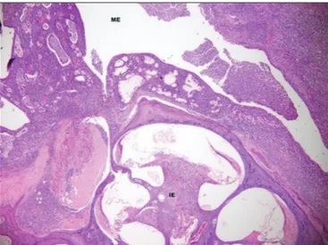
Figure 1. Group1 rats, acute suppurative inflammation in middle ear (ME); there is no involvement in inner ear (İE), routine histological characteristics (suppuration +++, fibrosis-).
Figure 2. Group 1, residual necrotic lamellous bone (LB) fragments due to destruction in middle ear (ME) bone wall, caused by infection at the recovery process. Overlying new woven bone (WB) tissue and thin fibrous wall are seen on the surface of degenerated epithelium. Suppuration continues in the lümen (suppuration +, fibrosis +).
was very mild. It was also found that inner ear involvement was more marked in groups that did not undergo treatment (Group 3) compared to the groups that underwent treatment (Group 1, Group 2) (Fig. 1). In the evaluation of osteoblas-tic and osteoclasosteoblas-tic activities, no difference was found be-tween Groups 1 and 2 (Fig. 2).
There was no significant difference between NAC+treatment group and only treatment group with respect to severity of suppuration and fibrosis, inner ear involvement or stage of infection (p > 0.05) (Tab. I). In group 3, no statistical com-parison was performed with groups 1 and 2 since exten-sive suppuration, fibrosis, and inner ear involvement was detected in all ears.
In the control group, which did not undergo any procedure, suppuration, fibrosis and inner ear involvement were not observed.
SEM findings
In SEM imaging, biofilm formation was established in all groups. There was no significant difference between Groups 1 and 2 in terms of biofilm formation (p > 0.05) (Tab. II).
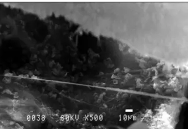
In the evaluation of SEM findings in Group 3, widespread biofilm areas were observed on the middle ear mucosa. As there was no treatment, together with polymorphonuclear leukocytes, many free bacteria, bacteria debris and ECM appearance were also observed (Fig. 3).
In SEM findings of Group 1 rats, widespread biofilm was observed along with polymorphonuclear leukocytes.
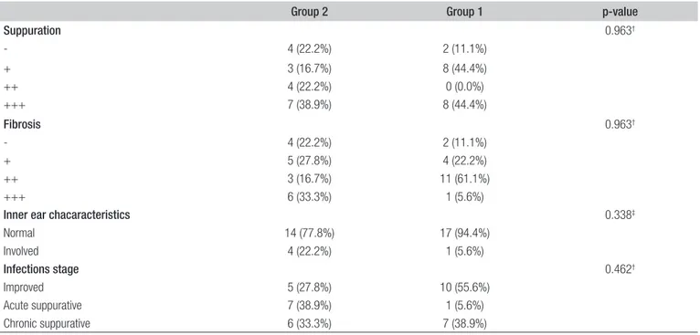
Table I. Histopathological findings of cases undergoing only treatment or NAC plus treatment.
Group 2 Group 1 p-value
Suppuration 0.963† - 4 (22.2%) 2 (11.1%) + 3 (16.7%) 8 (44.4%) ++ 4 (22.2%) 0 (0.0%) +++ 7 (38.9%) 8 (44.4%) Fibrosis 0.963† - 4 (22.2%) 2 (11.1%) + 5 (27.8%) 4 (22.2%) ++ 3 (16.7%) 11 (61.1%) +++ 6 (33.3%) 1 (5.6%)
Inner ear chacaracteristics 0.338‡
Normal 14 (77.8%) 17 (94.4%) Involved 4 (22.2%) 1 (5.6%) Infections stage 0.462† Improved 5 (27.8%) 10 (55.6%) Acute suppurative 7 (38.9%) 1 (5.6%) Chronic suppurative 6 (33.3%) 7 (38.9%)
† Mann Whitney U test; ‡ Fisher’s Exact test.
Table II. SEM findings in groups 1 and groups 2.
Group 2 Group 1 p-value
SEM 0.690†
+ 1 (20.0%) 2 (40.0%)
++ 4 (80.0%) 3 (60.0%)
+++ 0 (0.0%) 0 (0.0%)
† Mann Whitney U test.
SEM findings were similar in Group 2 to those in Group 1. Widespread biofilm formation was observed along with blood components (Fig. 4).
In control group without chronic otitis (Group 4), the nor-mal middle ear mucosa was observed without any biofilm formation (Fig. 5).
Discussion
To our knowledge, the present study is the first attempting to experimentally demonstrate the effect of NAC on bio-film formation in the middle ear and mastoid cells in COM, in terms of histopathological and SEM findings. In refrac-tory COM, in spite of medical treatment to which primary pathogen, P. aeruginosa, is sensitive, otorrhoea and inflam-mation cannot be improved. NAC also exerts an eradicat-ing effect on biofilm layers produced by various bacteria
in addition to its other benefits 9-12,16,17. The effect of NAC
on primary pathogen of COM, i.e. P. aeruginosa, has been
demonstrated in many in vivo and in vitro studies 12,16,17. For
this purpose, the efficacy of NAC has been investigated at different concentrations. In addition to other routes, NAC may also be administered locally. In the study by Özcan et
al. 18, in which the effect of NAC in myringosclerosis in rats
was investigated, 0.6 mg and 1.2 mg topical NAC was used, and it was demonstrated that it prevents the development of
myringosclerosis. In the study by El-Feky et al. 16, aiming
to determine the effect of the addition of NAC to cipro-floxacin treatment for biofilms on the surface of uretheral stents, 2-4 mg/ml NAC was used, and it was determined that the development of mature biofilms declined. Zhao et
al. 12 added NAC at 0.5 mg /ml and 1 mg /ml to
ciprofloxa-cin treatment and found that NAC decreased biofilm layer produced by P. aeruginosa and production of extrapolysac-charides. However, in that study, its effect on P.
aerugi-nosa was investigated in patients with respiratory tract
in-fections. Similar to this study, we used NAC at 0.5 mg/ml for eradication of biofilms. Herein, we found no significant difference between groups 1 and groups 2 with regard to se-verity of suppuration and fibrosis, involvement of inner ear, stage of infection or SEM findings (p > 0.05) (Figs. 1, 2). It may be thought that this is due to differences in experimen-tal animals used, variations in dose, the infectious process in which NAC was used and different organs. In previous studies, NAC was used in vivo and in vitro in pseudomonal infections, and not in treatment of COM, which may also explain the discrepant results.
It has been demonstrated in various studies that NAC
en-hances osteoblastic activity and bone regeneration 19.
How-ever, in the present study, in COM model, no difference was found between Group 1 and 2 in terms of osteoblastic and osteoclastic activity (Fig. 2).
There are few studies in the literature on the effect of NAC on COM treatment. In the only study carried out by Choe
et al. 20, it was established that NAC with ciprofloxacin was
effective in the treatment of refractory COM. Nevertheless, their study was performed with a small number of patients with no histopathological and SEM evaluation, and their conclusion was based upon solely clinical evaluation. Severe and destructive lesions developing in all of the ears undergoing experimental treatment procedures made it dif-ficult to evaluate the severity of histomorphological findings quantitatively, and hence to determine the grade of findings of infection and recovery. We hope that the present study will be a pioneer model that can guide future studies with biofilms and resistant infections. In future studies, lower mi-croorganism concentrations can be used in conjunction with higher dosages of NAC. This method may better reveal the efficacy of NAC in the process of biofilm formation in COM.
Figure 5. Control group SEM findings.
Conclusions
Although there are many studies demonstrating the effect of NAC on biofilms, very few of them examined its effi-cacy in COM. In our study, we found no additional effect of topical NAC to the treatment of COM evaluated histo-pathologically and with SEM in the rat model. We suggest that further studies are required using different concentra-tions of NAC with different experimental animals in order to reach more definitive conclusions.
References
1 Leone S, Molinaro A, Alfieri F, et al. The biofilm matrix of
Pseu-domonas sp. OX1 grown on phenol is mainly constituted by alginate oligosaccharides. Carbohydr Res 2006;341:2456-61. https://doi. org/10.1016/j.carres.2006.06.011
2 O’Toole GA, Kaplan, HB. Biofilm formation as microbial
develop-ment. Ann Rev Microbiol 2000;54:49-79. https://doi.org/10.1146/an-nurev.micro.54.1.49
3 Dohar JE, Hebda PA, Veeh R, et al. Mucosal biofilm formation on
middle-ear mucosa in a nonhuman primate model of chronic sup-purative otitis media. Laryngoscope 2005;115:1469-72. https://doi. org/10.1097/01.mlg.0000172036.82897.d4
4 Dodd S, Dean O, Copolov DL, et al. N-acetylcysteine for antioxidant
therapy: pharmacology and clinical utility. Expert Opin Biol Ther 2008;8:1955-62. https://doi.org/10.1517/14728220802517901
5 Berk M, Copolov DL, Dean O, et al. N-acetyl cysteine for
depres-sive symptoms in bipolar disorder a double-blind randomized place-bo-controlled trial. Biol Psychiatry 2008;64:468-75. https://doi.org/ 10.1016/j.biopsych.2008.04.022
6 Riise GC, Qvarfordt I, Larsson S, et al. Inhibitory effect of
N-acetyl-cysteine on adherence of Streptococcus pneumoniae and Haemophi-lus influenzae to human oropharyngeal epithelial cells in vitro. Respi-ration 2000;67:552-8. https://doi.org/10.1159/000067473
7 Stey C, Steurer J, Bachmann S, et al. The effect of oral N-acetylcysteine
in chronic bronchitis: aquantitative systematic review. Eur Respir J 2000;16:253-62. https://doi.org/10.1034/j.1399-3003.2000.16b12.x
8 Kauffman CA. Quandary about treatment of aspergillomas
per-sists. Lancet 1996;347:1640. https://doi.org/10.1016/s0140-6736(96)91481-6
9 Perez-Giraldo C, Rodriguez-Benito A, Moran FJ, et al. Influence
of N-acetylcysteine on the formation of biofilm by Staphylococcus epidermidis. J Antimicrob Chemother 1997;39:643-6. https://doi. org/10.1093/jac/39.5.643
10 Marchese A, Bozzolasco M, Gualco L, et al. Effect of fosfomycin
alone and in combination with N-acetylcysteine on E. coli biofilms. Int J Antimicrob Agents 2003;22:95-100. https://doi.org/10.1016/ s0924-8579(03)00232-2
11 Schwandt LQ, Van Weissenbruch R, Stokroos I, et al. Prevention of
biofilm formation by dairy products and N-acetylcysteine on voice prostheses in an artificial throat. Acta Otolaryngol 2004;124:726-31. https://doi.org/10.1080/00016480410022516
12 Zhao T, Liu Y. N-acetylcysteine inhibit biofilms produced by
Pseu-domonas aeruginosa. BMC Microbiol 2010;10:140. https://doi.org/ 10.1186/1471-2180-10-140
13 Dohar JE, Alper CM, Rose EA, et al. Treatment of chronic
sup-purative otitis media with topical ciprofloxacin. Ann Otol Rhinol Laryngol 1998;107(10 Pt 1):865-71. https://doi.org/ 10.1177/000348949810701010
14 Alper CM, Dohar JE, Gulhan M, et al. Treatment of chronic
sup-purative otitis media with topical tobramycin and dexamethasone. Arch Otolaryngol Head Neck Surg 2000;126:165-73. https://doi.org/ 10.1001/archotol.126.2.165
15 Saylam G, Tatar EC, Tatar I, et al. Association of adenoid surface
biofilm formation and chronic otitis media with effusion. Arch Oto-laryngol Head Neck Surg 2010;136:550-5. https://doi.org/10.1001/ archoto.2010.70
16 El-Feky MA, El-Rehewy MS, Hassan MA, et al. Effect of
ciprofloxa-cin and N-acetylcysteine on bacterial adherence and biofilm forma-tion on ureteral stent surfaces. Pol J Microbiol 2009;58:261-7.
17 Onger ME, Gocer H, Emir D, et al. N-acetylcysteine eradicates
Pseu-domonas aeruginosa biofilms in bone cement. Scanning 2016;38:766-70. https://doi.org/10.1002/sca.21326
18 Ozcan C, Görür K, Cinel L, et al. The inhibitory effect of topical
N-acetylcysteine application on myringosclerosis in perforated rat tympanic membrane. Int J Pediatr Otorhinolaryngol 2002;63:179-84. https://doi.org/10.1016/s0165-5876(01)00640-1
19 Yamada M, Tsukimura N, Ikeda T, et al. N-acetyl cysteine as an
os-teogenesis-enhancing molecule for bone regeneration. Biomaterials 2013;34:6147-56. https://doi.org/10.1016/j.biomaterials.2013.04.064
20 Choe WT, Murray MT, Stidham KR, et al. N-acetylcysteine as an