Prognostic factors for maximally or optimally cytoreduced
stage III nonserous epithelial ovarian carcinoma treated
with carboplatin/paclitaxel chemotherapy
Zeliha F. Cuylan
1, Mehmet M. Meydanli
1, Mustafa E. Sari
1, Ozgur Akbayir
2, Husnu Celik
3,
Murat Dede
4, Hanifi Sahin
1, Kemal Gungorduk
5, Esra Kuscu
6, Nejat Ozgul
7,
Tayfun Gungor
1and Ali Ayhan
6Departments of1Gynecologic Oncology, Zekai Tahir Burak Women’s Health Training and Research Hospital, Faculty of Medicine,
4
Obstetrics and Gynecology, Gulhane Training and Research Hospital, Faculty of Medicine, University of Health Sciences,
6
Division of Gynecologic Oncology, Department of Obstetrics and Gynecology, Faculty of Medicine, Baskent University,
7
Division of Gynecologic Oncology, Department of Obstetrics and Gynecology, Faculty of Medicine, Hacettepe University, Ankara,2Department of Gynecologic Oncology, Kanuni Sultan Suleyman Teaching and Research Hospital, Faculty of Medicine, University of Health Sciences, Istanbul,3Division of Gynecologic Oncology, Department of Obstetrics and Gynecology, Faculty of Medicine, Baskent University, Adana and5Department of Gynecologic Oncology, Tepecik Education and Research Hospital, Faculty of Medicine, University of Health Sciences, Izmir, Turkey
Abstract
Objective: To identify factors predictive of poor prognosis in women with stage III nonserous epithelial ovarian cancer (EOC) who had undergone maximal or optimal primary cytoreductive surgery (CRS) fol-lowed by six cycles of intravenous carboplatin/paclitaxel chemotherapy.
Methods: A multicenter, retrospective department database review was performed to identify patients with stage III nonserous EOC who had undergone maximal or optimal primary CRS followed by six cycles of car-boplatin/paclitaxel chemotherapy at seven gynecological oncology centers in Turkey. Demographic, clinico-pathological and survival data were collected.
Results: A total of 218 women met the inclusion criteria. Of these, 64 (29.4%) patients had endometrioid, 61 (28%) had mucinous, 54 (24.8%) had clear-cell and 39 (17.9%) had mixed epithelial tumors. Fifty-five (25.2%) patients underwent maximal CRS, whereas 163 (74.8%) had optimal debulking. With a median follow-up of 31.5 months, the 5-year progression-free survival (PFS) and overall survival (OS) rates were 34.8% and 44.2%, respectively. Bilaterality (hazard ratio [HR] 1.44, 95% CI 1.01–2.056; P = 0.04), age (HR 2.25, 95% CI 1.176–4.323; P = 0.014) and maximal cytoreduction (HR 0.34, 95% CI 0.202–0.58; P < 0.001) were found to be independent prognostic factors for PFS. However, age (HR 2.6, 95% CI 1.215–5.591; P = 0.014) and maximal cytoreduction (HR 0.31, 95% CI 0.166–0.615; P < 0.001) were defined as independent prognostic factors for OS.
Conclusion: The extent of CRS seems to be the only modifiable prognostic factor associated with stage III nonserous EOC. Complete cytoreduction to no gross residual disease should be the main goal of manage-ment in these women.
Key words:cytoreduction, epithelial ovarian cancer, non-serous histology, prognosis, stage III.
Received: November 7 2017. Accepted: March 23 2018.
Correspondence: Dr Zeliha F. Cuylan, Instructor-Gynecologic Oncology Fellow, Department of Gynecologic Oncology, Zekai Tahir Burak Women’s Health Training and Research Hospital, Zekai Tahir Burak Women’s Health Education and Training Hospital, Talatpasa Blv. 06230 Altindag/Ankara, Turkey. Email: tel.: +90 505 7071059; fax: +90 312 3214931, Email: zelihafi[email protected]
Introduction
Nowadays, it is well known that epithelial ovarian cancer (EOC) is not a homogenous disease.1 Rather, EOCs are represented by a number of molecularly distinct diseases broadly defined by histotype with different routes of spread, patterns of relapse, response to chemotherapy and prognosis.2
Primary cytoreductive surgery (CRS) and taxane-/ platinum-based adjuvant chemotherapy are the cor-nerstones of the initial treatment for all histological subtypes of EOC.3,4 Nevertheless, prognosis is vari-able, largely depending on the quality of primary CRS and the time of development of platinum resis-tance. Therefore, advanced EOC represents a hetero-geneous group regarding the outcome after initial management.5
Established prognostic factors for stage III EOC include age,6,7 performance status (PS),6 extent of residual disease (RD)6,7 and histology.7 Nevertheless, previous studies investigating the prognostic factors for advanced EOC suffer from several limitations, such as heterogeneous study populations (inclusion of patients with stage III and IV disease together),8–11 analyzing serous and nonserous malignant tumors simultaneously,6–15 small number of patients with nonserous EOC7,9–13 and the variation in adjuvant chemotherapy regimens.9,11,12,14 Although it has been well known for a long time that the quality of pri-mary CRS is the strongest predictor of outcome,16,17 the extent of RD after primary CRS was not an inclu-sion criterion in most of the previous studies, and patients with maximal, optimal and suboptimal CRS were generally analyzed simultaneously.6,8,10–12 It seems plausible to adjust the optimality of primary CRS and adjuvant chemotherapy for the evaluation of prognostic factors for stage III EOC. In addition, it is difficult to claim that older series do reflect the cur-rent practice in terms of primary CRS and adjuvant chemotherapy.12,13,15
Previous studies investigating the prognostic fac-tors for stage III EOC consistently analyzed serous and nonserous tumors together and included patients mostly with serous EOC.6,7,12,13 Therefore, prognostic factors for stage III nonserous EOC have not been delineated clearly. From the current perspective that different histotypes in EOC probably represent differ-ent disease differ-entities,18 we wondered whether the established prognostic factors for all histotypes6,7,12,13 are also valid when nonserous malignant tumors are analyzed as a separate group. Given the low
frequency of nonserous histotypes, we designed this multicenter retrospective study in order to shed some light on this issue with the aid of a well-defined homogenous study population. The purpose of the current study was to identify factors predictive of poor prognosis in women with stage III nonserous EOC who had undergone maximal or optimal pri-mary CRS followed by six cycles of intravenous (IV) carboplatin/paclitaxel chemotherapy.
Methods
Study design and eligibility
Medical records of women who underwent primary surgical treatment for EOC between January 2007 and December 2016 at seven gynecological oncology cen-ters in Turkey were retrospectively reviewed. The study protocol was approved by the local institutional review boards. All patients provided informed con-sent regarding research use of their medical informa-tion at admission.
The study population included women who had nonserous EOC (i.e., endometrioid, clear-cell, mucin-ous and mixed subtypes) with histopathologically proven stage III19disease. Women were included if they had undergone primary surgical treatment, including total hysterectomy plus bilateral salpingo-oopherectomy, with bilateral pelvic and para-aortic lymphadenectomy and other surgical procedures, resulting in maximal or optimal CRS. All patients had to have RD of 1 cm or less in order to be eligible. Patients who were cytoreduced to greater than 1 cm of RD were excluded. Because this study focused only on women with nonserous EOC, women with high-and low-grade serous carcinoma were excluded, as well as patients having no lymphadenectomy. We also excluded patients who received neoadjuvant che-motherapy, women with synchronous malignancies and those with incomplete medical records. A PS of 0 (normal activity) or 1 (symptomatic and fully ambu-latory) was also an eligibility criterion. Women with a PS≥ 2 were excluded. Patients with coexisting medi-cal comorbidities were excluded as this undoubtedly influences the decision-making process of a surgeon regarding whether to undertake an aggressive CRS.
Clinical information
Patient data were extracted from seven institutions with maintained EOC databases. After selecting the eligible cases, the following information was
abstracted from medical records: demographic charac-teristics, preoperative serum cancer antigen 125 (CA 125) level, date and type of surgical proce-dure, presence or absence of ascites, the status of peri-toneal cytology examination (negative, or positive), bilaterality, size of RD after surgery, stage of disease, time to recurrence, length of follow-up and survival. Tumor characteristics were abstracted from original pathology reports. Data were collected from centers with an online standardized form. All operations were performed by gynecological oncologists with the intent to achieve optimal cytoreduction. RD after pri-mary CRS was recorded according to the assessment by the surgeon. Lymphadenectomy was performed after completion of other cytoreductive procedures. All patients underwent detailed preoperative and sur-gical exploration to exclude primary colorectal and appendiceal carcinomas.
All pathological specimens from primary surgery were examined and interpreted by gynecological pathologists of the participating institutions who had experience in gynecological malignancies. Nonserous EOC was diagnosed after examination of permanent sections. Histological classification was performed with the criteria defined by the World Health Organi-zation (WHO).20,21Architectural grading was defined by standard International Federation of Gynecology and Obstetrics (FIGO) criteria. Clear-cell carcinomas were neither graded nor assigned as grade 3 in this study. Cases diagnosed as high-grade endometrioid were not removed from the endometrioid group.
The current study investigated cases with mixed nonserous histologies (including mucinous, clear-cell, endometrioid and transitional cell types) as a separate group and did not assign mixed tumors according to the dominant component. Mixed tumors were diag-nosed according to the WHO definition, in that more than one cell type was present, and the minority com-ponent accounted for at least 10% of the tumor. Mixed tumors containing serous component were excluded. For the purposes of this study, only pure tumors were classified as endometrioid, clear-cell or mucinous, whereas tumors with more than one cell type were classified as mixed. All tumors were staged according to the 2014 FIGO staging system.19 In patients treated before 2014, stage was determined retrospectively on the basis of surgical and pathologi-cal assessment.
The treatment policies were decided by the attend-ing physician or by the multidisciplinary tumor board at each participating institution. Adjuvant
chemotherapy was administered to all patients. The standard primary chemotherapy regimen included paclitaxel 175 mg/m2 plus carboplatin dosed at an area under curve of 5 or 6 every 21 days for six cycles. Targeted agents were not used to treat any of the patients during primary treatment.
Patients returned for follow-up evaluation every 3 months for the first 2 years, every 6 months for the next 3 years and annually thereafter. Computed tomography or magnetic resonance imaging was performed annually. Survival data were last calcu-lated on 31st December 2016. The survival status of the patients was determined as alive or dead at the time of the last follow-up. For all study subjects with a recorded death, this was confirmed by per-forming a social security death index search.
Definitions
Baseline PS was defined according to the Gynecologic Oncology Group (GOG) criteria, and only patients with a PS of 0 (normal activity) and 1 (symptomatic and fully ambulatory) were included in the current study in order to overcome the confounding effect of PS on the measured outcome.
Maximal cytoreduction was defined as no gross RD (microscopic RD) after primary CRS. Optimal cytore-duction was defined as less than or equal to 1 cm maximal diameter of the largest residual tumor nod-ule at the completion of the primary operation. Sub-optimal cytoreduction was defined as >1 cm of RD. Lymphadenectomy was defined as the perfor-mance of pelvic and para-aortic LN dissection at the same time. We defined pelvic lymphadenectomy as the removal of lymphatic tissue in the external, inter-nal and common iliac and obturator regions. Para-aortic lymphadenectomy was defined as removal of the lymphatic tissue over the inferior vena cava and aorta, beginning at the level of aortic bifurcation up to the left renal vessels.
Progression-free survival (PFS) was defined as the time, in months, from the date of primary surgery until the date of documented recurrence on the basis of clinical examination or radiological imaging; death from any cause, whichever occurredfirst; or the date of last contact for patients remaining alive without recurrent disease. Patients who had no active ovarian cancer at the last contact were censored in the PFS analysis. Overall survival (OS) was calculated as the time period, in moths, between the date of primary surgery to the date of death or the last contact. Sur-viving patients were censored at their last known
follow-up. As treatment after relapse was not uniform and varied among institutions that participated in the current study, the primary end-point was chosen as PFS.
Statistical analysis
Statistical analyses were performed using the statisti-cal software package SPSS version 23.0 (IBM Corp.,
Armonk, NY, USA). The data were expressed as median and range for continuous variables. Binary variables were reported as counts and percentages.
Survival analysis was based on the Kaplan–Meier method, and the results were compared using a log-rank test. Cox regression analysis was used to determine factors affecting PFS and OS, presented as hazard ratios (HRs) and 95% confidence intervals (CI), unadjusted or adjusted for all factors. All variables with a P value <0.05 in the univariate analysis were included in the multivariate analysis. A P value <0.05 was considered to indicate statistical significance.
Results
During the study period, 367 women with stage III nonserous EOC were treated at seven participating centers. We excluded 23 patients who had no lympha-denectomy, 17 women who received neoadjuvant che-motherapy and 9 women with incomplete medical records. One hundred patients who received subopti-mal debulking were also excluded. Therefore, the pre-sent analysis addresses the remaining 218 women. The rate of suboptimal CRS was found to be 31.4% (100/318) in the entire cohort. The inclusion process
of the patients and the suboptimal CRS rates with regard to the histotypes are demonstrated in Table 1.
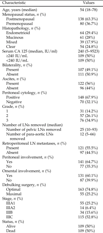
Of 218 women who met the inclusion criteria, 64 (29.4%) patients had endometrioid, 61 (28%) had mucinous, 54 (24.8%) had clear-cell and 39 (17.9%) had mixed epithelial tumors. The median age of the patients was 54 (range, 18–78) years, and the median duration of follow-up was 31.5 (range, 1–120) months. There were 55 (25.2%) women with stage IIIA1, 14 (6.4%) with stage IIIA2, 34 (15.6%) with stage IIIB and 115 (52.8%) with stage IIIC disease. Fifty-five (25.2%) patients underwent maximal CRS, whereas 163 (74.8%) had optimal debulking. Table 2 summa-rizes the clinicopathological characteristics of the study population.
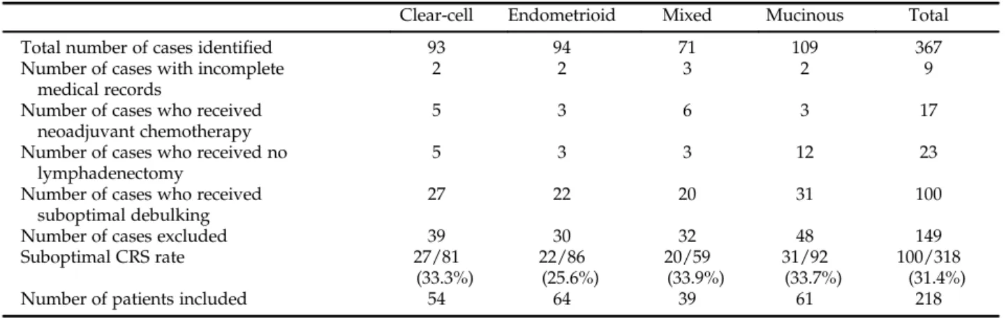
For the entire cohort, the 5-year PFS was 34.8%, with a median PFS of 28 months (95% confidence interval [CI] 20.58–35.42, Standard Error [SE]: 3.785). The median PFS for endometrioid, clear-cell, mixed epithelial and mucinous histotypes were 38, 24, 22 and 20 months, respectively (Fig. 1a).
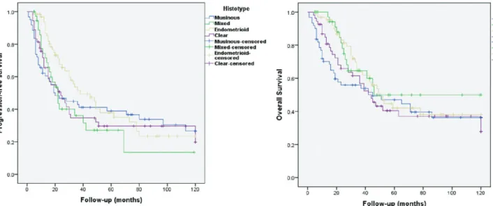
For 55 (25.2%) women undergoing maximal CRS, the median PFS was not reached yet, whereas the corresponding figure was found to be 22 months (95% CI 16.3–27.7 months) for 163 (74.8%) patients who received optimal debulking (P < 0.001) (Fig. 2). The median OS for women with maximal CRS was significantly longer than that of women undergoing optimal CRS (not reached yet vs 39 months [95% CI 30.6–47.4 months], respectively; P < 0.001) (Fig. 3).
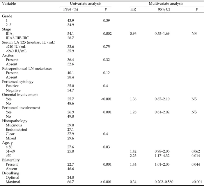
Univariate analysis demonstrated age (≤ 50 vs ≥70 years) (P = 0.039), bilaterality (P = 0.001), stage (stage IIIA1 vs others) (P = 0.002), omental
Table 1 The inclusion process of the patients and the suboptimal cytoreductive surgery rates with regard to the histotypes
Clear-cell Endometrioid Mixed Mucinous Total
Total number of cases identified 93 94 71 109 367
Number of cases with incomplete medical records
2 2 3 2 9
Number of cases who received neoadjuvant chemotherapy
5 3 6 3 17
Number of cases who received no lymphadenectomy
5 3 3 12 23
Number of cases who received suboptimal debulking
27 22 20 31 100
Number of cases excluded 39 30 32 48 149
Suboptimal CRS rate 27/81 (33.3%) 22/86 (25.6%) 20/59 (33.9%) 31/92 (33.7%) 100/318 (31.4%)
involvement (P < 0.001), peritoneal involvement (P = 0.001) and gross RD≤ 1 cm (P < 0.001) to be sig-nificant factors for decreased PFS (Table 2). At the end of multivariate analysis, bilaterality (HR 1.44, 95% CI 1.01–2.056; P = 0.04), age (≤ 50 vs ≥70 years) (HR 2.25, 95% CI 1.176–4.323; P = 0.014) and maximal cytoreduction (HR 0.34, 95% CI 0.202–0.58; P < 0.001) remained independent prognostic factors for PFS (Table 3).
The 5-year OS of the entire cohort was 44.2%, with a median overall survival of 47 months (95% CI 36.12–57.88, SE: 5.55). The median OS for endome-trioid, clear-cell, mixed epithelial and mucinous histo-types were 49 months, 44 months, not reached yet and 45 months, respectively (Fig. 1b).
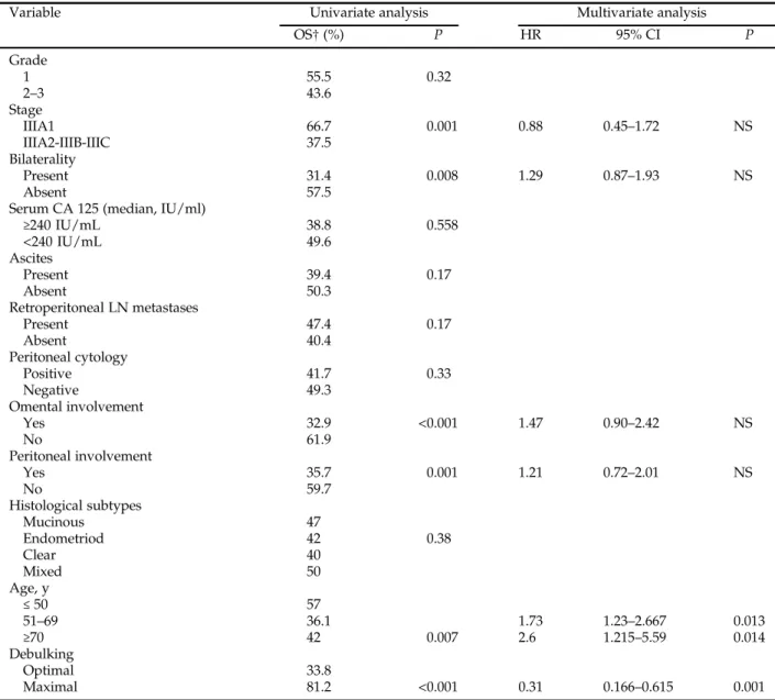
Univariate analysis demonstrated age (P = 0.007), bilaterality (P = 0.008), stage (stage IIIA1 vs others) (P = 0.001), omental involvement (P < 0.001), perito-neal involvement (P = 0.001) and gross RD≤ 1 cm (P < 0.001) to be significant factors for decreased OS (Table 3). At the end of multivariate analysis, age (51–69 years vs ≤50 years (HR 1.73, 95% CI 1.23–2.66; P = 0.013), (≤ 50 vs ≥70 years) (HR 2.6, 95% CI 1.215–5.591; P = 0.014) and maximal cytoreduction (HR 0.31, 95% CI 0.166–0.615; P < 0.001) remained independent prognostic factors for OS (Table 4). At the time of reporting, of 218 women with stage III nonserous EOC, 109 (50%) were dead, whereas 109 (50%) were alive.
Discussion
This study represents a retrospective analysis of 218 patients with stage III nonserous EOC who have undergone maximal or optimal CRS followed by six cycles of IV carboplatin/paclitaxel chemotherapy. Our results indicated age ≤ 50 years, achievement of maximal cytoreduction and unilaterality of the tumor as independent prognostic factors for prolonged PFS. Although the number of patients is relatively limited, to the best of our knowledge, this is the first study reporting on the prognostic factors for stage III nonserous EOC.
A large GOG study identified factors of poor prog-nosis in a similarly treated population of women with stage III EOC.6This study included 1895 patients with stage III EOC who had undergone primary CRS fol-lowed by platinum/paclitaxel chemotherapy. Age, PS, tumor histology and residual tumor volume were independent predictors of outcome in patients with stage III EOC.6However, non-serous histologies con-stituted 21.3% (n = 404) of their study population, whereas 35% of patients (n = 667) underwent subopti-mal CRS in that study.6
Landrum et al.7 have reported that histology, age and extent of RD are identified as statistically signifi-cant variables for OS in a cohort of 428 patients with stage III EOC undergoing optimal CRS followed by intraperitoneal paclitaxel/ platinum chemotherapy. It
Figure 1 The progression-free survival and overall survival curves of women with endometrioid, clear cell, mixed epithe-lial and mucinous histotypes in stage III nonserous epitheepithe-lial ovarian cancer.
should be noted that patients with clear-cell and mucinous histology comprised only 6.3% (n = 27) of their study population. However, our findings point-ing out age and extent of RD as independent
prognostic factors for OS are in agreement with those of Landrum et al.7
Extent of RD, PS and adjuvant chemotherapy regi-men were adjusted, and the confounding effects of these variables were minimized for the evaluation of
Figure 2 The progression-free survival curves of women who have undergone maximal and optimal cytoreduction in stage III nonserous epithelial ovarian cancer.
Figure 3 The overall survival curves of women who have undergone maximal and optimal cytoreduction in stage III nonserous epithelial ovarian cancer.
Table 2 Baseline characteristics of the patients
Characteristic Values
Age, years (median) 54 (18–78)
Menopausal status, n (%) Postmenopausal 138 (63.3%) Premenopausal 80 (36.7%) Histopathology, n (%) Endometrioid 64 (29.4%) Mucinous 61 (28%) Mixed 39 (17.9%) Clear 54 (24.8%)
Serum CA 125 (median, IU/ml) 240 (5–9523)
≥240 IU/mL 109 (50%) <240 IU/mL 109 (50%) Bilaterality, n (%) Present 107 (49.1%) Absent 111 (50.9%) Ascites, n (%) Present 122 (56%) Absent 96 (44%) Peritoneal cytology, n (%) Positive 148 (67.9%) Negative 70 (32.1%) Grade, n (%) 1 31 (14.2%) 2 57 (26.1%) 3 76 (34.9%)
Number of LNs removed (median)
Number of pelvic LNs removed 25 (10–93) Number of para-aortic LNs removed 12 (5–66) Retroperitoneal LN metastases, n (%) Present 121 (55.5%) Absent 97 (44.5%) Peritoneal involvement, n (%) Yes 141 (64.7%) No 77 (35.3%) Omental involvement, n (%) Yes 131 (60.1%) No 87 (39.9%) Debulking surgery, n (%) Optimal 163 (74.8%) Maximal 55 (25.2%) Stage, n (%) IIIA1 55 (25.2%) IIIA2 14 (6.4%) IIIB 34 (15.6%) IIIC 115 (52.8%) Status, n (%) Alive 109 (50%) Dead 109 (50%)
prognostic factors for stage III nonserous EOC in the current study. However, age appeared to be an inde-pendent prognostic factor for PFS and OS in multi-variate analysis, demonstrating that those patients≥ 70 years were 2.2 times more likely to have decreased PFS when compared to those ≤ 50 years. Landrum et al.7 have stated that patient age stands as a prognostic factor for survival independent of RD, PS, grade and stage. The authors suggested that better understanding of the changes in tumor biol-ogy or immune response in older patients may lead to new insights into the best treatment methods for
the rapidly increasing population.7 It should be noted that age at the time of diagnosis for patients with EOC has been consistently recognized as an independent prognostic factor in previous studies.7,8,12,22–24
It is increasingly appreciated that tumor cell type correlates with epidemiological risk factors, “BRCA 1 or 2” mutation status, differences in gene expression profile and genetic events during oncogenesis and response to chemotherapy.14Nevertheless, information on the prognostic significance of histology in the con-text of changing therapeutic standards is limited.5The
Table 3 Univariate and multivariate analyses for prognostic factors for progression-free survival
Variable Univariate analysis Multivariate analysis
PFS† (%) P HR 95% CI P Grade 1 43.9 0.39 2–3 34.9 Stage IIIA1 54.1 0.002 0.96 0.55–1.69 NS IIIA2-IIIB-IIIC 28.7
Serum CA 125 (median, IU/mL)
≥240 IU/mL 33.6 0.75 <240 IU/mL 35.9 Ascites Present 36.4 0.32 Absent 32.6 Retroperitoneal LN metastases Present 40.1 0.12 Absent 28.4 Peritoneal cytology Positive 35.0 0.4 Negative 34.7 Omental involvement Yes 25.7 <0.001 1.36 0.87–2.10 NS No 48.6 Peritoneal involvement Yes 26.9 0.001 1.28 0.81–2.02 NS No 49.0 Histopathology Mucinous 39.0 Endometriod 27.1 Clear 37.9 0.4 Mixed 29.6 Age, y ≤ 50 27.6 0.03 51–69 25.0 1.42 0.98–2.05 0.062 ≥70 2.25 1.17–4.32 0.014 Bilaterality Present 22.7 0.001 1.44 1.01–2.05 0.044 Absent 46.6 Debulking Optimal 24.8 Maximal 66.7 < 0.001 0.34 0.202–0.580 <0.001
prognostic significance of histotype is controversial in stage III EOC given the relative rarity of histological subtypes other than serous.25Most studies describing the role of tumor histology in prognosis have focused on mucinous and clear-cell tumors.5,26–30 However, histological subtype has been reported as an indepen-dent prognostic factor for stage III–IV EOC.9,10In con-trast, Chi et al.11and Bristow et al.31 showed that histological subtype was not a significant prognostic factor in earlier studies.
Winter et al.6 reported that mucinous or clear-cell histology was associated with a worse PFS and OS compared with serous carcinomas. In a meta-analysis
of 8704 women with stage III–IV EOC, it has been shown that the prognoses of women with mucinous tumors are worse than those with serous tumors, and the prognoses of patients with the clear-cell carcino-mas are unlikely to be better.8Nevertheless, histotype did not seem to have prognostic significance in women with stage III nonserous EOC in the current study. Our findings do not agree with previous reports showing the prognostic significance of histo-logical subtype in advanced EOC.6–10,12The prognos-tic significance of the tumor histology seems to be consistent when serous malignant tumors are com-pared with nonserous malignant tumors, whereas its
Table 4 Univariate and multivariate analyses for prognostic factors for overall survival
Variable Univariate analysis Multivariate analysis
OS† (%) P HR 95% CI P Grade 1 55.5 0.32 2–3 43.6 Stage IIIA1 66.7 0.001 0.88 0.45–1.72 NS IIIA2-IIIB-IIIC 37.5 Bilaterality Present 31.4 0.008 1.29 0.87–1.93 NS Absent 57.5
Serum CA 125 (median, IU/ml)
≥240 IU/mL 38.8 0.558 <240 IU/mL 49.6 Ascites Present 39.4 0.17 Absent 50.3 Retroperitoneal LN metastases Present 47.4 0.17 Absent 40.4 Peritoneal cytology Positive 41.7 0.33 Negative 49.3 Omental involvement Yes 32.9 <0.001 1.47 0.90–2.42 NS No 61.9 Peritoneal involvement Yes 35.7 0.001 1.21 0.72–2.01 NS No 59.7 Histological subtypes Mucinous 47 Endometriod 42 0.38 Clear 40 Mixed 50 Age, y ≤ 50 57 51–69 36.1 1.73 1.23–2.667 0.013 ≥70 42 0.007 2.6 1.215–5.59 0.014 Debulking Optimal 33.8 Maximal 81.2 <0.001 0.31 0.166–0.615 0.001
prognostic significance disappears when nonserous malignant tumors are studied as a separate group.
The volume of RD after primary CRS depends on the number and the size of tumor elements.12 In this retrospective study, RD status after primary CRS was recorded according to the assessment of the attending surgeon. We were not able to define the number of lesions left after primary CRS. However, the maxi-mum diameter of the greatest residual tumor nodule is a very crude estimate of RD, but it still provided valuable prognostic information in our study as it did in previous studies.7,12,17There was a statistically sig-nificant PFS and OS advantage for patients with no gross RD compared to those with macroscopic RD ≤1 cm in the current study. Our study has provided further information that the prognostic significance of the extent of RD stands even within a population of patients with≤ 1 cm of RD.
Our analysis has shown that the extent of RD is an independent predictor of OS, whereas the substage of the disease is not. Ourfinding are in line with that of Landrum et al.7who suggested that efforts to reduce the tumor burden to no gross RD might mitigate the impact of stage. In addition, it has been reported that residual tumor size was associated with an increased risk of death for advanced nonserous EOC, whereas taxane-based chemotherapy was not a prognostic factor.9
Several potential limitations of our study warrant consideration. First, the retrospective nature of the study cannot exclude any bias. Second, treatment after recurrence was not uniform and varied among institutions that participated in the study. Therefore, our findings associated with OS should be met cau-tiously. Third, our study was restricted by the lack of a central pathology review. Although a comprehen-sive central pathology review would be ideal, patients with nonserous EOC included in the current study do reflect the ‘real-world’ diagnosis and practice in our country. We used information from routine practice in order to assess prognoses of women with stage III nonserous EOC. These women were homogeneously diagnosed and treated in tertiary referral centers, and histopathological evaluation was performed by highly experienced gynecological pathologists. Despite the abovementioned limitations, our findings provide additional information to the body of knowledge on this topic.
The strength of the current study, compared with previous reports, is mainly the homogeneity of the patient population and the treatment, which provides a precise estimation of the magnitude of the effect
while controlling for confounder influences. The homogenous structure of our study seems to reduce the possibility of confounding and enhances the reli-ability of the prognostic effects of those have been estimated.
Ourfindings indicate that maximal cytoreduction is the only modifiable prognostic factor associated with stage III nonserous EOC. Maximal surgical effort seems to currently be the best option for the initial treatment of these women as PFS and OS seem to be superior whenever maximal cytoreduction has been achieved. We have to emphasize that women with no gross RD following primary CRS had survival rates that exceed any rates previously reported in stage III nonserous EOC treated with the current standard chemotherapy.
At any rate, the retrospective nature of our study does not permit us to draw definitive conclusions as similar previous studies. It should be noted that the results of the current study are limited only to patients who can be cytoreduced to 1 cm or less of RD.
We conclude that complete cytoreduction to no gross RD should be the main goal of management in women with stage III nonserous EOC, and it seems reasonable to perform maximal CRS whenever possi-ble in those patients. However, further exploration of molecular markers and biologic pathways to better characterize the behavior of these rare histotypes is warranted.
Acknowledgments
None.Disclosure
None declared.References
1. Prat J, Belhadj H, Berek J et al. Abridged republication of FIGO’s staging classification for cancer of the ovary, fallo-pian tube, and peritoneum. Eur J Gynaecol Oncol 2015; 36: 367–369.
2. Kobel M, Kalloger SE, Boyd N et al. Ovarian carcinoma sub-types are different diseases: Implications for biomarker stud-ies. PLoS Med 2008; 5: e232.
3. McGuire WP, Hoskins WJ, Brady MF et al. Cyclophospha-mide and cisplatin compared with paclitaxel and cisplatin in
patients with stage III and stage IV ovarian cancer. N Engl J Med 1996; 334: 1–6.
4. Bristow RE, Tomacruz RS, Armstrong DK, Trimble EL, Montz FJ. Survival effect of maximal cytoreductive surgery for advanced ovarian carcinoma during the platinum era: A meta-analysis. J Clin Oncol 2002; 20: 1248–1259.
5. Bamias A, Psaltopoulou T, Sotiropoulou M et al. Mucinous but not clear cell histology is associated with inferior sur-vival in patients with advanced stage ovarian carcinoma treated with platinum-paclitaxel chemotherapy. Cancer 2010; 116: 1462–1468.
6. Winter WE 3rd, Maxwell GL, Tian C et al. Prognostic factors for stage III epithelial ovarian cancer: A gynecologic oncol-ogy group study. J Clin Oncol 2007; 25: 3621–3627.
7. Landrum LM, Java J, Mathews CA et al. Prognostic factors for stage III epithelial ovarian cancer treated with intraperi-toneal chemotherapy: A gynecologic oncology group study. Gynecol Oncol 2013; 130: 12–18.
8. Mackay HJ, Brady MF, Oza AM et al. Prognostic relevance of uncommon ovarian histology in women with stage III/IV epithelial ovarian cancer. Int J Gynecol Cancer 2010; 20: 945–952.
9. Hosono S, Kajiyama H, Mizuno K et al. Comparison between serous and non-serous ovarian cancer as a prognos-tic factor in advanced epithelial ovarian carcinoma after pri-mary debulking surgery. Int J Clin Oncol 2011; 16: 524–532. 10. Bamias A, Sotiropoulou M, Zagouri F et al. Prognostic
evalu-ation of tumour type and other histopathological character-istics in advanced epithelial ovarian cancer, treated with surgery and paclitaxel/carboplatin chemotherapy: Cell type is the most useful prognostic factor. Eur J Cancer 2012; 48: 1476–1483.
11. Chi DS, Liao JB, Leon LF et al. Identification of prognostic factors in advanced epithelial ovarian carcinoma. Gynecol Oncol 2001; 82: 532–537.
12. Makar AP, Baekelandt M, Trope CG, Kristensen GB. The prognostic significance of residual disease, FIGO substage, tumor histology, and grade in patients with FIGO stage III ovarian cancer. Gynecol Oncol 1995; 56: 175–180.
13. Hoskins WJ, Bundy BN, Thigpen JT, Omura GA. The influ-ence of cytoreductive surgery on recurrinflu-ence-free interval and survival in small-volume stage III epithelial ovarian cancer: A gynecologic oncology group study. Gynecol Oncol 1992; 47: 159–166.
14. Gilks CB, Ionescu DN, Kalloger SE et al. Tumor cell type can be reproducibly diagnosed and is of independent prognostic significance in patients with maximally debulked ovarian carcinoma. Hum Pathol 2008; 39: 1239–1251.
15. Omura GA, Brady MF, Homesley HD et al. Long-term follow-up and prognostic factor analysis in advanced ovar-ian carcinoma: The gynecologic oncology group experience. J Clin Oncol 1991; 9: 1138–1150.
16. Hoskins WJ. The influence of cytoreductive surgery on progression-free interval and survival in epithelial ovarian cancer. Baillieres Clin Obstet Gynaecol 1989; 3: 59–71.
17. Hoskins WJ, McGuire WP, Brady MF et al. The effect of diameter of largest residual disease on survival after pri-mary cytoreductive surgery in patients with suboptimal residual epithelial ovarian carcinoma. Am J Obstet Gynecol 1994; 170: 974–979 discussion 9-80.
18. Vaughan S, Coward JI, Bast RC Jr et al. Rethinking ovarian cancer: Recommendations for improving outcomes. Nat Rev Cancer 2011; 11: 719–725.
19. Prat J. Oncology FCoG. Staging classification for cancer of the ovary, fallopian tube, and peritoneum. Int J Gynaecol Obstet 2014; 124: 1–5.
20. Kurman RJ, Carcangiu ML, Herrington CS, Young RH. Tumors of the ovary. In: WHO Classification of Tumours of Female Reproductive Organs, Fourth edn. Lyon: IARC, 2014; Chapter 1.
21. Tavassoli FA, Devilee P (eds). World Health Organization Classfication of Tumors. Pathology & Genetics of Tumours of the Breast and Female Genital Organs. Lyon: IARC (International Agency for Research on Cancer) Press, 2003; 153–158. 22. Duska LR, Chang YC, Flynn CE et al. Epithelial ovarian
car-cinoma in the reproductive age group. Cancer 1999; 85: 2623–2629.
23. Chan JK, Loizzi V, Lin YG, Osann K, Brewster WR, DiSaia PJ. Stages III and IV invasive epithelial ovarian carci-noma in younger versus older women: What prognostic fac-tors are important? Obstet Gynecol 2003; 102: 156–161. 24. Chan JK, Urban R, Cheung MK et al. Ovarian cancer in
younger vs older women: A population-based analysis. Br J Cancer 2006; 95: 1314–1320.
25. Bouchard-Fortier G, Panzarella T, Rosen B, Chapman W, Gien LT. Endometrioid carcinoma of the ovary: Outcomes compared to serous carcinoma after 10 years of follow-up. J Obstet Gynaecol Can 2017; 39: 34–41.
26. Karabuk E, Kose MF, Hizli D et al. Comparison of advanced stage mucinous epithelial ovarian cancer and serous epithe-lial ovarian cancer with regard to chemosensitivity and sur-vival outcome: A matched case-control study. J Gynecol Oncol 2013; 24: 160–166.
27. Pectasides D, Fountzilas G, Aravantinos G et al. Advanced stage mucinous epithelial ovarian cancer: The Hellenic cooperative oncology group experience. Gynecol Oncol 2005; 97: 436–441. 28. Miyamoto M, Takano M, Goto T et al. Clear cell histology as
a poor prognostic factor for advanced epithelial ovarian can-cer: A single institutional case series through central patho-logic review. J Gynecol Oncol 2013; 24: 37–43.
29. Chan JK, Teoh D, Hu JM, Shin JY, Osann K, Kapp DS. Do clear cell ovarian carcinomas have poorer prognosis com-pared to other epithelial cell types? A study of 1411 clear cell ovarian cancers. Gynecol Oncol 2008; 109: 370–376.
30. Lee YY, Kim TJ, Kim MJ et al. Prognosis of ovarian clear cell carcinoma compared to other histological subtypes: A meta-analysis. Gynecol Oncol 2011; 122: 541–547.
31. Bristow RE, Montz FJ, Lagasse LD, Leuchter RS, Karlan BY. Survival impact of surgical cytoreduction in stage IV epithe-lial ovarian cancer. Gynecol Oncol 1999; 72: 278–287.