A. O. Vet. Fak. Derg. Fac. Vet. Med., Univ. Ankara
28, (1-4): 50-71, 1981.
STUDms ON THE BIOLOGY OF PARAMPHISTOMUM
CERVI SCHRANK, 1790IN SHEEP IN THE DISTRICT OF ESKİŞEHİR ÇİFTELERSTATE FARM
Ayşe Burgu*
Eskişehir Çüteler Harası Yöresinde Koyunlarda Param-phistomum eervi Sehrank, 1790'nınBiyolojisi Üzerinde
Çalış-malar
Özet: Çiftl'ler Harası yöresindeki otlaklardan 406? Planorbis planorbis, 2041 Valvata macrostoma, 1471 Aplexa hypnorum, 167 Ly.mnaea truncatula, 17 Planorbis carinatus, 6 Lymnaea auricularia ve i Succiııea pfeifferi
top-lanmış ve P. cervi doğal enfeksiyonu yönünden bunların bakıları yapılmıştır. Yalnızca 4068 P. planorbis'ten 64'ünde
(%
1.57) P.cervi gelişimdii-nemlerine rastlanmıştır. Nisan ve kasım ayları arasında her qy enfekte P.pla-norbis'lere rastlanmış, maksimum enfeksiyon ekim ayında
(%
2.20)bulun-muştur.
P.cervi yumurtaları Çifteler Harası mezbahasında kesilen koyunlardan sağlanmıştır. Laboratuvarda S.pfeifftri dışında 6 çeşit sümüklüböcek, yumur-talardan gelişen P.cervi miracidium' ları ile enfekte edilmiştır. Yapay enfeksi-YQnlarda da yalnızca P.planorbis' ler duyarlı bulunmuş, diğer sümükliiböceklerde hiçbir gelişme olmamıştır. Sümükliiböceklerde enfeksiyon oranının, mimcidium sqyısına ve sümüklüböceklerin büyüklüğüne bağlı olduğu görülmüştür. P.pla-norbis' lerdeki P.cervi gelişim dönemleri incelenmiştir.
Onbeş kuzu i000 er P.cervi metaserkeri ile enfekte edilmiştir. Bu
hayvan-larda enfeksiyon oranı
%
19.6-77.0, prcpatent süre 102-142 gün olarak bulunmuştur.• Doç.Dr. Med.Vet. A.ü. Vet.Fak. Genel Parazitoloji ve He1mintoloji Birimı. Ankara-Turkey •.
Studies on the Biology of Paramphistomum ... sı
Suınmary: From the infected farm pastum 4068 Planorhis planorbis,
2041 Valvata macrostoma, 1471 Aplexa hypnorum, 167 Lymnaea truncatula,
17 Planorbis carinatus, 6 Lymnaea auricularia and i Succinea pftifferi were
cotlected and controlled for the natural P.cervi infection.
Only 64 out of 4068 P.planorbis (i. 57 %) were found infected with the larval stages of P.cervi. From April to November 1977, natural infectd P. planorbis were found every month with the maximum inftction in October (2.20
%).
P.cervi eggs were obtained from sheep at necropsy, in the Çifteler State Farm. Six different snail species with the exception of S.pfeifferi were exposed to P.cervi miracidia cultured in the laboratory.
Among the snail species only P.planorbis were found sıısceptible to P.cervi infection. No development has occurred in the other snails.
The infection rates were found related with the number of miracidia and the size of the snails. Difftrent d.welopment stages of P.cervi in P.planorbis were investigated.
Fifteen lambs were inftcted with 1000P.cervi metacercariae. The infte-tion rate in these animals wasfound to be between 19.6-77.0
%.
The prepa-tent period was found 102-142 days in the infected animals.Introduction
There are many referenees to the infeetions eaused by immature specimens of the Paramphistomidae family resulting in serious econo-mie losses and deaths in ruminants (I 6).
There are still gaps in our knowledge about the incidence, dis-tribution, treatment and particularly pathogenicity of various species ofparamphistomes in Turkey. Outbreaks ofparamphistomiasis among ruminants in Turkeyare not common, and serious disea~e signs and lossesduc to these parasites wcre not reeorded in both sheep and cattle. Paramphistomiasis is caused by massiye infcction of the smaIl İntesti-nes with İmmature paramphistomes, thus eggs are not recorded in the faeecs and the immature forms are not easy to detcct during the ali top-sics. Therefore, these handicaps may prcvent the portraye! of the actual situation of paramphistomiasis in mminants. Some slaughter-house examinations which werc conducted reccntly revealed that the distribution of paramphistomes in our ruminants were at a rate tha t one should not overlook.
52 Ayşe Burgu
In Turkey, the district of Eskişehir, Çifteler Statc Farm is one of
the areas where P.cervi is widespread in sheep. In this region, the oc-currence rate of these parasites in sheep is rccorded betwccn
57.7-100
%
(15).The life cycles of paramphistomes resemble one another in earh species, and one kind of snail species play the role of intermediate host, same as in most trematode biology.
However, these snail species show a variation according to the ,species of the paramphistomum and according to the geographical regions of the world (16, 17,24,25).
Bulinus syngenes, B.alluaudi in Kenya, B.truncatus in Iraq and Egypt, Planorbis planorbis in Bulgaria, Indoplanorbis exustus in India, P.planorbis, B. contortus and B,truncatus in Italy, P.planorbi~ and Anisus vortex İn Germany were noted as intermediate hosts for P.cervi (I, 3, 5, 6, 9, lO, 18). New analysis of the İntermcdiate host spectrum ofparamphistomes suggest that parasites whose intel'mediate hosts are Bulinid snails were not P.cervi (27, 28).
The purpose of this study is to shed some light on the biology of
P.cervi. .
Materials and Methods
Our studies were carried out both in the field and in the labora-tory.
Between April and November of 1977, every month, at lcast one time, this farm pasture and surrounding area were visited and cont-rolled.
The presence and types of freshwater snails in the area were de-termined and samples, were obtained and also seasonal changes were recorded. These snail specimens were identified according to the shell morphology by Dr. N.].Evans (14) from the British Museum. During the same period, in the Farm's slaughterhouse, the amounts of infected sheep wİth P.cervi were determined and the sample of P.cervİ eggs which were necessary for our laboratory studies, wcre obtained from mature parasites present in these İnfected sheep. The identification of these mature paramphistomes were made by Dr. O.Sey (26) from Hungary İn accordance with histological sections.
Studies on the Biology of Paramphistomum ... S3
In the laboratory, snails were eontrolled und er the stcromieros-eope for determination ,of natural infeetion. Naturally infeeted snaİ1s were separated and the others were held one ~onth in the laboratory and at the end of this period, were eontrollcd again in the same manner and the final natural infeetion situation of snails were notcd. Speeies of snails whieh were naturaUy infeeted and their numbers were reeorded and the infeetion rates were determined monthly.
Mature worms whieh werc colleeted from the infected slaughtered sheep in the Farm's slaughterhouse wcre transferred into the labora-tory in the saline solution (0.9 %) on the same day. Thcse viable ma-ture worms were plaeed in the ineubalar (37°C.) nightly and the next day eggs containing water were separated from theparasites by means of a mesh and were washed 2-3 times by distilled water.
In the laboratory, factors influeneing the development of P.eervi eggs such as temperature, light and variable mediums (distilled water, spring water, tap water = ehlorinatcd water and physiologieal saüne solution) were investigated. Fu!1y developed egg eultures were plaeed under light souree in a 30 cm. distance to seeure the miraeidial hatch-ing. The movement an'd survival time of miracidia were investigated.
In the laboratory six diffcrent snail speeies of various sizes with the exception of S.pfeifferi were infected by new!y hatehed P.cervi miracidia İn varİous numbers. In the laboratory-infection of snails, 5, ıo and 15 miraddia were used.These miracidia were taken one by one by means of a pipet and plaeed into the haemagglutination plate holes and soon one snail was put into eaeh hole. Thus snails were kcpt to-gether with the desired number of miracidia for eight hours. In these infeetion cxpcrim~nts, the cffieaey of snail size, number of miraddia used and the infection time were investigated and the infection rate was dctermined.
In the infceted snails, earliest detcction of different development stages such as a sporoeyst, rcdia, and eercarİa were reeorded and their location in the snaİ1 tissue were notcd.
In the snails, these developing stagcs were revealed by means of snail dis'leetions without using any stain. Only half suspensed India ink was uscd to cause the cerearial mueoid-fin to be visible and eh-loroform was uscd to secuı'e eerearial inactivity.
Cercarial shedding of infected snails in the boxes were preveı;ı-ted by means of a black clotI~cover. When shedding of new eereariae
54 Ayşe Burgu
were needed, snails were exposed to light. Thus, sııails were placcd into the petri dish lined with a plastic sheet and put und er the light source (75 watt) with a 30 cm. distance and enc\ystment was seeured on the plastic sheet. Eneystment process was observed and influcııcing fac-tor5 were recorded. Metacercariae on the plastic sheet were kept among the wet filter paper in the refrigcrator.
Infestation experiments on sheep were conduc!ed on 15 Karaman lambs which were 5-6 months old. Faecal samples from each lamb were checkcd and none were found infected. Three out of 15 lambs were infected with metaeercariae which were obtained from the na-turany infectcd P.planorbis and, the others were infected with me-tacercariae from the experimentall'y infected P.planorbis. Each lamb received 1000 P.cervi metacercariae that were 3 to 20 days old and
which were viable.
The presence of Paramphistomum eggs in the sheep faeccs werc establühed by petri-sedimentation technique. After the first detection ofP.cervi eggs in the faeces ofinfected animals, autopsies were perfor-med on these sheep within one week.
At the autopsies, rumen, reticulum, omasus and anterİor parts of the small intestines were controlled and the collected parasites were counted and the infcetian rate was det~rmined by comparing the number of mctacercariae given into these animals.
Results
A- Field Studies
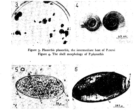
From the area pastures (Fig. 1,2) 4068 Planorbis planorbis, 2041 Valvata macrostarna, 1471 Aplexa hypnorum, 167 Lymnaea trun-ca tu la, 17 Planorbis carinatus, 6 Lymnaea auricularia, I Succinea pfeifferi snails were coIIected and werc checkcd for the natural P.cervi infection. Among these snails natural P.cervi infection was detected only in P.planorbis (Fig. 3, 4). Sixtyfour out of 4068 (1.57 %) P. planorbis was found to be naturaIIy infectcd bctween April and No-vember of 1977. Infected P.planorbis were detected each and every month; infection was found to be Icast prevalent in Apd) (0.75 %),
and more prevalent in October (2.20
%).
Infcction was detcrminedin the middle and big size P.planorbis between April to August. In the small size P.planorbis, along with the big and middlc size P.pla-norbis, infections werc dctermined after the beginning of September.
Studies on the Biology of Paramphistomum ... 55
While the infection of P.planorbis was 1.57
%,
P.cervi occurence rate found among slaughtercd sheep, during this same time span, was 80.76%.
B- Laboratory Studies
1- Charactcl'istics of P.cel'vi Eggs: P.cervi eggs (Oval, operculated,
smooth-surfaccd and white-greyish in colour) that were obtained from the mature parasites of infected sheep, were measured 121-169 microns in length and 68-95 microns in width. At the posterior pole of the eggs, a small button-like projection was generally asymmetri-cally situated (Fig. 5).
In the centre of the egg, embryo was seen in an early
segmenta-tion stage (which comrnonly consisted of 4 blastomers) and surroun-ded by yolk ceIls.
At the operculated anterior pole, egg and operculum connection secured by means of tecth that were sİtuated at the rims were detec-ted and this structure became visible in the miracidium-developed or hatched eggs (Fig. 6).
2- Facto1'S 11!/luencing the Development of P.cervi Eggs: In the
labo-ratory, factors influencing the development of P.cervi eggs such as temperature, light and variable water mcdiums were investigateci and tempcrature was found to be the most effective factor.
The development of P.cervi eggs in variable water mediums did not make any differencc. In variable water mediums having the same temperatures, hatching of miracidia occured on the same pe-riods.
A very smaIl percentage of eggs developed atı 7OC. The develop-ment process did not occur all at the same time. Therefore miracidia began to hatch on the
27
th dayand this process continued nearly one month without being in a mass manner.Eggs hatched on the 13th day at 26-27°C., and on the 12th day at 29 oC. At these tempcratures, a mass-manner hatching of miracidia occured one or two days after their first detection and continued as long as one week in a decreasing frequency.
At the temperatures of 32°C. and 35 oC., one or two miracidia were scen on the 9 th day but a mass-manner hatching of eggs occur-red on the i i th and ıo th days respectively and continued for 3-4
56 Ayşe Burgu
days. At these temperatures, higher than 30°C. an appreciable per-centage of eggs failed to hatch. At 42 oC., P.cervi eggs did not develop. Light does not seem to have any effect on the development of the mi-racidium. In the eggs incubated in the dark or light, miracidia deve-loped at the same period, if the temperatures were equaI.
P.eervi eggs were preserv'ed one month in the nJrigerator (at 4oC.) without any development, and miracidia hatched on the 11 th day after they were placed in the incubator at 29 oC.
Miracidia did not hatch from the egg euhures which their water freezed or dried totaııy. When the miracidium-devcloped eggs were placed into the refrigerator to prevent early hatehing (For obtaining miracidia on time), miracidiaemergedlateandnevcrhatchedin mass numbers after they were transferred into the laboratory conditions.
3- Development in the P.ccrvi Eggs: The transpareney of the P. eervi egg shell permitted the observation of the development. At the
26-27°C., within the first 4 days no marked changes üceurred in the
eggs except the segmentian of the embryo. On the 6 th day of the deve-lopment; length of the embryo has increased and the yolk edls deerea-sed. At this stage embryo measured an average of 47X 79 microns. On the 9 th day of development the embryo measured an average of ıı6 X63 mierons and it was located along the one side of the egg. On the 11 th day of the development, yolk cells wholly disappeared and the developed miracidium was seen in a limited aetivity. At this stage opereular line became visible in the egg,. On the 13 th day, miraeidium measured an average of 185X65 microns and, filled the egg by iıs entire lcngth. The miraeidium was very active and from time to time İts anterior part pressed the opereulum.
Aetivity of miracidium suceessively increased till the hatehing 2,ild eggs' opereulum either eompletcly feIl away from the rest of the shell or pushed outwards in a door-like fashion (Fig. 6).
4- Factors Injluencing the Hatching of Miracidiae: At first, the
".n-terior part and soon other parts of the miraeidium were introdueed through the opereular opening and emerged by thcir rapid body extentions and contraetions. At this phase, the eilial movements were seen more vigorously espeeially on the part of the body stiIl within the egg.
• Light was found to be more effeetive faetor for influeneing tht lıatching. When the completcly-miracidium-developed eggs' eulture
Studies on the Biology of Paramphistol1lum ... 57
were placcd under the light source, miracidia emerged after 15-20
minutes of exposure to light. With contrabt to this, when the eggs that were in the same stage were kcpt in total darkness, hatching procedurc was delayed markedly.
In the egg cultures on diffcrent temperatures hatching did not occur on the same day nor did they terminate on the same day. For example, some eggs hatched on 13 th day at 26-27 oC., others hatehed as Iate as one week.
P.cervi miracidium was in the shape of a torpido, covered with cilia and carricd no eyespots (Fig. 7). Their size were mcasured to be 215-253 x49-65 microns.
Newly hatched miracidia swam more activcly in the distilled water, spring water compared to tap water and principally in phy-siological saline solution.
At the 26-27°C. tcmperatures, all miracidia survived for 4.and 8 hours. Sixtcen hours after hatehing, only i110 of the mir3cidia werc found to survive but they had lost thcir activity.
5- Factors Iııj!ııeııcing the Infectian of the Snai!s: In the laboratory,
six different snail specics of various sizes with the exception of S.pfeif-feri were infected by P. cervi miracidia in various numbers and, cnly in the case of P.planarbis, development stages were deteeted.
Infection rate among small and middle sized P.planorbis is higher while their morta1ity ratc was found highcr toa.
Infection rate of P.planorbis which were infected witlı 5, ıo and 15 P.cervi miracidia was found to be 37.22, 53.37, 57.69
%
respecti-vely. It was also observed that the infection rate increased with the number of miracidium used. But, when the snails placcd into the egg cultme which contained large p,roportions ofmiracidia, snails' reaction and exercctian were increased. Thus, it was also observed that ton many miraddia had a negative effect on the P.ptmorbis infectiom.At the infection of P.planorbis with 5 miracidia performed in August, infeetion rate was found to be 37
%
and in Novembcr 34%.
Thus, no important seasonal variations were reeorded in the infection ratc. However, the development of larva i stages in the snails (From, sporocyst to emerging cercariae) was slower in November as campared to August, when they were shedding cercariae as of the 35 th day of infection. Since ccrcariae shedding of P.planorbis infected inNovem-5(\ Ayşe Burgu
.
bel' were markedly delayed and the dark pigmented cercariae of P. cervi were detected in the ~;nailtissue stilI on the 74 th day of infection.
6- Penetration c!f thc Snails by Miracidia and the Dcvelopmcnt of LarMl Stages: The transparency of the shell and mantle tissue in the young snails facilitated the observation of miracidial penetration.
Most miracidia become quite ~gitated when theyare in the vi-cinity of th~ maiL. However, same exhibit no attraction to the snail and same are attracted to the snail [aeces for a long time. Agitatcd miracidiae swim a short eliptical course around the snails and they do not leave the head, foot and tentade of the snails alone. Eventu-ally same miracidia entered from the mantle cavity and soan swam out of the snail again.
It was scen that miracidium penetrated the P.planorbis only through the mantle cavity and not from the head, foot or tentaculum.
The miracidium within the mantle cavity apply İts anterior part to the posterior mantle waıı and usuaııy within 5 minutes the anterior part is complet~ly embedded in the snail tissue. At ıhis stage, the snail reacts vigorously and this snail reaction sametimes was found to be sufficient to discharge the miracidium out of the snail. The complete process of p,~netration lasted about 15-20 minutes or more.
a) Sporocyst: Development of lan'al stages in the snail was ob-ser ved by the disscction of snails. Location and identification of spo-rocy~ts, especiaIly in the early stages of development were difficuIt to detect, but this diffieulty was reduecd in the young snails due to their scant amount of mail tissue.
At the beginning of penctration, no morphological changes were observed in the miracidium and the attaching miracidium could become free <ınd couıd swim out again but unbalanced and sJow in manner.
The miracidium embcdded its anterior part in the snail tissue, was eovered eompletely with cilia and its body showed marked swel-ling differentiations (Fig. 8).
The transformatian of miracidium into sporocyst was not imme-diatc. One to 2hours after the infection of the snail by the miracidium,
it was observcd that the miracidium was shcrter and its body was divided into two parts with a constriction and it!: cilia at the anterior part stili persisted (Fig. 9).
Studies on the Biology of Paramphistomum ... 5'1
Eariiest deteetion of sporoeysts, saeeuler in shape, was two days after the infeetion (Fig. ro).
Eight days after the infeetion the sporocysts were dongatcd or curved in shape and the developing rediae İn it were observed (Fig.
ıı).
Eadiest deteetion of mature sporocyst in the P.planorbis was twelve days after the infeetion. In this sporoeyst i i rediac wcre eoun-ted whose pharynx and İntestines eould be identified and was mca5ured to be 190oX8io mierons. (Fig. 12). Sporocysts were detceted mainly around the intestines and in the mande tissu~.
b) Rediae: Earliest detection of rediae, free of sporoeysts, were
12-13 days after the infection. The£e young rcdiae wcre opaquc,
sligh_tlycurved in shaJ?e and had a limited activity (Fig. i3). In the young rediae the pharynx and the inh~stiiıes were "isible and they contained embryo balls from whieh cercariac or daughfer rediae would develop (Fig. 14). It was ob~:;rved that a week was necessary for the birth pore to become vi~ible. RediCi.ewere defce(ed around the intestines, hepato-pancreas and in the man tle tissue of the P. planorbis.
The developing rediae showed great variations in ~ize and the ma turc rediae measured 700- 115° microns in lengh t and 125-25°
microns in width.
c) Daııgkter rediae: The daughter rediae were obsen'cd in P. planorbis in different months of the year. The produetion of daughter rediae were less compared to the rcgular produetion of cereariae. Daughtcr rediae were cbseıved after 40 days of the infcction. it was observcd that, the loealization in the snail tissuc was an important faetor rather than the scasonal factors for the produetion of daughter rediae. Mostly daughter rcdiae were developed from the rediae whieh
\
were situated at the lateral part of the mantIc tissuc of the snail and they wcre easily identified at the first external circle of the snail befare its disseetion.
Developing daughter rediae were distinguished from the deve--- laping eereariae by their pharynx, their whitish-grey colour and the ir
longitudinal arrangement in the body of the rcdiae (Fig. i5, 16). d) Factors Inftuencing tke Emergency of Cercariae: Early fi'~e imma-turc cereaı iac werc deteeted in the snail tissuc after 24 days of the
in-60 Ayşe Burgu
fection. Thus, immature cercariae wcreliberated from rediae before thcir complete development. Like the rediae the developing ccrcariae show great variability in size at the beginning. In the young cereariae,
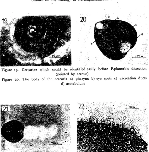
the body was markedly small, eye spots were prominent and pigment was onlyaccumulated around thcm. The tail was short and wide rather than long (Fig. 17).
W'ithin the next few days, the immature cercariae gradually inc-reased in size and the tail beeame longer. The eye spots were very prominent and dark. The pigment around them were like irreguler blotehes and soon pigmentation coV(~red all the body in rows and branche~ (Fig. 18).
The mature cercariae were large, quite active and dark pigmen-te'd so, they were easily recognized before the P.planorbis dissection (Fig. ıg). Their body was 300-480 mierons long and 180-325 microm wide while the tail was 425-510 microns long and 75-80 microns wide. The mature cercaria is a vigorously swimming organism and its body made relaxation and contraction movements. it has a pear-shaped body, and its posteriar part is wider than its anterior (Fig. 20).
The tail is cylindrical and tapers graduaIly towards the posterior cnd. "Vhen the c;;rcariae were placed into half diluted India ink sus-pension, the mucoid-fin, which is on and around the tail, (Fig. 21)
becamc visiblc. In the matUl'e ccreariae the mucoid [in were seen comtant in slıape but in the immatUl'e cereariae, this structure was not reeorded espeeiaııy betüre emergeney. Cercariae swimming di-rection is mostly circular but they mayaıso swim straight or in zig-zag lines. They rise graduaIly to the water surfaee and graduaııy des-cend again and cover a long distance in a very short time.
it was seen that light was a great factor in shedding cercariae. Infected snails kept in the dark started shedding cercariae again after being exposed to liglıL
Dnder natural day light, snails continued to shcd many hours but in smaIl numbers. Dnder ;,trong iııumination, large numbers cr cercarİae emcrgcd during the second or third hour af ter exposnre of the snails to the light. Snails that had released eercarİae wıder strong iııuminatİon, required a rest period at lrast for about 2 or 3 days.
During the experiments, cereariae were eollected from the snails onlyone day in a week in orda to regulate met.:1.cercarial age confor-mity and to obtain them İn high numbers.
Studies on the Biology of Paramphistomum ... 61
Some infeeted P.planorbis continued to shed eenariae İn the la-boratory more than 13 months and some snails wcre frec of the infee-tion after i i months.
7- Fadors InflueTteing on the Encystınent of Cereariae: Shortly after
reIeasing most ecrcariae commence eneystation and become meta-eercariac (Fig. 22). Dndcr thc light saul'ce and, in the prescnee of vegetation in th~ water most eercariac eneyst011 the vegetatio,ı within
15-20 mİnutcs.
Cereariac leaving the snails formed into metaeereariac-mostly on the sidcs of a pctri dish and rarely at the bottom-lined with a plas-tic sheet-within 25-30 minutes. Thus, it was seen that the eneystation was slightly delayed in the abscnee of vegctation. Most metaeereariae wcre scen near the surfaee of the water on the plastie sheet and espe-eiaııy on its eurved parts.
When the green leavcs were piaeed between plastie sheet and petri dish, cereariae eneysted on the same par ts as cxplained above, thus they had no effect on the eneystation.
When the petri dishes were pIaeed under the light at a distanee less than 30 cm., most of the metaeereariae were not eneysted due to the inercasing temperature of water in the petri dish.
In our studies during the eolleetion of metacereariae, same me-taeereariae were ingested by P. pIanorbis. But these were diseharged by snail faeeal material and on their examination, they wcre found viable.
During the eneystment eerearia attaehes wjth its ventral surfaee, and the makrial for eystation begin to seerete from the pores aıı over the body (Fig. 23), whiIe the taiI mavement inercases. Tail breaks off the body of the eerearia-at the the earIicst-wjthin 3-5 mİnutes, but free tail motion eontinues for several hours. By the cnd of20 minutes,
a eyst formes around the ecrearia. At first, the eyst wall was soft and fragile, but become hard after 2-3 days.
Metaeerearia was in the form of a halfsphere and was surrounded with a thiek layer of a eyst walI. Thcse metaeereariae were measured 210-255 mierons in diameter (Fig. 24).
Newly formed metaeercariae lost most of their aetivity 2 weeks
after eneystation but stilI same of their aetivity can be observed a very long time on thcir eareful examination under the strong light
mie-62 Ayşe Burgu
roscope. Alsa eyc-spots of metacereariae become invisible 2 weeks
after eneystation.
, One fifth of the mctacaeariae kept among wct filter paper in the refrigerator kcpt their viability for about six months. Deaths star. ted after the second month. In the dead metaeereariae, embryo was ~;ecnto shrivel up and turn gray but the eyst wall persisted unchangcd.
8- Experimental Iıifeetion of Lambs with P.eervi Metaeereariae: Fif-teen lambs were cach infected with 3-20 days old 1000 P.cervi me-tacercariae. No clinical symptoms were observed after the administ-tration of such adosc.
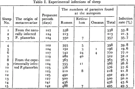
Prepatent period was found to be 102-142 day s in the infected lambs and at the end of thcse periods autopsies wcre performed within one week. Parasites were dctected in the rumen, less frequentIy in the rctieulum and in the omasus. No parasites were deteeted in the abo-masus and in the smaIl intestines. A minimum of 196 and a maximum of 77° P.eervi werc found from each infeeted animaL. The infectian rate was determined to be r9.6-77
%
(Table I).Parasites wcrc kept alive 7-8 hours in a warm (37°C.) saline so-lutian and their lenghts and widths were measured to be 3.5-5.5 mm. and r.8-2.2mm.respeetively afterfixation with 5
%
formalin.Tablo i. Experimental infections of sheep
The numbers of parasites found Prepatent at the autopsics
Sheep The origin of periods Reticu- Infcetian No. metacercariae (days) Rumen lum Omasus Total rate (%)
--- --- --- ---- ---
----i From the natu- 107 538 538 53.8 2 raııy infected 124- 213 213 21.3 3 P. planorbis 124- 350 7 357 35.7 ---4 102 393 5 398 39.8 5 104- 192 3 i 196 19.6 6 107 74-2 24- 4- 77° 77.0 7 Lig 282 4° 322 32.2
8 From the expe- 121 363 363 36.3 9 rimentaııy infec- 124- 355 ii 366 36.6
ıo ted P.planorbis 125 376 376 37.6
II 125 210 8 218 21.8
12 125 492 492 4-9.2
13 127 500 500 5o.0
14- 14-1 436 4-36 43.6
Studies on the Biology of Paramphistomuın ...
Discussion
63
lt is stated that the intel'mediate host of P.cervi varies according to the geographical regions. However, Planorbid type snails play the main role ofan intel'mediate host (r, 3, S, 6, g, LO, 18,27,28). In our
study P.planorbis were recorded as a intel'mediate host for P.cervi. Heavy infestation of the snails with varİous paramphistomes are reported mainly in the Iate summer and autumn months and the İn-cidence varics betwcen 3 to 7S
%
in the infected arcas (4, 5, 6, g, ıg, 30). In our study naturaIly infectcd P.planorbis were found each and every month between April and November; infection was found to be lcast prevalent in April (0.7S%)
and most prevalent in Octabel'(2.20
%).
Infection was determined in the middle and big size P.planorbis between April to August. In the smaIl size P.planorbis along with the big and middle size P.planorbis, infections were de-termined after the beginning of Scptembel'. These results have shown that the sheep could contract the infectian from the infected pa:ıtures as soan as they begin to graze in the spring months. These results have alsa shown that the snails were infected much earlier and were able to endure the winter months while they were aıready infected.
Güralp (I S), reported that the Çifteler State Farm and the sur-raunding area is one of the areas where P.cervi is widespread in sheep in Turkey. Alsa wc found that the occurence of P.cervi in sheep was an average of 80.76
%,
contrary to this, the infectian rate of P.pla-norbis was at an average of i.S7%.
it was noted that the main factor for the development of P.cervi eggs was temperaturc rather than light P2, 21,23). Similar results were obtained from our studies and alsa the development of P.cervi eggs in variable mediums did not show any differentiation provided that all the water mediums had the same temperature.
Light is an important factor which influence the hatching. P. cervi miracidia are 240 microns in size and same miracidia of
Pa-ramphistomum species kcpt their viability longer in lakes and pools compared witIı other media. Alsa it is note that tap water is a good medium than distilled water and saline solution (o.g %) for same Paramphistomum miracidia (7, 2i, 23). In our study light was found
to be a most effectiye factor for hatching and it was seen that newly emerged miracidia swam more active in distilled water and spring water. Their size was measurcd to be 215-253X49-6S microus.
Ayşe Burgu
It was recorded that too many miraddia had a' negative effect on the experimental infection of snails and the young (little or me-dium sizcd) snails were suitable for infcetian (13, 23). During the experiments it was observed that the infection rate increased with the number of miracidium used. But contrast to this, it was seen that the large proportions of miracidia had a negative effeeL On the sub-ject of snail size, it was seen that the infeetion rate among the smaIl and middle sized P.pJanorbis is highcr while their mortality rate is higher too.
Generally it was aeeepted that the miracidia lost their ciliadu-ring the penctration and entered the snail host tissue as a young spo-rocyst. On the observation of miracidial penetratian into the snails, both mcchanic~l and enzymatic effeets together play a role (21, 23). The transformatian of the miraeidium into a sporocyst is a gradual process and no cIear line can be drawn betwecn the invading miraci-di um and young sporoeyst (8). In our stumiraci-dies persistence of eilium of the anterior part of the miraeidium was alsa observed, 1-2 hours af ter
the infection of snails.
On the development of P.cervi in the snail host, a very limited avaible information is present, but our observation are in conformity with the papers of Kraneburg (2ı).
In same Paramphistomum species, the oceurenee of daughter rediae were reeorded only in Autumn months (20). In our experi-m~nts, however, the daughter rediae were observcd in P.pJanorbis in
different months of the year. Yet, the production of daughter rcdiae were less compared to the regular produetion of cereariae and they were observed after the 40 tlı day ofinfeetion. In spitc of the fact that there were no records on the literatuI'e, in our studyalsa it was obser-ved that, the localization in the snail tissue was alsa an important factor for the production of daughter rediae. Beeause the daughter rediae developed mainly from the rediae where they developed in the external circle of the snail mantıc tissuc. Thcrefore it is possible to detect the rediae whieh carricd daughtcr rediae from these external lateral parts of the mantle tissue of P.planorbis before its dissection on the examination under the stcromicroscopc.
It has been rceordcd that the ıight was the most important fac-tar for cercarial shedding of infected snails and the snails eould con-tinue to shcd abaut 1-10 months (21, 23, 29). We obtained similar
Studies on the Bİology of Paramphİstomum ... 65
results and wc saw that the infected P.planorbis continı.led to'shed cercariae in the laboratory more than 13 months and some snails were free of the infection after 11 months.
Paramphistomum cercariae can be encysted on the vegetation and other subjects in the water and they can stay viable for about 5 months when theyare preserved in the refrigcratQr (1, 3, 21). In our stud;e.>encystation occured on the plastic sheet which was plaeed in the petri dish and especial1y around its folds. It was also observed that one fifth of the metacercariae kept among wet fİlter paper in the refri-gerator with a temperature of 4 oC. kept. their viability for about 6 months, deaths started after the second mantlı ..
In the P.cervi infection prepatent period was ):eported 1.0be 129 days in goats, I03-1 15 days in cattle and sheep and the infection ra1.e was 45.82 % in goats, 40.6 % in sheep and 44.8 %in cattle (2,22); In our study it was determined that the prepatent period was
I02-142 days and the İnfection rate was 19.6-77 % in lambs.
References
1- Abdel Ghani, A.F. (I 960): The cercaria and the metacercaria of
Paramphistomum cervi. Agric.Rcs.Rev., Cairo, 38, 237-243.
2- Abdel Ghani, A.F. (1960): Experimental infeetion of animals with
Faseiola and Paramphistomı.m ~yst. Agric.Res.Rev., Cairo, 38, 275
-28
7.
.
3- Altaif, K.I., S.N.Al-Abbassy, I.M.Al-Saqur and A.K.Jawad
(1978): Experimental studies on the suitability of aquatic snails as intermediate hosts for ParampJıistomum cervi in Iraq. Ann.trop.Med:
Parasit., 72, 151-155. . ,
4- Arfaa, F. (1962): A study on Paramphistomum microbothrium in
Khu-zistan S. W. Iran. Annls Parasit.hum.comp., 37, 519-555.
5- Arru, E. e S.Deiana (1962): Sull' andamento stagionale della paramfistomosi intestinale (Paramphistomum eervi) dei caprini e degli
ovini in Sardr:gna. Atti.Soc.itaI.Sci.vet.,16, 231-232.
6- Arru, E. e S.Deiaiıa (1969): eli ospiti intermediti Paramphisto-mum (P.cervi) in Sardegna.Atti Soc.ital. Sci.vet., 23, 909-912. 7- Colvin, Jr. H.J. (1962): The longevity of miracidia of
Paramphis-tomum mierobothrioides in various media. Proc. La Acad. Sci., 25,
66 Ayşe Burgu
8~' Daw~ B. (1960):, The penetration of Fasciola' hepatica into Limnea
tTUncatula and of F.gigantica ii/to L.aııricıılaria. Trans.Soc.trop.Mcd.
Hyg., 54, 9-10•
'9- Deiana, S. (1953): L'infestione del Bulinus contortus 'da cercarie di Schistosoma bovis (Sonsino, 1876) e del Paramphistomıım an'i (Sch-rank, 1790) in diverse stagionidell' anno. BioI. sper., 5, 1939~i940.
İ0- Dinnik, J.A~ (195i ): An intermediate host of the. common .s.tomaclz
fluke, Paramphistomum cervi (Schmnk), in Kenya. E.Afr.agric.
J.,
26, 124-125.11- Dinnik, j.A. and N.N.Dinnik (1957): Development
~f
Paramp-histonıum sııkari Dinnik, 1954 (Tremaloda: Pf!wmphislomidae) ir, a snail host. P<ı.rasitoIogy, 47, 209-216.~2- Dinnik, j.A. (I 958): Identification of liD-erf/1Ike and stomac1ı f/ııkt! eggs recoveredfromfaeces of inJested al/imals. BulI. cpizoot.Dis. Ali'.,
6, 135-138. "
13- Dinnik j.A. and N.N.Dinnik (1962): The growıh of Paramp-histomum microboıhrium Fischoeder to matııril] and iis longeuil)' in (aU/e.
BulI. epizoot.Dis.Afr., 10, 27-31. 14- Evans, N.j. (19n): Kişisel habeı1eşme.
15-
Güralp, N. (1974): Helm intoloji. Vet. Fak. Y"-yın. Ankara [;11iv., No. 307/208.16- Horak, I.G. (1971): Paramphistomiasis of domestic ruminants. Adv.
Para~it., 9, 33-72. '
i
7- Kamburov, P.tl
976): On the species of paramphistomidsoccurriııgin Europe. Third Int. Symposium,' Kosice, Theses of reports.
~
18~ K~ın1;Jur~v,.P., I. Vassilev and P. Saınnaliev (197°): The . species composition oj Paramphistomidae Fischoeder, ı9°i, in Bulgaria.
KhelmintoIogiya, 'Sof., i, 19-22 (Ref: Helminth. Abst., 1978,
, 47; 3497).
19- Kanyar, R.D. and R.T. Varshney (1963): Amphistomiasis in sheep and goals in UUar Pradesh. Indian ].vet.Sci., 33, 94-98". 20-' Kıselev, N.P. (1967): The biolog)' of Paramphistomı;m ichikawai
Fııkui. Veterinariya, Moscow 44, 5 1-53 (Ref: HeIminth. Abst., 1969, 38, 1035)'
21- Kranebu~g, W. (19n): Beitriige zıtr Biologie und Pathogenitiit des einheimischen Pansenegels Paramphistomum cervi. I.
Erıtwicklur,gssla-Studies on the Biology ofParamphistomum ... 61
dien in der Aııssenwelt und im ,(wisehenwirt. B'~rI.Münch. tierarzt!. "\Vschr., go, 316-320. '
22- Kraneburg, W. und
J.
Boch (1978): Beitriige zur Biologie vndPathogenitiit des einheimisıhen Pansenegels Paramphistomum eervi. :3. E1ıtwieklung in Rind, Sehaf und Reh. BerI. Münch. tierllrzt!. ,)'Vsc1:ır.,.
91, 71-75.
23- Lengy,
J.
(I960): Study of Paramphistomum microbothtium Fischo.1der1901, A rumen parasite of eaU/e in [srael. BuII.Rcs.Coun.Israel, B 9,
7I~I3°'
24- Sey, O. (1974): On the speeies of Paramphistomum of eattle and sheep in Iwngaı)'. Acta vet.hung., 2,1, 19-37.
25- Sey, O. (1975): Some remarks on tlze European speeies of
Paramphis-tomum havin,f{ mierobothrium type
rif
genital atrium. Second European1fulticolloquy of Pm"asit" Trogir, Summaries, 76. 26- Sey, O. (1977): Kişisel haberleşme.
27- Sey, O. (1977): E.ı:amination of amphistomes (Trematoda: Paramp-histomata) parasitizin/!, in Eg)'P1ion ruminanls. P?rasit. hung., l0,
.47-5° (Ref: Hdminth, At)st., 1979, 48, 1633).
28- Sey, O. a,nd E. Arru (1977): A review of speeies of
Paramphisto-mum FiscllOeder, i90i oeeııning in Sardinian domes/ie mminants. Riv. Parassit., 38, 295-30r.
29- Varnıa, A.K. (1961): Observations on the biology and pathogeniei~y' of Cotylophoron eOlJ'lopho1'llm (FiscllOeder, 1901) .f.Hdminth., 35, 161--168.
30- Zharikov I.S. (I964): Ecologieal and parasitologieal study of motlıı-ses of the ge1H1sPlanorhis in the ,Hinsk region. Sb. nauch. Trud.
bclo-russk. n;ı.uchno-.issled. vet.Imt., 99-102 (Ref: HcIminth. Abst., 1965,34, 1734).
69 Ayşe Burgu
Figure'1. The lrough and iıs conıinuation of wel area in Çifteler Sıate Farm, where snails wcre colIecıed
Figure 2. Canals for irrigation carrying intermediate hosl snails in Çifteler
4
O;S ",m' ~-.-~
Figure 3. Planorbis planorbis, the intermedialc host of P.cervi Figure 4. The shcll morphology of P.pl~norbis
Fıgure 5. The P.cervi egg a) operculum b) slight projection cL embryo
Figure 6. The miracidium-dcveloped egg a) operculum and its connection with egg secured by means of lectlı b) slight projection c) miracidium
Studies on the Biology of Paramphistomum ... 69
, Figure 7. The miracidium
Figure 8. The miraeidium at the beginning of the penetralion process
F:gurc g. The miracidium one hour after the infection Figure ıo. The sporocyst two days after the infection
Figure iı.The sporocyst eight days after the infeelion
7Ô Ayşe Burgu
i
Figure ı3. The young redia 14 days after the infcetian a) pharynx b) embryo baIls Figure 14. Theredia containing the develaping cercariae .22days after thejnfection
a) pharynx b) develaping ccrcariae
Figure 15, The ant,erjor part of the mature rcdia containing the develaping cercariae a) eye spots of cercaria
Figure ıG. The anterior part of the mature redia containing the develaping daughter redia. a) pharynx of daughter redia
•\
.." J...•....
~.~
~L.
Figure ı7. The smaIl and short taiIcd young cercariae Figure ı8. Cercariae in different dcvelopmcnt stages
Studies on the Biology of Paramphistomum ... 71
Figure ıg. Cercariae wlıich could. be identified easily before P.planorbis dissection (pointed by arrows)
Figure 20. The body of the cercaria a) pharynx b) eye spots e) excreation ducts d) acetabulum
Figure 21. The mucoid fin around the tail 'of the cercariae Fıgme 22. Cystogenous secretion during the process of encystment
Figure 23. P.cerv; metacercariae on the lettuce leave . Figure 24. The metacercariae