Contents lists available atScienceDirect
Neuroscience Letters
journal homepage:www.elsevier.com/locate/neulet
Research article
Isoflurane versus sevoflurane for early brain injury and expression of
sphingosine kinase 1 after experimental subarachnoid hemorrhage
Orhan Altay
a, Hidenori Suzuki
a, Bilge Nur Altay
c, Vahit Calisir
d, Jiping Tang
a, John H. Zhang
a,b,*
aDepartments of Physiology, Loma Linda University School of Medicine, Loma Linda, USA bNeurosurgery, Loma Linda University School of Medicine, Loma Linda, USA
cLokman Hekim University School of Medicine, Ankara, Turkey
dDepartment of M.T. Management Engineering-Statistical Analysis Iskenderun Technical University, Iskenderun, Turkey
A R T I C L E I N F O Keywords:
Subarachnoid hemorrhage Early brain injury Isoflurane Sevoflurane Sphingosine kinase-1
A B S T R A C T
The first step to treat aneurysmal subarachnoid hemorrhage (SAH) is aneurysmal obliteration under general anesthesia but not treat the SAH itself and the secondary effects. However, the identification of anesthetics with properties that help to attenuate post-SAH brain injury can be useful for improving outcomes of SAH patients. We examined whether 2% isoflurane and 3% sevoflurane posttreatment are protective against early brain injury (EBI) after SAH. This study used 87 8-week-old male CD-1 mice. We induced SAH by endovascular perforation in mice. Animals were randomly divided into 4 groups: sham-operated (n = 16), SAH + vehicle-medical air (n = 26), SAH + 2% isoflurane (n = 22), and SAH + 3% sevoflurane (n = 23). Neurobehavioral function, brain water content and Western blotting were evaluated at 24 h. The expression of sphingosine kinase (SphK), cleaved caspase-3 and cyclooxygenase-2 (COX2) was determined by Western blotting. Cell death was examined by terminal deoxynucleotidyl transferase-mediated uridine 5′-triphosphate-biotin nick end-labeling staining. Both 2% isoflurane and 3% sevoflurane significantly improved neurobehavioral function, and brain edema at 24 h after SAH and attenuated cell death, associated with an increase in SphK1, a decrease in cleaved caspase-3 and COX2. The neuroprotective effects were similar between 2% isoflurane and 3% sevoflurane. These findings suggest that both 2% isoflurane and 3% sevoflurane significantly inhibited EBI by suppressing post-SAH apoptosis and brain inflammation possibly via the SphK1-related pathway.
1. Introduction
Subarachnoid hemorrhage (SAH) is a life-threatening disease with a risk of sudden death. A majority of deaths occur within the first 48 h post-ictus owing to the impact of the initial bleeding [1], while most of ruptured cerebral aneurysms have been successfully managed with surgical clipping or endovascular coiling [2]. However, the interven-tions treat the aneurysm but not treat SAH itself and the secondary effects. After insult, cumulated subarachnoid blood, elevation of in-tracranial pressure and reduced cerebral perfusion initiate an acute injury cascade such as microvascular disturbance, apoptosis, in-flammatory reaction (cytokine/chemokine production), leading to early brain injury (EBI), one of the important causes of unfavorable outcomes after SAH [3].
The vast majority of SAH patients are exposed to volatile anesthetics used for neuroanesthesia such as isoflurane and sevoflurane during the aneurysmal obliteration and in the intensive care unit. Isoflurane and
sevoflurane are noncombustible halogenated (fluorinate) and lipophilic molecules, and isoflurane has been shown to increase membrane fluidity and activate sphingomyelin hydrolysis [4,5]. These volatile anesthetics have been reported to have short-term neuroprotective ef-fects against focal and global ischemia/hypoxia [6,7]. The identifica-tion of anesthetics that help to attenuate post-SAH brain injury is clinically highly relevant. Suitable anesthetic drugs should show a rapid recovery profile and be free of complications. In this regard, sevo-flurane is more preferable than isosevo-flurane because it has lower irritation of the respiratory tract than isoflurane [8,9]. But, sevoflurane's poten-tial for toxicity due to its relatively high rate of metabolism and its reaction with carbon dioxide absorbents has been a source of con-siderable concern [9].
Isoflurane is neuroprotective and the most studied one among the volatile anesthetics [10–13]. We recently demonstrated that one-hour 2% isoflurane administration at 1 h after SAH prevented post-SAH neuronal apoptosis, blood–brain barrier (BBB) disruption, and brain
https://doi.org/10.1016/j.neulet.2020.135142
Received 29 October 2019; Received in revised form 2 June 2020; Accepted 5 June 2020
⁎Corresponding author at: Department of Physiology, Loma Linda University School of Medicine, Risley Hall, Room 223, Loma Linda, CA, 92354, USA.
E-mail address:[email protected](J.H. Zhang).
Available online 06 June 2020
0304-3940/ © 2020 Elsevier B.V. All rights reserved.
inflammation through sphingosine-related pathway including sphin-gosine kinase (SphK) expression and sphinsphin-gosine-1-phosphate (S1P) receptor activation in mice [7,14,15]. S1P is generated from ceramide, an integral component of plasma membranes, which is produced via the pathways including the de-novo synthesis and the sphingomyelinase-mediated hydrolysis of sphingomyelin, by the sequential action of ceramidase and SphK [16]. Many factors can alter SphK activity and regulate subsequent S1P levels [16,17]. S1P regulates diverse biological processes including cell survival and anti-inflammatory effects after binding to S1P receptor-1 to 5 [17]. Cyclooxygenase-2 (COX2) an in-ducible isoform is thought to mediate inflammatory events in response to inflammatory stimuli, and is a critical factor in the cytotoxicity as-sociated with inflammation [18]. The interaction of SphK and COX2 is also reported [19]. Another group reported that isoflurane increased SphK1 mRNA expression and SphK activity in both in vivo murine model of renal ischemia-reperfusion injury as well as an in vitro model [20]. In the literature, posttreatment with sevoflurane has also de-monstrated that it is protective against focal cerebral ischemia and reperfusion injury in rats [21,22]. However, it remains unknown whether the impacts of sevoflurane or isoflurane have superiority over each other in SAH, and what biological processes are associated with sevoflurane’s effects on SAH brain.
In this study we tested two hypotheses; 1) sevoflurane posttreat-ment attenuates post-SAH EBI through antiapoptosis and anti-in-flammation in mice; and 2) the signaling pathways of sevoflurane-mediated protection involve SphK expression like isoflurane.
2. Materials and methods
2.1. Experimental design and animal groups
The animal and ethics review committee at Loma Linda University approved all protocols. Eighty-two 8-wk-old male CD-1 mice (30–38 g; Charles River, Wilmington, MA) were used for the study.
To examine whether sevoflurane attenuated EBI after SAH and to compare the effects with those of isoflurane, animals were randomly divided into 4 groups, and evaluated at 24 h: sham-operated+30% O2+70% medical air (O2-medical air; sham group, n = 16), SAH + O2 -medical air (vehicle group, n = 23), and SAH + 2% isoflurane + O2 -medical air (2% isoflurane treatment group, n = 21), and SAH + 3% sevoflurane + O2-medical air (3% sevoflurane treatment group, n = 22).
2.2. Mouse SAH model
SAH endovascular monofilament perforation model was produced as described previously [14]. Briefly, animals were anesthetized with an intraperitoneal injection of ketamine/xylazine (100/10 mg/kg), and all animals breathed spontaneously throughout the procedures. A sharpened 4-0-monofilament nylon suture was advanced through the internal carotid artery (ICA) to perforate the anterior cerebral artery. In the sham surgery, the filament was advanced 5 mm through the ICA without perforating the artery. Body temperature was kept constant (37.5 ± 0.5 °C) during the operation.
2.3. Drug administration
One hour after SAH induction, 2% isoflurane or 3% sevoflurane (Baxter, Deerfield, IL) was continuously administered with a properly calibrated vaporizer in an anesthetic chamber (the lid with a tough, reliable clip-top, 240 × 140 × 120 mm) for 1 h with 30% O2and 70% medical air.
2.4. Severity of SAH
The severity of SAH was blindly evaluated using the SAH grading
scale by Sugawara et al [23] at sacrifice. Briefly, using high-resolution pictures of the base of the brain depicting the circle of Willis and basilar arteries, cisterns including the arteries were divided into six segments, and each segment was allotted a grade from 0 to 3 depending on the amount of subarachnoid blood clot in the segment: grade 0, no sub-arachnoid blood; grade 1, minimal subsub-arachnoid blood; grade 2, moderate blood clot with recognizable arteries (the circle of Willis and basilar arteries); and grade 3, blood clot obliterating all the arteries within the segment. The animals received a total score ranging from 0 to 18 after adding the scores from all six segments. The grading system is rather subjective but has high reproducibility and consistency be-tween investigators [23]. Four mice with SAH grading scores ≤7 in-dicating mild SAH were excluded from the study, because the mice with mild SAH had no significant brain injury as measured by neurological scores and brain water content (BWC) in our previous studies [7,14,15].
2.5. Mortality and neurological scores
We calculated mortality at 24 h after SAH. Neurological score was blindly evaluated at 24 h after SAH as previously described [7,14,15]. The evaluation consisted of six tests that were scored 0–3 or 1–3. The six tests included: spontaneous activity; symmetry in the movement of all four limbs; forelimbs outstretching; climbing; side stroking; and response to vibrissae (whisker stimulation). Animals were given a score of 3–18 in 1-number steps (higher scores indicate greater function).
2.6. BWC
Brains were quickly removed and separated into the left and right cerebral hemispheres, cerebellum, and brain stem, and weighed (wet weight) at 24 h (n = 6 per group) after surgery. Next, brain specimens were dried in an oven at 105 °C for 72 h and weighed again (dry weight). The percentage of BWC was calculated as ([wet weight-dry weight]/wet weight) X100% [7,14,15].
2.7. Western blotting
The left cerebral hemisphere (perforation side) at 24 h after SAH was isolated and used (n = 5 per group). Western blotting was per-formed as previously described [7] using the following primary bodies: SphK1 (1:250, AP7237C, Abgent, San Diego, CA), anti-caspase-3 (1:200, sc-7148, Santa Cruz Biotechnology, Santa Cruz, CA) and anti−COX2 (1:1000, ab15191, Abcam, Cambridge, MA) anti-bodies, followed by goat anti-rabbit secondary antibodies (sc-2004, Santa Cruz Biotechnology).
2.8. Terminal deoxynucleotidyl transferase-mediated uridine 5′-triphosphate-biotin Nick End-labeling (TUNEL) staining
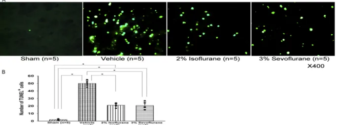
TUNEL staining was performed as previously described [7]. Briefly, animals were euthanized 24 h after surgery (n = 5 per group). The mouse brains were fixed by cardiovascular perfusion with phosphate-buffered saline and then 10% paraformaldehyde, and post-fixed in 10% paraformaldehyde followed by 30% sucrose (weight/volume) for 3 days. Ten-micron-thick coronal sections at the level of bregma 1 mm (caudally) were cut on a cryostat (LM3050S; Leica Microsystems, Bannockburn, Ill). TUNEL staining with an in-situ cell death detection kit (Roche, Mannheim, Germany) was performed. TUNEL-positive cells in the left (perforation side) basal cortex were counted in two fields per case at ×400 magnification and expressed as the mean number of TUNEL-positive cells with a fluorescence microscope [7]. The mor-phology in the basal cortex of the same model was reported before in detail [24].
2.9. Statistics
Data were expressed as mean ± SD. After confirming that each population being compared followed a normal distribution using Shapiro-Wilk W tests, statistical differences were analyzed using one-way analysis of variance (ANOVA) followed by the Tukey test. Differences in mortality were tested using Fisher’s exact tests. P < 0.05 was considered statistically significant.
In the statistical analysis, we calculated the power of the tests. The
number of animals per group necessary to reach the desired power of 0.800 was in the range of 4 to 6.
3. Results
3.1. Mortality and SAH grade
The mortality rate was not significantly different among the vehicle, 2% isoflurane and 3% sevoflurane treatment groups (30.4%, 7 of 23 mice; 23.8%, 5 of 21 mice; and 27.3%, 6 of 22 mice, respectively). No sham-operated mice died. There was no significant difference in SAH grade among the vehicle and the treatment groups at 24 h (p = 0.435, Fig. 1A).
3.2. Neurological score and BWC
In this study, neurological score was not deteriorated in the sham group, and significantly lower in the vehicle group compared to the 2% isoflurane (P = 0.007) and 3% sevoflurane (P = 0.036) groups (ANOVA;Fig. 1B). BWC in the left hemisphere was significantly higher in the vehicle group compared to the sham group, and was significantly improved in the 2% isoflurane (P < 0.001) and 3% sevoflurane (P = 0.001) groups (ANOVA;Fig. 1C).
3.3. Effects of isoflurane and sevoflurane on SphK1-related antiapoptotic pathway
Western blot analyses showed that SphK1 expression was sig-nificantly increased in the 2% isoflurane and the 3% sevoflurane treatment groups compared with the sham (P = 0.001, P < 0.001, re-spectively) and the vehicle (P < 0.001, P < 0.001, rere-spectively) groups (ANOVA;Fig. 2A). The SphK1 expression was not different between the 2% isoflurane and 3% sevoflurane treatment groups (P = 0.499). Al-though expression of cleaved caspase-3 was significantly increased in the vehicle group compared with the sham group (P < 0.001), 2% isoflurane and 3% sevoflurane significantly reduced cleaved caspase-3 expression compared to the vehicle group at 24 h after SAH (P < 0.001, P < 0.001, respectively; ANOVA; Fig. 2B). There was no difference observed in cleaved caspase-3 levels between the 2% isoflurane and 3% sevoflurane treatment groups (P = 0.999).
Consistent with Western blot results, immunofluorescence analyses revealed that TUNEL-positive cells in the left basal cortex were more prominent in the vehicle group compared to the treatment groups (Fig. 3A, B), although the positive cells were hardly observed in the sham group.
3.4. Effects of isoflurane and sevoflurane on COX2 expression
To determine whether 2% isoflurane and 3% sevoflurane have a different effect on inflammation, we investigated the expression of COX2. The expression of COX2 in the vehicle group was significantly higher than the sham group (P < 0.001) and both treatment groups (P = 0.003, P = 0.004, respectively). There were no differences in COX2 expression levels between the 2% isoflurane and 3% sevoflurane treatment groups. (P = 0.999;Fig. 4).
4. Discussion
Apoptosis and inflammatory reaction are key pathologic manifes-tations of EBI after SAH, causing poor outcomes [3,7,14,15]. Apoptosis is an active and complex process triggered by protein-regulated pro-grammed cell death, which typically shows shrinkage of the cell body and cytoplasm with membrane blebbing [25]. The presence of activated caspase-3 reflects the execution of the apoptotic program in damaged neuroglial cells [25]. After endothelial and neuroglial apoptosis, BBB dysfunction may allow greater influx of blood-borne cells and
Fig. 1. Subarachnoid hemorrhage (SAH) grade indicating the severity of SAH
(A), neurological score (B), and brain water content (C) at 24 h post-SAH. Values are mean±SD; *P<0.05, ANOVA.
substances into brain parenchyma, thus amplifying inflammation, leading to further parenchymal damage and edema formation after SAH. COX2 mediates inflammatory events in response to inflammatory stimuli. We reported that 2% isoflurane post-treatment reduces apop-totic cell death, brain inflammation and EBI after SAH [7,14,15]. However, it has been unknown whether sevoflurane has protective ef-fects against apoptosis, inflammation and EBI after SAH.
Volatile anesthetics have been shown to be protective against
ischemic brain injury when used as pre-treatment and post-treatment [10–13,21,22]. However, pre-treatment is clinically feasible only when the occurrence of brain ischemia or SAH is predictable. In reality, the onset of most of cerebral insults occurs outside the hospital and is not predictable. So, we administered the treatment after SAH. The fol-lowing different mechanisms underlying beneficial effects of isoflurane or sevoflurane have been proposed in brain tissues of ischemic injuries: modulation of excitotoxicity [26,27] or mitochondrial adenosine tri-phosphate-sensitive potassium channels [13,22], induction of inducible nitric oxide synthase [11], and activation of sphingosine-related pathway including SphK1 and S1P receptors [7]. SphK1 phosphorylates sphingosine and decreases pro-apoptotic sphingosine levels to form S1P in macrophages, vascular endothelium and neuroglial cells, and the product S1P activates a potent antiapoptotic and anti-inflammatory signaling cascade [27–30], in addition to promoting cell growth and survival [31]. The present study showed that 2% isoflurane and 3% sevoflurane post-treatment improved neurological score and brain edema, associated with increased SphK1 expression and decreased ex-pression of activated caspase-3 and COX2. That is, 1 -h 2% isoflurane and 3% sevoflurane administration at 1 -h post-SAH equally attenuated EBI at 24 h, and the antiapoptotic and anti-inflammatory effects might need at least partly sphingosine-related pathway activation including SphK1. However, increased SphK1 expression is not necessarily equivalent to increased SphK1 activity and therefore an increase in the potentially protective product S1P: thus, further studies are needed to measure SphK1 activity and S1P levels and to determine the direct relationships with caspase-3 and COX2.
Minimum alveolar concentration (MAC) is a measurement of the concentration of inhaled anesthetic needing to prevent oriented movement in 50% of subjects exposed to a noxious stimulus. MAC is the most common method used to estimate the potency of an inhaled an-esthetic, but heterogeneity in research methodology can result in widely disparate values. Multiple studies have been published in an effort to define the MAC, but results among publications vary greatly [32]. So, we needed to consider choosing the concentration and dura-tion of isoflurane and sevoflurane after SAH for this study. Various articles reported that isoflurane inhibited neuronal injury dose-depen-dently, which was maximal at 2% [13]. The other study reported that 1% isoflurane caused spatial learning impairment and neurodegenera-tion by increasing apoptosis, while 1.5 or 2% isoflurane had no adverse effects in mice [26]. In cats, 3% isoflurane broke down the BBB [33]. On the other hand, 2.4% (1 MAC) sevoflurane conferred neuroprotec-tive effects in ischemic preconditioning experiment [7]. Post-conditioning 1.0 MAC and 1.5 MAC of sevoflurane not only reduced infarct volume but also improved learning and memory, while 0.5 MAC sevoflurane failed to show neuroprotective effects, indicating that the neuroprotective effect is dose-dependent [21]. In addition, adminis-tration of 3% sevoflurane for 60 min was effective against EBI after SAH [34]. Thus, we determined neuroprotective effects of 2% isoflurane and 3% sevoflurane treatment one hour after SAH induction in this study, since it is clinically relevant.
Another issue is whether isoflurane and sevoflurane affect cerebral blood flow (CBF) after SAH. Isoflurane anesthesia dose-dependently increases CBF while decreases metabolisms [35], and 3% isoflurane significantly increased BBB permeability associated with capillary di-latation in normal animals [33]. Two MAC of isoflurane (2.8%) and sevoflurane (4.8%) were demonstrated to increase CBF compared with controls [36], although autoregulation of CBF was not impaired during 1 MAC of sevoflurane anesthesia in rats [37]. We can assume that isoflurane and sevoflurane have no or very little effect on CBF given the sustained increase in intracranial pressure, which persists in en-dovascular perforation model that mimics clinical mechanisms of an-eurysm rupture [38]. However, effects of isoflurane and sevoflurane on CBF in post-SAH brain remain undetermined, and it should be ad-dressed in future studies.
In conclusion, this study demonstrated that isoflurane and
Fig. 2. Representative Western blots and quantitative analysis of sphingosine
kinase 1 (SphK1) (A), and cleaved caspase-3 (B) in the left cerebral hemisphere at 24 h after subarachnoid hemorrhage. The protein band density values are calculated as a ratio of that of β-actin. Values are mean±SD; *P<0.05, ANOVA.
sevoflurane posttreatment have neuroprotective effects at least at 24 h post-SAH, and that increased SphK1 expression may mediate the ef-fects. This study suggested that isoflurane and sevoflurane are good candidates for suppressing EBI after SAH. This is because the first step for intensive care of aneurysmal SAH patients is aneurysmal oblitera-tion under general anesthesia. Further studies are warranted to elabo-rate the dose and duration of sevoflurane treatment and to test the combination with other neuroprotectants for EBI after SAH, as well as to clarify the protective mechanisms.
Funding
This study is partially supported by NIH NS060936 to JT and NS053407 to JHZ.
Declaration of Competing Interest
The authors report no conflicts of interest. References
[1] J.P. Broderick, T.G. Brott, J.E. Duldner, T. Tomsick, A. Leach, Initial and recurrent bleeding are the major causes of death following subarachnoid hemorrhage, Stroke 25 (1994) 1342–1347.
[2] J.B. Bederson, E.S. Connolly Jr., H.H. Batjer, R.G. Dacey, J.E. Dion, M.N. Diringer, J.E. Duldner Jr., R.E. Harbaugh, A.B. Patel, R.H. Rosenwasser, American Heart Association, Guidelines for the management of aneurysmal subarachnoid hemor-rhage: a statement for healthcare professionals from a special writing group of the Stroke Council, American Heart Association, Stroke 40 (2009) 994–1025. [3] M. Fujii, J. Yan, W.B. Rolland, Y. Soejima, B. Caner, J.H. Zhang, Early brain injury,
an evolving frontier in subarachnoid hemorrhage research, Transl. Stroke Res. 4 (2013) 432–446.
[4] K.M. Lochhead, R.A. Zager, Fluorinated anesthetic exposure “activates” the renal cortical sphingomyelinase cascade, Kidney Int. 54 (1998) 373–381.
[5] B. Antkowiak, How do general anaesthetics work? Naturwissenschaften. 88 (2001) 201–213.
[6] R.S. Payne, O. Akca, N. Roewer, A. Schurr, F. Kehl, Sevoflurane-induced pre-conditioning protects against cerebral ischemic neuronal damage in rats, Brain Res. 1034 (2005) 147–152.
[7] O. Altay, Y. Hasegawa, P. Sherchan, H. Suzuki, N.H. Khatibi, J. Tang, J.H. Zhang, Isoflurane delays the development of early brain injury after subarachnoid he-morrhage through sphingosine-related pathway activation in mice, Crit. Care Med. 40 (2012) 1908–1913.
[8] M.S. Scheller, A. Tateishi, J.C. Drummond, M.H. Zornow, The effects of sevoflurane on cerebral blood flow, cerebral metabolic rate for oxygen, intracranial pressure, and the electroencephalogram are similar to those of isoflurane in the rabbit, Anesthesiology 68 (1988) 548–551.
[9] C.M. Duffy, B.F. Matta, Sevoflurane and anesthesia for neurosurgery: a review, J. Neurosurg. Anesthesiol. 12 (2000) 128–140.
[10] H.P. Zhang, L.B. Yuan, R.N. Zhao, L. Tong, R. Ma, H.L. Dong, L. Xiong, Isoflurane preconditioning induces neuroprotection by attenuating ubiquitin-conjugated pro-tein aggregation in a mouse model of transient global cerebral ischemia, Anesth. Analg. 111 (2010) 506–514.
[11] P. Zhao, Z. Zuo, Isoflurane preconditioning induces neuroprotection that is in-ducible nitric oxide synthase-dependent in neonatal rats, Anesthesiology 101 (2004) 695–702.
[12] M. Kawaguchi, H. Furuya, P.M. Patel, Neuroprotective effects of anesthetic agents, J. Anesth. 19 (2005) 150–156.
[13] J.J. Lee, L. Li, H.H. Jung, Z. Zuo, Postconditioning with isoflurane reduced ischemia-induced brain injury in rats, Anesthesiology 108 (2008) 1055–1062. [14] O. Altay, H. Suzuki, Y. Hasegawa, B. Caner, P.R. Krafft, M. Fujii, J. Tang,
J.H. Zhang, Isoflurane attenuates blood-brain barrier disruption in ipsilateral hemisphere after subarachnoid hemorrhage in mice, Stroke 43 (2012) 2513–2516. [15] O. Altay, H. Suzuki, Y. Hasegawa, R.P. Ostrowski, J. Tang, J.H. Zhang, Isoflurane on
brain inflammation, Neurobiol. Dis. 62 (2014) 365–371.
[16] M. Maceyka, S.G. Payne, S. Milstien, S. Spiegel, Sphingosine kinase, sphingosine-1-phosphate, and apoptosis, Biochim. Biophys. Acta 1585 (2002) 193–201. [17] K.K. Dev, F. Mullershausen, H. Mattes, R.R. Kuhn, G. Bilbe, D. Hoyer, A. Mir, Brain
sphingosine-1-phosphate receptors: implication for FTY720 in the treatment of multiple sclerosis, Pharmacol. Ther. 117 (2008) 77–93.
[18] C. Iadecola, K. Niwa, S. Nogawa, X. Zhao, M. Nagayama, E. Araki, S. Morham, M.E. Ross, Reduced susceptibility to ischemic brain injury and
N-methyl-D-Fig. 3. positive cells (green) in the ipsilateral basal cortex at 24 h after SAH. Representative immunofluorescence images (A). Quantitative analysis of
TUNEL-positive cells (B). Values, mean±SD; *P<0.05, ANOVA.
Fig. 4. Representative Western blots and quantitative analysis of
cycloox-ygenase-2 (COX-2) in the left cerebral hemisphere at 24 h after subarachnoid hemorrhage. The protein band density values are calculated as a ratio of that of β-actin. Values are mean±SD; *P<0.05, ANOVA.
aspartate-mediated neurotoxicity in cyclooxygenase-2-deficient mice, Proc. Natl. Acad. Sci. U.S.A. 98 (2001) 1294–1299.
[19] A.J. Snider, K.A. Orr Gandy, L.M. Obeid, Sphingosine kinase: role in regulation of bioactive sphingolipid mediators in inflammation, Biochimie 92 (2010) 707–715. [20] M. Kim, M. Kim, N. Kim, V.D. D’Agati, C.W. Emala Sr., H.T. Lee, Isoflurane mediates
protection from renal ischemia-reperfusion injury via sphingosine kinase and sphingosine-1-phosphate-dependent pathways, Am. J. Physiol. Renal Physiol. 293 (2007) F1827–1835.
[21] J.K. Wang, L.N. Yu, F.J. Zhang, M.J. Yang, J. Yu, M. Yan, G. Chen, Postconditioning with sevoflurane protects against focal cerebral ischemia and reperfusion injury via PI3K/Akt pathway, Brain Res. 1357 (2010) 142–151.
[22] S. Adamczyk, E. Robin, M. Simerabet, E. Kipnis, B. Tavernier, B. Vallet, R. Bordet, G. Lebuffe, Sevoflurane pre- and post-conditioning protect the brain via the mi-tochondrial KATP channel, Br. J. Anaesth. 104 (2010) 191–200.
[23] T. Sugawara, R. Ayer, V. Jadhav, J.H. Zhang, A new grading system evaluating bleeding scale in filament perforation subarachnoid hemorrhage rat model, J. Neurosci. Methods 167 (2008) 327–334.
[24] F. Nakano, L. Liu, F. Kawakita, H. Kanamaru, Y. Nakatsuka, H. Nishikawa, T. Okada, M. Shiba, H. Suzuki, Morphological characteristics of neuronal death after experimental subarachnoid hemorrhage in mice using double immunoenzy-matic technique, J. Histochem. Cytochem. 67 (2019) 919–930.
[25] A.H. Wyllie, J.F.R. Kerr, A.R. Currie, Cell death: the significance of apoptosis, Int. Rev. Cytol. 68 (1981) 251–307.
[26] M. Kawaguchi, H. Furuya, P.M. Patel, Neuroprotective effects of anesthetic agents, J. Anesth. 19 (2005) 150–156.
[27] M. Kudo, M. Aono, Y. Lee, G. Massey, R.D. Pearlstein, D.S. Warner, Effects of vo-latile anesthetics on N-methyl-D-aspartate excitotoxicity in primary rat neuronal-glial cultures, Anesthesiology 95 (2001) 756–765.
[28] J.E. Hughes, S. Srinivasan, K.R. Lynch, R.L. Proia, P. Ferdek, C.C. Hedrick, Sphingosine-1-phosphate induces an antiinflammatory phenotype in macrophages,
Circ. Res. 102 (2008) 950–958.
[29] M. Tauseef, V. Kini, N. Knezevic, M. Brannan, R. Ramchandaran, H. Fyrst, J. Saba, S.M. Vogel, A.B. Malik, D. Mehta, Activation of sphingosine kinase-1 reverses the increase in lung vascular permeability through sphingosine-1-phosphate receptor signaling in endothelial cells, Circ. Res. 103 (2008) 1164–1172.
[30] A.M. Whetzel, D.T. Bolick, S. Srinivasan, T.L. Macdonald, M.A. Morris, K. Ley, C.C. Hedrick, Sphingosine-1 phosphate prevents monocyte/endothelial interactions in type 1 diabetic nod mice through activation of the S1P1 receptor, Circ. Res. 99 (2006) 731–739.
[31] N.C. Hait, C.A. Oskeritzian, S.W. Paugh, S. Milstien, S. Spiegel, Sphingosine kinases, sphingosine 1-phosphate, apoptosis and diseases, Biochim. Biophys. Acta 1758 (2006) 2016–2026.
[32] A.L. Quasha, E.L. Eger 2nd, J.H. Tinker, Determination and applications of MAC, Anesthesiology 53 (1980) 315–334.
[33] S. Te´trault, O. Chever, A. Sik, F. Amzica, Opening of the blood-brain barrier during isoflurane anaesthesia, Eur. J. Neurosci. 28 (2008) 1330–1341.
[34] M. Sorar, O. Altay, Subarachnoid hemorrhage and sevoflurane, Turk. Neurosurg. (2018),https://doi.org/10.5137/1019-5149.JTN.24788-18.1.
[35] T. Maekawa, C. Tommasino, H.M. Shapiro, J. Keifer-Goodman, R.W. Kohlenberger, Local cerebral blood flow and glucose utilization during isoflurane anesthesia in the rat, Anesthesiology 65 (1986) 144–151.
[36] C. Lenz, A. Rebel, K. van Ackern, W. Kuschinsky, K.F. Waschke, Local cerebral blood flow, local cerebral glucose utilization, and flow-metabolism coupling during se-voflurane versus isoflurane anesthesia in rats, Anesthesiology 89 (1998) 1480–1488.
[37] H. Lu, C. Werner, K. Engelhard, M. Scholz, E. Kochs, The effects of sevoflurane on cerebral blood flow autoregulation in rats, Anesth. Analg. 87 (1998) 854–858. [38] J.Y. Lee, O. Sagher, R. Keep, Y. Hua, G. Xi, Comparison of experimental rat models
of early brain injury after subarachnoid hemorrhage, Neurosurgery 65 (2009) 331–343.