Published online 2016 January 2. Research Article
A Multicenter Evaluation of Blood Culture Practices, Contamination Rates,
and the Distribution of Causative Bacteria
Mustafa Altindis,
1,*Mehmet Koroglu,
1Tayfur Demiray,
2Tuba Dal,
3Mehmet Ozdemir,
4Ahmet
Zeki Sengil,
5Ali Riza Atasoy,
1Metin Doğan,
4Aysegul Copur Cicek,
6Gulfem Ece,
7Selcuk Kaya,
8Meryem Iraz,
9Bilge Sumbul Gultepe,
9Hakan Temiz,
10Idris Kandemir,
11Sebahat Aksaray,
12Yeliz Cetinkol,
13Idris Sahin,
14Huseyin Guducuoglu,
15Abdullah Kilic,
16Esra Kocoglu,
17Baris
Gulhan,
18and Oguz Karabay
191Department of Clinical Microbiology, Faculty of Medicine, Sakarya University, Sakarya, Turkey 2Department of Clinical Microbiology, Training and Research Hospital, Sakarya University, Sakarya, Turkey 3Department of Clinical Microbiology, School of Medicine, Yildirim Beyazit University, Ankara, Turkey
4Department of Clinical Microbiology, Meram Medical Faculty Hospital, Necmettin Erbakan University, Konya, Turkey 5Department of Medical Microbiology, Medical Faculty, Medipol University, Istanbul, Turkey
6Department of Clinical Microbiology, School of Medicine, Recep Tayyip Erdogan University, Rize, Turkey 7Department of Clinical Microbiology, School of Medicine, Izmir University, Izmir, Turkey
8Department of Clinical Microbiology, School of Medicine, Izmir Katip Celebi University, Izmir, Turkey 9Department of Clinical Microbiology, School of Medicine, Bezmi Alem University, Istanbul, Turkey 10Department of Clinical Microbiology, Diyarbakir Training and Research Hospital, Diyarbakir, Turkey 11Department of Clinical Microbiology, School of Medicine, Dicle University, Diyarbakir, Turkey 12Department of Clinical Microbiology, Haydarpasa Numune Hospital, Istanbul, Turkey 13Department of Clinical Microbiology, School of Medicine, Ordu University, Ordu, Turkey 14Department of Clinical Microbiology, School of Medicine, Duzce University, Duzce, Turkey 15Department of Clinical Microbiology, School of Medicine, Yuzuncuyil University, Van, Turkey
16Department of Clinical Microbiology, School of Medicine, Gulhane Military Medical School, Ankara, Turkey 17Department of Clinical Microbiology, School of Medicine, Abant Izzet Baysal University, Bolu, Turkey 18Department of Clinical Microbiology, School of Medicine, Erzincan University, Erzincan, Turkey 19Department of Infection Diseases, School of Medicine, Sakarya University, Sakarya, Turkey
*Corresponding author: Mustafa Altindis, Department of Clinical Microbiology, Faculty of Medicine, Sakarya University, Sakarya, Turkey. Tel: +90-2642957277, Fax: +90-2642956629,
E-mail: [email protected]
Received 2015 May 13; Revised 2015 September 3; Accepted 2015 September 22.
Abstract
Background: The prognostic value of blood culture testing in the diagnosis of bacteremia is limited by contamination.
Objectives: In this multicenter study, the aim was to evaluate the contamination rates of blood cultures as well as the parameters that affect the culture results.
Materials and Methods: Sample collection practices and culture data obtained from 16 university/research hospitals were retrospectively evaluated. A total of 214,340 blood samples from 43,254 patients admitted to the centers in 2013 were included in this study. The blood culture results were evaluated based on the three phases of laboratory testing: the pre-analytic, the analytic, and the post-analytic phase. Results: Blood samples were obtained from the patients through either the peripheral venous route (64%) or an intravascular catheter (36%). Povidone-iodine (60%) or alcohol (40%) was applied to disinfect the skin. Of the 16 centers, 62.5% have no dedicated phlebotomy team, 68.7% employed a blood culture system, 86.7% conducted additional studies with pediatric bottles, and 43.7% with anaerobic bottles. One center maintained a blood culture quality control study. The average growth rate in the bottles of blood cultures during the defined period (1259 - 26,400/year) was 32.3%. Of the growing microorganisms, 67% were causative agents, while 33% were contaminants. The contamination rates of the centers ranged from 1% to 17%. The average growth time for the causative bacteria was 21.4 hours, while it was 36.3 hours for the contaminant bacteria. The most commonly isolated pathogens were Escherichia coli (22.45%) and coagulase-negative staphylococci (CoNS) (20.11%). Further, the most frequently identified contaminant bacteria were CoNS (44.04%).
Conclusions: The high contamination rates were remarkable in this study. We suggest that the hospitals’ staff should be better trained in blood sample collection and processing. Sterile glove usage, alcohol usage for disinfection, the presence of a phlebotomy team, and quality control studies may all contribute to decreasing the contamination rates. Health policy makers should therefore provide the necessary financial support to obtain the required materials and equipment.
Keywords:Blood Specimen Collection, Phlebotomy, Blood-Borne Pathogens, Bacteriological Techniques
Copyright © 2016, Ahvaz Jundishapur University of Medical Sciences. This is an open-access article distributed under the terms of the Creative Commons Attribu-tion-NonCommercial 4.0 International License (http://creativecommons.org/licenses/by-nc/4.0/) which permits copy and redistribute the material just in noncom-mercial usages, provided the original work is properly cited.
1. Background
Bacteremia is a common cause of morbidity and mortality in hospitalized patients (1). The early and accurate identifica-tion of the causative organism is therefore necessary for pa-tient survival. The blood culture test is considered to be the “gold standard” in the diagnosis and treatment of bactere-mia. However, the prognostic value of blood culture testing is limited by contamination (2, 3). A blood culture contami-nant is defined as a microorganism isolated from a blood culture that was introduced into the culture during either specimen collection or processing and that was not patho-genic for the patient from whom the blood was collected (2). The most common contaminant microorganisms are coagulase-negative staphylococci and other skin flora spe-cies such as Viridans streptococci, Corynebacterium spespe-cies other than C. jekieum, Bacillus species, and
Propionibacte-rium acnes (4). The Standards of the American Society for
Microbiology and the Clinical and Laboratory Standards Institute (CLSI) state that acceptable contamination rates should be no higher than 2 to 3% (5, 6). Patients with con-taminated blood cultures often receive unnecessary an-tibiotics, and additional laboratory tests are needed to determine the cause of the positive blood culture test. Con-taminated blood cultures also lead to an increased length of hospital stay, increased costs, increased work load, and the unnecessary removal of central intravenous lines (7, 8).
High quality blood culture results are dependent on evaluation during the three phases of laboratory testing: the pre-analytic, the analytic, and the post-analytic phase. Recently, the use of sensitive automated blood culture systems with rich culture media has led to increased con-tamination rates (2, 9, 10).
2. Objectives
In this multicenter study, we aimed to evaluate the contamination rates of blood cultures, as well as the pri-mary parameters affecting the culture results, through-out the entire process from the collection of the blood culture to the interpretation of the results in different tertiary care hospitals in Turkey.
3. Materials and Methods
In this study, sample collection practices and culture data obtained from 16 university/research hospitals were retro-spectively analyzed in 2013. A total of 214,340 blood samples collected from 43,254 patients who were admitted to the cen-ters in 2013 were included in the analysis. The study cencen-ters were: Sakarya University Training and Research Hospital, Sa-karya; Necmettin Erbakan University Meram Medical Faculty Hospital, Konya; Medipol University Medical Faculty, Istan-bul; Recep Tayyip Erdogan University, Rize; Izmir University Medical Faculty, Izmir; Izmir Katip Celebi University Medical Faculty, Izmir; Bezmi Alem University Medical Faculty, Istan-bul; Diyarbakir Training and Research Hospital, Diyarbakir; Dicle University Medical Faculty, Diyarbakir; Haydarpasa Numune Hospital, Istanbul; Ordu University Medical
Fac-ulty, Ordu; Duzce University Medical FacFac-ulty, Duzce; Yuzun-cuyil University Medical Faculty, Van,; GATA Medical Faculty, Ankara; Abant Izzet Baysal University Medical Faculty, Bolu; and Erzincan University Medical Faculty, Erzincan. All neces-sary forms, including the daily practices of the centers, were completed by each individual center and then collected at Sakarya University Training and Research Hospital, Sakarya (Figure 1).
The blood culture bottles were incubated in BactecTM BD 9120 and 9240 (Becton Dickinson, MD, USA), BacT/ALERT (bioMerieux, Durham, NC, USA), and VERSATEK blood cul-ture (TREK Diagnostic Systems, Cleveland, Ohio) systems at 37°C for 7 - 10 days. After growth, the culture samples were inoculated onto 5% sheep blood agar (Oxoid Ltd., Basing-stoke, UK) and the plate was incubated at 36.8°C for 18 - 24 hours. Isolate identification was performed using the BD PhoenixTM 100 (Becton Dickinson, MD, USA) and VITEK 2 (bioMérieux, Marcy l’Etoile, France) fully automated mi-crobiology systems and conventional methods. The blood culture results were evaluated based on the three phases of laboratory testing: the pre-analytic, the analytic, and the post-analytic phase. The evaluated parameters included patient variables, specimen variables, collection, handling, and processing in the pre-analytic phase; the performance of selected laboratory tests in the analytic phase; and test reporting variables, recording, reporting, and interpreting in the post-analytic phase (9).
4. Results
4.1. Pre-Analytic Phase Evaluation
The blood samples from the patients were obtained through either the peripheral venous route (64%) or an intravascular catheter (36%). Povidone-iodine (60%) or alcohol (40%) was applied to disinfect the skin prior to blood sampling (Table 1).
Across all the centers, our analysis reveals that 62.5% of them do not have a dedicated phlebotomy team; in 93.7% of them blood is drawn while wearing gloves; 73.3% of them cleanse the bottle stoppers; and the term set is recognized as aerobic bottles obtained from two differ-ent arms (80%) or one aerobic bottle plus one anaerobic bottle both obtained from the same arm (20%) (Table 1).
4.2. Analytic Phase Evaluation
We determined that 68.7% of the centers employed the BacT/ALERT (bioMérieux, Durham, NC, USA) blood culture system. Further, 86.7% of the centers conducted additional studies with pediatric bottles, 43.7% with anaerobic bottles, and 66.6% with fungal bottles. All of the laboratories have established critical value reporting, although only one (7.1%) of them maintains a blood culture quality control study.
4.3. Post-Analytic Phase Evaluation
which the relevant device gave the initial growth signal; 80% of the centers carried out Gram staining upon the de-tection of a signal, while 80% did not perform Gram stain-ing for the bottles with no recorded signal. As a result of the assessments, the average growth rate in blood culture bottles sent for testing during the defined period (1259 - 26400/year) was calculated to be 32.3%. Out of the growing microorganisms, 67% were described as causative agents, while 33% were referred to as contaminants. The contami-nation rates reported by the centers ranged from 1% to 17%. The average growth time for the bacteria that were accept-ed as causative agents was 21.4 hours, while it took an aver-age of 36.3 hours for contaminant bacteria to grow (Table 1). The most common pathogens that grew in the blood cul-tures were identified as, in decreasing order, Escherichia coli
(22.45%), coagulase-negative Staphylococci CoNS (20.11%),
Enterococci spp. (9.41%), Klebsiella spp. (9.18%), Staphylococ-cus aureus (7.87%), Pseudomonas aeruginosa (7.46%), Acineto-bacter baumannii (6.44%), methicillin-resistant
coagulase-negative Staphylococci (5.88%), and other members of the Enterobacteriaceae family (5.20%) (Table 2). The most frequently identified contaminant bacteria were CoNS (44.04%), Diphtheroid bacilli (32.13%), Streptococcus spp. (6.81%), and others (17.03%).
The opinion of the physician, the number of positive blood culture bottles, and any inflammation marker lev-els (such as white blood cell count, procalcitonin, and CRP) were all considered when determining whether a particular bacterial growth represented a causative agent or a contamination in all of the centers.
Figure 1. Location of Centers Participating in the Study
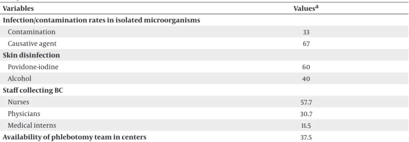
Table 1. The Ratios Related to the Collection and Processing of Blood Cultures in 16 University or Research and Training Hospitals in Turkey in 2013
Variables Valuesa
Infection/contamination rates in isolated microorganisms
Contamination 33 Causative agent 67 Skin disinfection Povidone-iodine 60 Alcohol 40 Staff collecting BC Nurses 57.7 Physicians 30.7 Medical interns 11.5
Availability of pediatric bottles in centers 86.7
Availability of anaerobic bottles in centers 66.6
Average growth rate in the bottles 32.3
The average growth time for causative agents, h 21.4
The average growth time for contaminant agents, h 36.3
Route of BC collection
Intravenous catheter 36
Peripheral venipuncture 64
Hospital classification
University hospital 56.3
Training and research hospital 43.7
Hospitals with ≥ 500 beds 56.25
Report of growing signal time to clinicians 40
Overall rate of glove usage in the centers 93.75
Number of bottles for diagnosis ( ≥ 2) 77.8
Using conventional identification methods 28.6
Quality control application 7.1
Sample rejection criteria
Insufficient blood samples 30.8
Registration errors 69.2
Fungal blood culture assessment 66.7
Abbreviation: BC, Blood cultures. aData are presented as percentage.
Table 2. Distribution of Species (%) Isolated From Blood Cultures in 16 Different Hospitals in Turkey in 2013
Microorganisms Valuesa Escherichia coli 22.45 CoNS 20.11 MRCoNS 5.88 Enterococcus spp. 9.41 Klebsiella spp. 9.18 Staphylococcus aureus 7.87 Pseudomonas aeruginosa 7.46 Acinetobacter baumannii 6.44
Other members of Enterobacteriaceae 5.2
Abbreviations: BC, Blood cultures; CoNS, Coagulase-negative staphylococci; MRCoNS, Methicillin-resistant coagulase-negative staphylococci. aData are presented as percentage.
5. Discussion
Bloodstream infections are a significant cause of mor-tality and morbidity in any hospital setting. The reported mortality rate worldwide due to bloodstream infections is between 30% and 55% (11-14). Increasing the reliability of blood culture results and reducing contamination rates are both related to the pre-analytic, analytic, and post-analytic phases of laboratory testing (15-17). Of the three phases, the most errors occur during the pre-analytic phase, and most such errors are related to specimen col-lection, specimen handling, and patient variables (18).
According to the literature, the collection of specimens from intravenous catheters is associated with higher blood culture contamination rates (19). Using a direct venous puncture to a peripheral vein is therefore rec-ommended for obtaining higher specificity and posi-tive predicposi-tive power (19). In a systematic review, venous puncture was suggested as the most appropriate method to decrease blood culture contamination (4). In the cur-rent study, of all the blood culture samples, 64% were collected from peripheral venous blood, while 36% were
collected from intravascular catheters. The collection of specimens from intravenous catheters may hence be the reason for our high contamination rates.
In the present study, povidone-iodine (60%) and alcohol (40%) were used for skin disinfection prior to blood collec-tion. It has been reported that alcohol is superior to prod-ucts without alcohol when it comes to skin disinfection prior to blood collection due to alcohol’s quick drying time (20, 21). Many researchers have stated that alcohol alone is sufficient, since it is more cost-effective and time-effective than isopropyl alcohol in combination with povidone-io-dine (19-21). Our high contamination rates may be related to the reference for using povidone-iodine in the centers. It is suggested to be necessary to wait at least 3 minutes af-ter the application of povidone-iodine for the emergence of an antiseptic effect. The contamination rates may there-fore be due to an unwillingness to comply with the wait-ing period. Mimoz et al. indicated that chlorhexidine re-duced the incidence of blood culture contamination more than povidone-iodine. They suggested that skin prepara-tion using alcoholic chlorhexidine was more efficacious in reducing the contamination of blood cultures than skin preparation using aqueous povidone-iodine (22). Based on our findings, it is suggested that the use of alcohol should be increased in our hospital setting.
On the other hand, our study indicated that the ratio of wearing gloves and decontaminating the blood cul-ture bottles prior to use were lower in our centers. Blood culture bottle tops may be nonsterile even if they are covered with a lid, since the sterility of the top varies by manufacturer. Although Bekeris et al. found no correla-tion between blood culture contaminacorrela-tion and the clean-ing of culture bottle tops (23), the clinical laboratory standards institute recommends that culture bottle tops be cleaned with 70% isopropyl alcohol (6). Based on our results, it is concluded that routine sterile gloving may decrease blood culture contamination and that cleaning culture bottle tops may also decrease the contamination rates. In the current study, the blood samples were col-lected by nurses, doctors, and interns. This was necessary because some 62.5% of the centers included in this study had no phlebotomy team. Blood culture contamination is lower when experienced and specialized staff collect the blood samples and so a dedicated phlebotomy team should ideally draw the blood samples for culture (15-19).
Various sensitive blood culture systems and blood cul-ture bottles were used in the Turkish centers. The use of sensitive automated blood culture systems with rich culture media has led to increased contamination rates. In the literature, the most common organisms that indi-cate a contaminated specimen were CoNS,
Propionibacte-rium spp., Micrococcus spp., coryneform-type bacilli, Lac-tobacillus spp., Bacillus spp., and Viridans streptococci (4).
The most common contaminant bacteria in the present study were coagulase-negative Staphylococcus sp., coryne-form-type bacilli, and Streptococcus sp., which is similar to findings in the literature. In addition, only one of the
units included in our study had a blood culture quality control study. This data revealed the need to seriously reconsider the applications of blood cultures during the laboratory stage.
In our study, the mean detection time for bacteria con-sidered to be a causative microorganism was 21.4 hours, while for contaminants it was 36.3 hours. Both the litera-ture and the data obtained in our study showed that clin-ically significant isolates were related to a shorter detec-tion time (15). Therefore, the detecdetec-tion time should serve as an important guiding factor in the determination of contaminants and causative agents.
5.1. Conclusion
Improving blood collection techniques, establishing a phlebotomy team, encouraging venous sampling, and taking more than one blood culture sample can all con-tribute to reducing the rate of contamination during the pre-analytical phase. It will be appropriate to record time-to-detection values of the blood cultures as well as the number of bottles and detected blood-borne patho-gen. During the post-analytical phase, the clinical find-ings concerning the patients, the number of positive blood culture bottles, and any inflammation markers (i.e., white blood cell count, procalcitonin, and CRP lev-els) play an important role in determining whether the isolated bacteria is a causative agent or a contaminant.
Footnote
Authors’ Contribution:Study concept and design:
Mustafa Altindis; acquisition of data: Mustafa Altindis, Mehmet Koroglu, Tuba Dal, Tayfur Demiray; analysis and interpretation of data: Mustafa Altindis, Mehmet Koroglu, and Tayfur Demiray; drafting of the manuscript: Tuba Dal, Mustafa Altindis, and Tayfur Demiray; critical revision of the manuscript: Mustafa Altindis and Oguz Karabay; sta-tistical analysis: Tuba Dal; administrative, technical, and material support: Mustafa Altindis, Tayfur Demiray and Ali Riza Atasoy; study supervision: Mustafa Altindis.
References
1. Tekin R, Dal T, Pirinccioglu H, Oygucu SE. A 4-year surveillance of device-associated nosocomial infections in a neonatal intensive care unit. Pediatr Neonatol. 2013;54(5):303–8. doi: 10.1016/j.ped-neo.2013.03.011. [PubMed: 23643153]
2. Hall KK, Lyman JA. Updated review of blood culture contami-nation. Clin Microbiol Rev. 2006;19(4):788–802. doi: 10.1128/ CMR.00062-05. [PubMed: 17041144]
3. Tekin R, Dal T, Bozkurt F, Deveci O, Palanc Y, Arslan E, et al. Risk fac-tors for nosocomial burn wound infection caused by multidrug resistant Acinetobacter baumannii. J Burn Care Res. 2014;35(1):e73– 80. doi: 10.1097/BCR.0b013e31828a493f. [PubMed: 23799478] 4. Snyder SR, Favoretto AM, Baetz RA, Derzon JH, Madison BM, Mass
D, et al. Effectiveness of practices to reduce blood culture con-tamination: a Laboratory Medicine Best Practices systematic re-view and meta-analysis. Clin Biochem. 2012;45(13-14):999–1011. doi: 10.1016/j.clinbiochem.2012.06.007. [PubMed: 22709932] 5. Nijssen S, Florijn A, Top J, Willems R, Fluit A, Bonten M. Unnoticed
spread of integron-carrying Enterobacteriaceae in intensive care units. Clin Infect Dis. 2005;41(1):1–9. doi: 10.1086/430711. [PubMed:
15937755]
6. Weinstein MP, Doern GV. A Critical Appraisal of the Role of the Clinical Microbiology Laboratory in the Diagnosis of Blood-stream Infections. Journal of Clinical Microbiology. 2011;49(9 Supplement):S26–9. doi: 10.1128/jcm.00765-11.
7. Aycan IO, Celen MK, Yilmaz A, Almaz MS, Dal T, Celik Y, et al. [Bac-terial colonization due to increased nurse workload in an inten-sive care unit]. Rev Bras Anestesiol. 2015;65(3):180–5. doi: 10.1016/j. bjan.2014.05.004. [PubMed: 25990495]
8. Shahangian S, Snyder SR. Laboratory medicine quality indica-tors: a review of the literature. Am J Clin Pathol. 2009;131(3):418–31. doi: 10.1309/AJCPJF8JI4ZLDQUE. [PubMed: 19228647]
9. Chukwuemeka IK, Samuel Y. Quality assurance in blood culture: A retrospective study of blood culture contamination rate in a tertiary hospital in Nigeria. Niger Med J. 2014;55(3):201–3. doi: 10.4103/0300-1652.132038. [PubMed: 25013249]
10. Hawkins R. Managing the pre- and post-analytical phases of the total testing process. Ann Lab Med. 2012;32(1):5–16. doi: 10.3343/ alm.2012.32.1.5. [PubMed: 22259773]
11. Rodriguez-Creixems M, Alcala L, Munoz P, Cercenado E, Vicente T, Bouza E. Bloodstream infections: evolution and trends in the microbiology workload, incidence, and etiology, 1985-2006. Medicine (Baltimore). 2008;87(4):234–49. doi: 10.1097/ MD.0b013e318182119b. [PubMed: 18626306]
12. Nasrolahei M. Evaluation of Blood Cultures in Sari Hospitals. MJIRC. 2005;8(1):25.
13. Kalantar E, Motlagh M, Lordnejad H, Beiranvand S. The preva-lence of bacteria isolated from blood cultures of iranian chil-dren and study of their antimicrobial susceptibilities. Jundisha-pur J Nat Pharm Products. 2008;3(1):1–7.
14. Barati M, Taher MT, Abasi R, Zadeh MM, Barati M, Shamshiri AR. Bacteriological profile and antimicrobial. Arch Clin Infect Dis. 2009;4(2):87–95.
15. Balikci A, Belas Z, Eren Topkaya A. [Blood culture positivity: is it pathogen or contaminant?]. Mikrobiyol Bul. 2013;47(1):135–40. [PubMed: 23390910]
16. Engel C, Brunkhorst FM, Bone HG, Brunkhorst R, Gerlach H, Grond S, et al. Epidemiology of sepsis in Germany: results from
a national prospective multicenter study. Intensive Care Med. 2007;33(4):606–18. doi: 10.1007/s00134-006-0517-7. [PubMed: 17323051]
17. Riedel S, Bourbeau P, Swartz B, Brecher S, Carroll KC, Stamper PD, et al. Timing of specimen collection for blood cultures from fe-brile patients with bacteremia. J Clin Microbiol. 2008;46(4):1381–5. doi: 10.1128/JCM.02033-07. [PubMed: 18305133]
18. Willems E, Smismans A, Cartuyvels R, Coppens G, Van Vaeren-bergh K, Van den Abeele AM, et al. The preanalytical optimiza-tion of blood cultures: a review and the clinical importance of benchmarking in 5 Belgian hospitals. Diagn Microbiol Infect Dis. 2012;73(1):1–8. doi: 10.1016/j.diagmicrobio.2012.01.009. [PubMed: 22578933]
19. Mermel LA, Allon M, Bouza E, Craven DE, Flynn P, O'Grady NP, et al. Clinical practice guidelines for the diagnosis and manage-ment of intravascular catheter-related infection: 2009 Update by the Infectious Diseases Society of America. Clin Infect Dis. 2009;49(1):1–45. doi: 10.1086/599376. [PubMed: 19489710] 20. Baron EJ, Miller JM, Weinstein MP, Richter SS, Gilligan PH,
Thomson RB, et al. A guide to utilization of the microbiology laboratory for diagnosis of infectious diseases: 2013 recom-mendations by the Infectious Diseases Society of America (IDSA) and the American Society for Microbiology (ASM)(a). Clin Infect Dis. 2013;57(4):e22–e121. doi: 10.1093/cid/cit278. [PubMed: 23845951]
21. Maiwald M, Chan ES. The forgotten role of alcohol: a systematic re-view and meta-analysis of the clinical efficacy and perceived role of chlorhexidine in skin antisepsis. PLoS One. 2012;7(9):e44277. doi: 10.1371/journal.pone.0044277. [PubMed: 22238652] 22. Mimoz O, Karim A, Mercat A, Cosseron M, Falissard B, Parker F, et
al. Chlorhexidine compared with povidone-iodine as skin prepa-ration before blood culture. A randomized, controlled trial. Ann Intern Med. 1999;131(11):834–7. [PubMed: 10610628]
23. Bekeris LG, Tworek JA, Walsh MK, Valenstein PN. Trends in blood culture contamination: a College of American Patholo-gists Q-Tracks study of 356 institutions. Arch Pathol Lab Med. 2005;129(10):1222–5. doi: 10.1043/1543-2165(2005)129[1222:TIBCCA ]2.0.CO;2. [PubMed: 16196507]