© 2019 Turkish Journal of Plastic Surgery | Published by Wolters Kluwer - Medknow
140
Abstract
Case Report
I
ntroductIonCombination of the cervical spine fractures with maxillofacial trauma have serious outcomes because of the high potential for mortality and neurologic morbidity. Therefore, the incidence of maxillofacial fractures complicated by spinal trauma, is of significant concern to the craniomaxillofacial surgeon. In this case report, authors describe the stages of diagnosis and treatment of such a patient.
c
aser
ePortAn 8-year-old male child was admitted to our emergency room with maxillofacial and spinal trauma after a car crash. He was conscious, oriented, and cooperative. His blood pressure was 106/62 mmHg (normal). His pulse was 120 bpm (high) and temperature was 36.6°C (normal). He had a jaw and neck pain. His jaw and lower face were swollen. A hard neck collar was placed immediately. Malocclusion was detected during his examination. He had a neck spasm and limited neck movement. After the patient was stabilized, his computed tomography (CT) scans were taken.
Through CT scans, he was diagnosed with a mandibular double-fracture pattern combined within body and the parasymphisis region complicated with an odontoid fracture. Since he had malocclusion, we decided that it would be best to perform early open reduction and fixation.
Neurosurgeons’ decision was to treat him nonoperatively for the cervical fracture. This treatment required immobilization of the neck until the fracture was healed. Therefore, the mandible fixation had a very high risk of neurodamaging in case of a minimal displacement of the neck during the operation. He was meticulously entubated with a fiberoptic laryngoscope by a senior member of the anesthesiolgy staff. Patients neck collar was kept intact during this procedure Figure 1. Intubation is very risky for neck trauma patients; therefore, it is very important for the anesthesiology team to be as experienced, skilled, and careful as the plastic surgery team. While intubating, it was important not to move his head or neck at all. An arch bar was used to gain proper maxillary-mandibular occlusion before fixating a stable plate. Circummandibular wires were used to hold the arch bar in position. Nonabsorbable plates were preferred due to excessive prices of the absorbable plates. Rigid fixation was accomplished with two-to-four-hole 12.0-mm spaced, mini, straight plates (1.9 thickness) (Trimed® Ankara, Turkey) and eight screws (2.0 mm diameter/7 mm length) via intraoral approach [Figure 2]. The plates were positioned at the inferior lower border of the mandible not to
Mandibular fractures are the most common fractures of facial bones.If the fracture is detected and the occlusion is affected, surgery is the best option. Motor vehicle accidents and fall injuries are the most common reasons for combined injuries of maxillofacial and cervical spine (c-spine). The most common scenario is the cooccurrence of midfacial trauma and upper c-spine injuries. Despite the low incidence of the cooccurrence of spinal injuries and maxillofacial trauma, it is crucial to have guidelines that assess and manage this situation because of its severe outcomes.
Keywords: Cervical spine injuries, mandibular fracture, midfacial trauma, odontoid fracture
Address for correspondence: Dr. Mustafa Keskin,
Department of Plastic, Reconstructive and Aesthetic Surgery, Istanbul Medipol University, Istanbul, Turkey. E‑mail: [email protected]
Access this article online
Quick Response Code:
Website:
http://www.turkjplastsurg.org DOI:
10.4103/tjps.tjps_93_18
This is an open access journal, and articles are distributed under the terms of the Creative Commons Attribution‑NonCommercial‑ShareAlike 4.0 License, which allows others to remix, tweak, and build upon the work non‑commercially, as long as appropriate credit is given and the new creations are licensed under the identical terms.
For reprints contact: [email protected]
How to cite this article: Bahadirli N, Hanci M, Keskin M. A case of a
mandibular body fracture complicated with odontoid fracture. Turk J Plast Surg 2019;27:140-2.
A Case of a Mandibular Body Fracture Complicated with
Odontoid Fracture
Nilufer Bahadirli, Mustafa Hanci, Mustafa Keskin
Department of Plastic, Reconstructive and Aesthetic Surgery, Istanbul Medipol University, Istanbul, Turkey
Bahadirli, et al.: Mandibular and odontoid fracture
141 141
Turkish Journal of Plastic Surgery ¦ Volume 27 ¦ Issue 3 ¦ July-September 2019 141 harm the permanent tooth buds due to the patient’s young age.
The arch bar was left to keep the occlusion after the operation and was removed 1 month later. There were no early or late complications after the surgery.
d
IscussIonMandibular fractures are the most common fractures of facial following nasal bone fractures.[1] When a mandible is fractured, the occlusion of a patient is usually affected. If the fracture is detected and the occlusion is affected, surgery is the best option. The treatments of these fractures are important for the patient to restore proper speech, mastication, and swallowing.[1] For the optimal timing of the surgery, concomitant injuries and their type and severity should be taken into account. Multidisciplinary modality should be considered. Early reduction and fixation reduce the risk of nonunion, malunion, osteomyelitis, and malocclusion.[2]
Motor vehicle accidents and fall injuries are the most common reasons for combined injuries of maxillofacial and cervical spine (c-spine).[3] The most common scenario is the cooccurrence of midfacial trauma and upper c-spine injuries. The most common c-spine that is involved is C2. One half of the neck’s motion (flexion, extension, and especially rotation)
is achieved by atlanto-axial junction. For this reason, C2 is the most common c-spine to be affected in a trauma patient. A strong ligament limits the rotation of the neck. This restriction is lost when the odontoid is fractured. The anterolisthesis or retrolisthesis of the C1–C2 complex in relation to C2 body may result in serious neurological complications. Odontoid fractures at the junction of dens and the body are the most common types (Type II) of axis fractures. It is important to note that up to 40% of the odontoid fractures are fatal at the time of the accident. Odontoid fractures are not necessarily treated operatively.[4] Nonoperative immobilization methods are frequently used. The treatment options are considered and decided by a neurosurgeon for cervical fractures.
When the trauma is complicated and the airway is difficult to obtain, the examination and imaging of the patient can be challenging. Urgent recognition of a cervical injury is crucial. Careful intubation of the patient beforehand can be the only option for those cases. Therefore, it is very important for a plastic surgeon to be aware and cautious about a cervical injury when examining a patient with maxillofacial trauma. Furthermore, in case of an operation, neurological damage is usually underestimated and immobilization is not performed adequately as it should be. Cervical immobilization devices should not be removed until spine injury has been excluded.[5] The mortality rate of maxillofacial injuries that are complicated with cervical injuries is relatively high (8%).[6] 10%–25% of the patients with facial fractures are reported to have delayed diagnosis of spinal fractures.[7] There are reported cases in which the spinal injury was detected only after the maxillofacial reduction. As a result, all patients with a facial trauma should be considered to have spinal injuries up front. Despite the low incidence of the cooccurrence of spinal injuries and maxillofacial trauma,[5] it is crucial to have guidelines that assess and manage this situation because of its severe outcomes. Figure 2: (Above left) Inferior three‑dimensional view of the mandibular
fracture preoperatively, (above right) front three‑dimensional view of the mandibular fracture preoperatively, (below left) sagittal computed tomography view of the odontoid fracture preoperatively, (below right) panoramic radiograph of the patient postoperatively
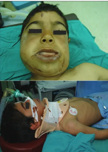
Figure 1: (Above) Preoperative view of the patient (below) operational
positioning of the patient
Bahadirli, et al.: Mandibular and odontoid fracture
Turkish Journal of Plastic Surgery ¦ Volume 27 ¦ Issue 3 ¦ July-September 2019
142
It is very important to know about and foresee the outcomes of a multitraumatic patient especially for maxillofacial surgeons because, in case of an operation, the neck is often mobilized for anesthesiology procedures and also during the reduction of the facial bones.[5] If it is necessary to operate the patient because of her/his facial trauma, it is crucial to immobilize the neck with neurosurgeons and anesthesiologists and then despite its technical difficulties operate with caution without moving the neck at all.
Declaration of patient consent
The authors certify that they have obtained all appropriate patient consent forms. In the form the patient(s) has/have given his/her/their consent for his/her/their images and other clinical information to be reported in the journal. The patients understand that their names and initials will not be published and due efforts will be made to conceal their identity, but anonymity cannot be guaranteed.
Financial support and sponsorship
Nil.
Conflicts of interest
There are no conflicts of interest.
r
eferences1. Koshy JC, Feldman EM, Chike-Obi CJ, Bullocks JM. Pearls of mandibular trauma management. Semin Plast Surg 2010;24:357-74. 2. Elahi MM, Brar MS, Ahmed N, Howley DB, Nishtar S, Mahoney JL,
et al. Cervical spine injury in association with craniomaxillofacial
fractures. Plast Reconstr Surg 2008;121:201-8.
3. Jamal BT, Diecidue R, Qutob A, Cohen M. The pattern of combined maxillofacial and cervical spine fractures. J Oral Maxillofac Surg 2009;67:559-62.
4. Jaiswal AK, Sharma MS, Behari S, Lyngdoh BT, Jain S, Jain VK. Current management of odontoid fractures. Indian J Neurotrauma 2005;2:3-6.
5. Roccia F, Cassarino E, Boccaletti R, Stura G. Cervical spine fractures associated with maxillofacial trauma: An 11-year review. J Craniofac Surg 2007;18:1259-63.
6. Reich W, Surov A, Eckert AW. Maxillofacial trauma – Underestimation of cervical spine injury. J Craniomaxillofac Surg 2016;44:1469-78. 7. Beirne JC, Butler PE, Brady FA. Cervical spine injuries in patients with
facial fractures: A 1-year prospective study. Int J Oral Maxillofac Surg 1995;24:26-9.