Pomegranate extract attenuates unilateral ureteral
obstruction‑induced renal damage by reducing oxidative
stress
Alper Otunctemur, Emin Ozbek, Suleyman Sami Cakir1, Emre Can Polat2, Murat Dursun3, Mustafa Cekmen4, Adnan Somay5, Nurver Ozbay5 Departments of Urology, Okmeydani Training and Research Hospital, Istanbul, 1Bayburt State Hospital, Bayburt, 2Istanbul Medipol University, Istanbul, 3Bahcelievler State Hospital, Istanbul, 4Department of Biochemistry, Kocaeli University, Kocaeli, 5Department of Pathology, Fatih Sultan Mehmet Training and Research Hospital, Istanbul, Turkey.
Original Article
INTRODUCTION
Obstructive nephropathy is an important cause of end stage renal disease in children and adults. It results in a progressive and permanent loss in renal function that is characterized by interstitial inflammation and tubulointerstitial fibrosis. The acute phase of obstructed kidney in unilateral ureteral obstruction (UUO) is characterized by dramatic changes in
Access this article online Quick Response Code:
Website:
www.urologyannals.com
DOI:
10.4103/0974-7796.150488
Address for correspondence:
Dr. Alper Otunctemur, Department of Urology, Okmeydani Training and Research Hospital, 34384, Sisli, Istanbul, Turkey. E-mail: [email protected]
Received: 15.05.2014, Accepted: 24.06.2014
Aims: Ureteral obstruction may cause permanent kidney damage at late period. We know that the
pomegranate extract (PE) play a strong role on removal of free oxygen radicals and prevention of oxidative stress. In the current study study, we evaluated the effect of PE on kidney damage after unilateral ureteral obstruction (UUO).
Settings and Design: A total of 32 rats were divided into four groups. Group 1 was a control, Group 2 was
a sham, Group 3 was rats with UUO and Group 4 was rats with UUO that were given PE (oral 100 µL/day). After 14 days, rats were killed and their kidneys were taken and blood analysis was performed.
Subjects and Methods: Tubular necrosis, mononuclear cell infiltration, and interstitial fibrosis scoring were
determined histopathologically in a part of kidneys; nitric oxide (NO), malondialdehyde (MDA), and reduced glutathione (GSH) levels were determined in the other part of kidneys.
Statistical Analysis Used: Statistical analyses were performed by the Chi-square test and one-way analysis
of variance.
Results: There was no difference significantly for urea‑creatinine levels between groups. Pathologically,
there was serious tubular necrosis, mononuclear cell infiltration and fibrosis in Group 3, and there was significantly decreasing for tubular necrosis, mononuclear cell infiltration and fibrosis in Group 4 (P < 0.005). Furthermore, there was significantly increasing for NO and MDA levels; decreasing for GSH levels in Group 3 compared the other groups (P < 0.005).
Conclusions: We think that the PE prevents kidney damage by decreasing oxidative stress in kidney. Key Words: nitric oxide, pomegranate extract, reactive oxygen species, renal damage, ureter obstruction
Otunctemur, et al.: Pomegranate extract attenuates renal damage glomerular filtration rate, renal blood flow, and interstitial
edema.[1,2]
On the other hand, the chronic phase of the obstructed kidney is characterized by development of hydronephrosis, renal atrophy, interstitial fibrosis, and renal dysfunction.[3]
Reactive oxygen species (ROS) are a recently recognized mechanism in the pathogenesis of UUO in experimental studies.[4-6] Increased lipid peroxidation (LPO) has been
reported in renal cortexes of UUO animals. It has been shown that oxidative stress in UUO contributes to the development of tubulointerstitial lesions and renal fibrosis. Various factors with complex cellular and molecular interactions have also been proposed as possible causes that lead to tubulointerstitial lesions and renal fibrosis.[7]
Pomegranate extract (PE) and its derivatives have been used for centuries to confer health benefits in a number of inflammatory diseases. Edible parts of pomegranate fruit represent 52% of total fruit weight, comprising 78% juice and 22% seeds.[8]
Fresh juice is rich in Vitamin C, and polyphenolic compounds such as anthocyanins, punicalagin, and ellagic and gallic acid.[9,10] PE is a rich source of potent polyphenolic, flavonoid
antioxidants (anthocyanins). The soluble polyphenol content in PE varies within the limits of 0.2‑1.0% depending on the variety and includes mainly anthocyanins that have been shown to possess anti-atherogenic properties. Anthocyanins were shown to be effective inhibitors of LPO, production of nitric oxide (NO), and inducible nitric oxide synthase activity in different model systems.[11] Pomegranate has become more
popular because of the attribution of important physiological properties, such as anticancer,[12,13] and cholesterol-lowering,
cardioprotective[14]. Many investigators have reported that
pomegranate and it’s derivatives have free radical scavenger and potent antioxidant activity.[15‑17] It has also been shown
that pomegranate can suppress nuclear factor-κB (NF-κB) activation through a novel mechanism in vascular endothelial cells.[18,19]
Nitric oxide acts as an intercellular messenger and regulates cellular functions such as vasorelaxation and inflammation. NO has an important role in the elimination of pathogens and tumor cells; however, overproduced NO is oxidized to ROS, resulting in the disruption of cell signaling and uncontrolled systemic inflammation.[20,21] Malondialdehyde (MDA) is one
of the important markers of LPO.[22] Excessive MDA produced
as a result of tissue injury and DNA damage could combine with free amino groups of proteins, resulting in the formation of MDA-modified protein adducts. Glutathione (GSH) is the major intracellular antioxidant with multiple biological functions, including the maintenance of the thiol moieties
of proteins and the reduced from of many other biologically active molecules.[11]
As a result of these informations, in this study we investigated the possible inhibitory effects of pomegranate against the UUO induced oxidative stress and renal injury in rat models.
SUBJECTS AND METHODS Drugs and animals
Pomegranate extract was dissolved in distilled water and administered via nasogastric gavage. The average of 2.5 mL diluted PE contains 100 µL PE, which are equivalent to 2.8 µmol total polyphenols per day.
Male Wistar albino rats (200‑250 g) were housed in clean plastic cages in a temperature and humidity-controlled facility with a constant 12 h light/dark cycle with free access to food and water. The use of animals and the experimental protocol were approved by the Institutional Animal Care and Use Committee and animals were treated in accordance with the Guide for the Care and Use of Laboratory Animals of Research Council.
Experimental design
One week after acclimatization, UUO was induced. Briefly after induction of general anesthesia by intraperitoneal injection of thiopental (100 mg/kg), the abdominal cavity was exposed via midline incision and the left ureter was ligated at 2 points with 3‑0 silk. The sham‑operated rats had their ureters manipulated, but not ligated. All rats were given amikacin sulfate (6 mg/kg, intramuscularly route) before operation.[19]
After a quarantine period of 7 days, 32 rats were randomly divided into four groups, each consisting of eight animals as follows: Rats in Group 1 were control; rats in Group 2 were sham operation; rats in Group 3 underwent unilateral ureteral ligation and received no treatment. Rats in Group 4 were subjected to unilateral ureteral ligation and received PE (100 µ/L) for 14 days. After 15 days, rats were killed and their kidneys were taken and blood analysis was performed. Tubular necrosis, mononuclear cell infiltration and interstitial fibrosis scoring were determined histopathologically in a part of kidneys; NO, MDA and reduced glutathione (GSH) levels were determined in the other part of kidneys. Urea, creatinine levels were investigated in a blood analysis.
Biochemical assays
Twenty-four hours after the administration of last doses of PE, on 15th day, rats were anesthetized by intraperitoneal injection
of ketamine and sacrificed. Renal cortical tissues were separated into two parts for biochemical analysis and light microscopic examination. Blood samples were also taken by cardiac puncture
to assess the serum levels of urea and creatinine concentrations. In frozen tissues biochemically MDA, end product of LPO, reduced glutathione (GSH), nonenzymatic antioxidant, and total nitrite, a stable product of NO, were evaluated as a means of oxidative stress.
Renal impairment was assessed by serum urea and creatinine levels, as well as by the kidney histology. Serum urea and creatinine levels were determined with an autoanalyzer (Beckman Coulter Synchron L × 20, Ireland) by using commercial Becman Coulter diagnostic kits. Kidney tissue (300 mg) was homogenized in icecold tamponade containing 150 mM KCL for determination of MDA. MDA levels were assayed for products of LPO. MDA referred to as thiobarbituric acid reactive substance, was measured with thiobarbituric acid at 532 nm using a spectrofluorometer, as described previously.[23]
GSH was determined by a spectrophotometric method, which was based on the use of Ellman’s reagent.[24]
Total nitrite (NOx) was quantified by the Griess reaction[25] after
incubating the supernatant with Escherichia coli nitrate reductase to convert NO3 to NO2. Griess reagent (1 ml 1% sulfanilamide, 0.1% naphtyl‑ethylenediamine hydrochloride, and 2.5% phosphoric acid; Sigma Chemical Co., St. Louis, MO, USA) was then added to 1 ml of supernatant. The absorbance was read at 545 nm after a 30‑min incubation. The absorbance was compared with the standard graph of NaNO2, obtained from the reduction of NaNO3(1‑100 lmol/l). The accuracy of the assay was checked in two ways; the inter- and –intra-assay coefficients of variation were 7.52 and 4.61%, respectively. To check conversion of nitrate to nitrite (recovery rate), known amounts of nitrate were added to control plasma samples; these samples were deproteinized and reduced as above.
Histopathological examinations
Histopathological evaluation of the kidney tissues was done. Paraffin-embedded specimens were cut into 6-µm thickness and stained with hematoxylin and eosin stain for light microscopic examination using a conventional protocol[26] (Olympus, BH-2,
Tokyo, Japan). A semi-quantitative evaluation of renal tissues was accomplished by scoring the degree of severity according to previously published criteria.[27] All sections of kidney samples
were examined for tubular necrosis. Briefly, minimum of 50 proximal tubules associated with 50 glomeruli were examined for each slide and an average score was obtained. Severity of lesion was graded from 0 to 3 according to the percentage of the tubular involvement. Slides were examined and assigned for severity of changes using scores on a scale in which (0) denotes no change; grade 1 – changes affecting <25% tubular damage (mild); grade 2 – changes affecting 25‑50% of tubules (moderate); Grade 3 – changes affecting >50% of tubules (severe).
Histopathological evaluation was performed on left kidney tissues. Paraffin-embedded specimens were cut into 5-mm thick sections and stained with hematoxylin and eosin and Masson’s trichrome for examination under a light microscope (BH-2; Olympus, Tokyo, Japan). To evaluate leukocyte infiltration, the widening of interstitial spaces with focal leukocyte infiltration was assessed in five randomly chosen sections prepared from each kidney sample. For each section, the average number of leukocytes per 0.28 mm2 was calculated
from these leukocyte-infiltrated foci using a high-power microscopic field (×400).
To estimate the grade of interstitial fibrosis, the interstitial area that was stained green with Masson’s trichrome was evaluated as a percentage of the total examined area in five randomly chosen sections prepared from each kidney sample using an image analyzer (Leica; Leica Micros Imaging Solutions, Cambridge, UK). For each section, interstitial space widening with focal leukocyte infiltration and interstitial fibrosis was assessed in high‑power fields (×400) to quantify the results. The Banff classification of kidney pathology was used for scoring the degree of mononuclear cell infiltration and interstitial fibrosis. The score was graded from 0 to 3, depending on the severity of histological characteristics.[19,28]
Statistical analyses
Results of all groups were shown as mean values ± standard deviation. Statistical analyses of the histopathologic evaluation of the groups were carried out by the Chi-square test and biochemical data were analyzed by the one-way analysis of variance. The significance between two groups was determined by the Dunnett’s multiple comparison tests, and P < 0.05 was accepted as statistically significant value.
RESULTS
Biochemical variables in plasma and tissue
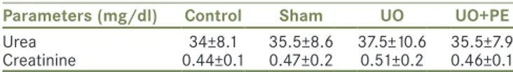
There was no difference significantly for urea-creatinine levels between groups [Table 1].
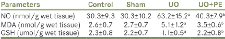
Tissue MDA levels significantly increased in Group 3 compared with Groups 1, 2, and 4 (P < 0.05). Rats with PE administration (Group 4) showed reduced levels of LPO as measured by MDA levels [Table 2]. UUO also induced a significant increase in the tissue NO levels that have been Table 1: Effects of UO alone and its combination with PE on plasma urea, creatinine levels in rats
Parameters (mg/dl) Control Sham UO UO+PE Urea 34±8.1 35.5±8.6 37.5±10.6 35.5±7.9 Creatinine 0.44±0.1 0.47±0.2 0.51±0.2 0.46±0.1 Values are expressed as mean±SD for eight rats in each group. SD: Standard deviation, UO: Ureteral obstruction, PE: Pomegranate extract
Otunctemur, et al.: Pomegranate extract attenuates renal damage prevented by PE [Table 2]. The unilateral ureteral ligation was
accompanied by a marked reduction in GSH level in the kidney tissues of rats (P < 0.05), and treatment with PE partially elevated the GSH levels [Table 2].
Histopathologic examinations results
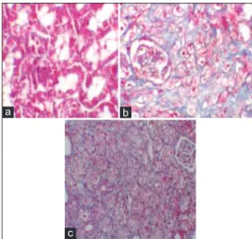
Histopathological examination of kidney showed that no pathologic findings in the control group [Figure 1a]. In rats with UUO, there were mild and severe tubular necrosis in the proximal tubules compared control and sham groups [Figure 1b and c]. In rats treated with UUO + PE, despite the presence of mild tubular degeneration and tubular necrosis are less severe, and glomeruli maintained a better morphology when compared with UUO group [Figure 1d].
Histopathologic examination was normal in rats with only sham operation (Group 2) and in rats with no operation (Group 1). Severe leukocyte infiltration was observed in the periglomerular and peritubular interstitium of the kidneys of the rats in Group 3 with UUO [Figure 2a and b]. Quantitative analysis of the focal leukocyte infiltration area in the interstitium showed that leukocyte infiltration was significantly reduced in
rats that received UUO + PE (Group 4) [Figure 2c]. UUO caused a significant interstitial fibrosis in rats that received no treatment (Group 3), and the percentage area of interstitial fibrosis in the kidney of rats with UUO that received no treatment was significantly greater than that of rats with UUO that received PE (Group 4) [Figure 3a-c]. These changes are summarized in Table 3.
DISCUSSION
In our study confirmed through a quantitative survey the protective role of PE on renal tissue damage after the induction of UUO in rats. Our results showed that the obstructed kidney had significantly higher tissue MDA, NO levels, and lower GSH levels along with more fibrosis. The current data demonstrate that UUO structural and functional alterations in the kidney with a concomitant increase in proinflammatory cytokines in the blood. PE, on the other hand, reduced the severity of injury, depressed the concentration of these cytokines and increased the antioxidative capacity.
Pomegranate extract is rich in antioxidants of the polyphenolic class that includes tannins and anthocyanins. These antioxidants are more potent, on a molar basis, than many other antioxidants, including Vitamins C and E, coenzyme Q‑10, and lipoic acid. The antioxidant levels in PE were found higher than levels in other natural juices, such as blueberry, cranberry, and orange, as well as in red wine.[11] Pomegranate has become more popular
because of the attribution of important physiological properties, such as anticancer, cholesterol-lowering, cardioprotective. The present study demonstrated ameliorative effects of PE, a phenolic antioxidant, on UUO-induced nephrotoxicity, in line with the consideration that oxygen-free radicals are important mediators of UUO-induced acute renal failure.
Table 2: Effect of PE on the levels of MDA, GSH, and NO in each rat group
Parameters Control Sham UO UO+PE
NO (nmol/g wet tissue) 30.3±9.3 30.3±10.2 63.2±15.2a 40.3±7.9b
MDA (nmol/g wet tissue) 2.6±0.7 2.7±0.7 5.1±1.2a 3.5±0.6b
GSH (umol/g wet tissue) 2.3±0.8 2.2±0.7 1.1±0.5a 2.2±0.8b
Values are expressed as mean±SD for eight rats in each group.
aSignificantly different from sham, bSignificantly different from UO group
(P<0.05). NO: Nitric oxide, MDA: Malondialdehyde, GSH: Glutathione, SD: standard deviation, UO: Ureteral obstruction, PE: Pomegranate extract
Figure 1: (a) Normal tubulus and glomerules in kidney kortex H and
E ×100 (control group). (b) Normal tubulus and glomerules in kidney kortex H and E ×100 (sham group). (c) Severe tubular total necrosis, tubular degeneration and epithelial vacuolization in the proximal tubules H and E ×400 (unilateral ureteral obstruction [UUO] group). (d) Mild epithelial vacuolization in the proximal tubules and normal glomerules H and E ×100 (UUO + PE treated group)
d c
b a
Figure 2: (a) Normal kidney morphology in a sham group. (b) Leukocyte
infiltration was observed in the peritubular interstitium of the unilateral ureteral obstruction. (c) Leukocyte infiltration was reduced in the PE-treated group (H and E, ×400)
c
b a
Apoptotic cell death has been reported to play an important role in UUO-induced renal damages.[29] The lack of investigation
on whether PE has affected the apoptotic cell death because of UUO may be a limitation of this study. However, in a recent study, curcumin and melatonine which is an antioxidant and anti-inflammatory agent like PE, has been reported to prevent UUO-mediated apoptotic cell death and reduce the UUO related renal damage. Although we believe that PE can reduce the UUO-induced renal damage by a similar mechanism that prevents apoptosis-related cell death, there is a need for further study on that subject for verifications.
The pathogenesis of renal fibrosis caused by UUO involves infiltration of the kidney by inflammatory cells including monocytes, activation, and possible transformation of intrinsic renal cells, and interactions between infiltrating and resident cells. NF-κB is activated during renal obstruction, and inhibition of NF-κB activity has been demonstrated to prevent renal fibrosis induced by obstruction.[30,31] In the present study,
in agreement with these findings, PE treatment prevented renal fibrosis in UUO rats. ROS are a recently recognized mechanism
in the pathogenesis of UUO in experimental studies.[4-6] Hence
here we measured the MDA, GSH, and NO, as a means of oxidative stress. Our findings corroborate those of earlier studies demonstrating that an enhanced endogenous oxidative stress has a major role in the severity of UUO-induced acute renal failure.[32,33] MDA, a stable lipid hydroperoxide, provides
an index of the peroxidation of lipids (LPO) in biological tissues.[34]
In the present study, we found increased MDA levels in UUO group and as protective effect of PE lower MDA levels in group determined by UUO + PE. The GSH antioxidant system is considered the most notable cellular protective mechanism. GSH has a very important role in protecting against oxygen-free radical damage by providing reducing equivalents for several enzymes, as well as scavenging hydroxyl radicals and singlet oxygen. Its depletion is a common consequence of increased formation of ROS[35] like UUO-induced nephrotoxicity. In
group given UUO + PE, we found increased GSH levels. However, our study shown that PE effect NO levels protectively in similar to some previous studies with different antioxidant agents.[36,37] These findings strongly indicate that PE is
important in protecting the kidney from UUO-induced injury through improvement in oxidant status.
In this study, the histopathological examination of kidneys showed severe and extensive damage in UUO rats, which have tubular necrosis and edema. This could be due to the formation of highly reactive radicals as a consequence of oxidative stress caused by UUO. The kidneys of the control group showed normal histological features, but the UUO group revealed more extensive and marked tubular necrosis. On the other hand, the tubules from rats of the UUO + PE group were nearly normal in histological appearance except for a slight desquamation and atrophy of the tubular epithelial cells. Similar changes were also reported by some studies who demonstrated structural changes in renal tissue of gentamicin-treated animals and its reversal by various agents.[37‑39]
CONCLUSION
The results reported here indicate that PE exerts a preventative effect on UUO-induced kidney damage in rats by reducing oxidative stress. We therefore propose that PE supplementation therapy can be used for kidney protection in patients with UUO, such as ureteral stones. However, further animal and clinical studies are needed to confirm our suggestion.
REFERENCES
1. Vaughan ED Jr, Sorenson EJ, Gillenwater JY. The renal hemodynamic response to chronic unilateral complete ureteral occlusion. Invest Urol 1970;8:78‑90.
Table 3: Semi‑quantitative analysis of tubular necrosis, interstitial fibrosis, mononuclear cell infiltration in control, Sham, UO, and UO+PE treated rats
Groups n Tubular
necrosis Interstitial fibrosis cell infiltrationMononuclear
0 1 2 3 0 1 2 3 0 1 2 3
Control 8 8 0 0 0 8 0 0 0 7 1 0 0 Sham 8 8 0 0 0 8 0 0 0 7 1 0 0 UOa 8 0 1 4 3 0 2 3 3 0 2 2 4
UO+PEb 8 2 4 1 1 3 4 1 0 2 5 1 0
UO: Ureteral obstruction, PE: Pomegranate extract, Score‑0: No degeneration, 1: Mild degeneration, 2: Moderate degeneration, 3: Severe degeneration. aStatistical significant difference from the Sham group, bStatistical significant difference from the UO group and P<0.05
Figure 3: (a) Normal kidney morphology in a sham group. (b) Severe
fibrosis was observed in the peritubular interstitium of the unilateral ureteral obstruction. (c) Mild fibrosis was reduced in the PE-treated group (masson and trichrome ×400)
c
b a
Otunctemur, et al.: Pomegranate extract attenuates renal damage 2. Schreiner GF, Harris KP, Purkerson ML, Klahr S. Immunological aspects of acute ureteral obstruction: Immune cell infiltrate in the kidney. Kidney Int 1988;34:487‑93. 3. Gulmi FA. Pathophysiology of urinary tract obstruction. In: Walsh PC, Retik AB, Vaughan ED, Wein AJ, Kavoussi LR, Novick AC, et al., editors. Campbell’s Urology. Philadelphia: W.B. Saunders; 2002. p. 434‑43. 4. Ricardo SD, Ding G, Eufemio M, Diamond JR. Antioxidant expression in experimental hydronephrosis: Role of mechanical stretch and growth factors. Am J Physiol 1997;272:F789‑98.
5. Saborio P, Krieg RJ Jr, Kuemmerle NB, Norkus EP, Schwartz CC, Chan JC. Alpha‑tocopherol modulates lipoprotein cytotoxicity in obstructive nephropathy. Pediatr Nephrol 2000;14:740‑6. 6. Kawada N, Moriyama T, Ando A, Fukunaga M, Miyata T, Kurokawa K, et al. Increased oxidative stress in mouse kidneys with unilateral ureteral obstruction. Kidney Int 1999;56:1004‑13. 7. Klahr S. Urinary tract obstruction. Semin Nephrol 2001;21:133‑45. 8. Tsuda T, Horio F, Osawa T. Cyanidin 3‑O‑beta‑D‑glucoside suppresses nitric oxide production during a zymosan treatment in rats. J Nutr Sci Vitaminol (Tokyo) 2002;48:305‑10. 9. Ellman GL. Tissue sulfhydryl groups. Arch Biochem Biophys 1959;82:70‑7. 10. de Nigris F, Williams‑Ignarro S, Lerman LO, Crimi E, Botti C, Mansueto G, et al. Beneficial effects of pomegranate juice on oxidation‑sensitive genes and endothelial nitric oxide synthase activity at sites of perturbed shear stress. Proc Natl Acad Sci U S A 2005;102:4896‑901. 11. Aviram M, Dornfeld L, Kaplan M, Coleman R, Gaitini D, Nitecki S, et al. Pomegranate juice flavonoids inhibit low‑density lipoprotein oxidation and cardiovascular diseases: studies in atherosclerotic mice and in humans. Drugs Exp Clin Res 2002;28:49‑62.
12. Kaplan M, Hayek T, Raz A, Coleman R, Dornfeld L, Vaya J, et al. Pomegranate juice supplementation to atherosclerotic mice reduces macrophage lipid peroxidation, cellular cholesterol accumulation and development of atherosclerosis. J Nutr 2001;131:2082‑9. 13. Houghton DC, Plamp CE III, DeFehr JM, Bennett WM, Porter G, Gilbert D. Gentamicin and tobramicin nephrotoxicity. A morphologic and functional comparison in the rat. Am J Pathol 1978;93:137‑52. 14. Buege JA, Aust SD Microsomal lipid peroxidation. Methods Enzymol 1978;52:302‑10. 15. Chen WC, Chen HY, Wu HC, Wu MC, Hsu CD, Tsai FJ. Vascular endothelial growth factor gene polymorphism is associated with calcium oxalate stone disease. Urol Res 2003;31:218‑22. 16. Esen T, Akinci M, Aytekin Y, Altug T, Hekim N. The effects of citrate, polyphosphates and pridoxin on intratubular crystallization in hyperoxaluric rats. Turk J Urol 1990;16:365‑370. 17. Selvam R. Calcium oxalate stone disease: role of lipid peroxidation and antioxidants. Urol Res 2002;30:35‑47. 18. Itoh Y, Yasui T, Okada A, Tozawa K, Hayashi Y, Kohri K. Preventive effects of green tea on renal stone formation and the role of oxidative stress in nephrolithiasis. J Urol 2005;173:271‑5. 19. Ozbek E, Ilbey YO, Ozbek M, Simsek A, Cekmen M, Somay A. Melatonin attenuates unilateral ureteral obstruction‑induced renal injury by reducing oxidative stress, iNOS, MAPK, and NF‑kB expression. J Endourol 2009;23:1165‑73.
20. Huang HS, Ma MC, Chen CF, Chen J. Changes in nitric oxide production in the rat kidney due to CaOx nephrolithiasis. Neurourol Urodyn 2006;25:252‑8.
21. Aviram M, Dornfeld L, Rosenblat M, Volkova N, Kaplan M, Coleman R, et al. Pomegranate juice consumption reduces oxidative stress, atherogenic modifications to LDL, and platelet aggregation: Studies in humans and in atherosclerotic apolipoprotein E‑deficient mice. Am J Clin Nutr 2000;71:1062‑76.
22. Schubert SY, Neeman I, Resnick N. A novel mechanism for the inhibition of NF‑kappaB activation in vascular endothelial cells by natural antioxidants. FASEB J 2002;16:1931‑3.
23. Villegas I, Martı×n AR, Toma W, et al. Rosiglitazone, an agonist of peroxisome proliferator‑activated receptor c, protects against gastric ischemia–reperfusion damage in rats: Role of oxygen free radicals generation. Eur J Pharmacol 2004;505:195‑203.
24. Wasowicz W, Nève J, Peretz A. Optimized steps in fluorometric determination of thiobarbituric acid‑reactive substances in serum: importance of extraction pH and influence of sample preservation and storage. Clin Chem 1993;39:2522‑6. 25. Beutler E. Glutathione in red blood cell metabolism. A Manual of Biochemical Methods. New York: Grune and Stratton; 1975. p. 112‑4. 26. Sun Y, Oberley LW, Li Y. A simple method for clinical assay of superoxide dismutase. Clin Chem 1988;34:497‑500. 27. Allen CT. Hematoxylin and eosin. In: Prophet EB, Mills B, Arrington JB, Sobin LH, editors. Laboratory methods in histochemistry. Washington, DC: Armed Forced Institute of Pathology, American Registry of Pathology; 1992. p. 53. 28. Kinugasa F, Noto T, Matsuoka H, Urano Y, Sudo Y, Takakura S, et al. Prevention of renal interstitial fibrosis via histone deacetylase inhibition in rats with unilateral ureteral obstruction. Transpl Immunol 2010;23:18‑23. 29. Hashem RM, Soliman HM, Shaapan SF. Turmeric‑based diet can delay apoptosis without modulating NF‑kappaB in unilateral ureteral obstruction in rats. J Pharm Pharmacol 2008;60:83‑9. 30. Cheung RT, Tipoe GL, Tam S, Ma ES, Zou LY, Chan PS. Preclinical evaluation of pharmacokinetics and safety of melatonin in propylene glycol for intravenous administration. J Pineal Res 2006;41:337‑43. 31. Pignone AM, Rosso AD, Fiori G, Matucci‑Cerinic M, Becucci A, Tempestini A, et al. Melatonin is a safe and effective treatment for chronic pulmonary and extrapulmonary sarcoidosis. J Pineal Res 2006;41:95‑100. 32. Martínez‑Salgado C, López‑Hernández FJ, López‑Novoa JM. Glomerular nephrotoxicity of aminoglycosides. Toxicol Appl Pharmacol 2007;223:86‑98. 33. Walker PD, Shah SV. Gentamicin enhanced production of hydrogen peroxide by renal cortical mitochondria. Am J Physiol 1987;253:C495‑9. 34. Kuhad A, Tirkey N, Pilkhwal S, Chopra K. Effect of Spirulina, a blue green algae, on gentamicin‑induced oxidative stress and renal dysfunction in rats. Fundam Clin Pharmacol 2006;20:121‑8. 35. Abdel‑Raheem IT, Abdel‑Ghany AA, Mohamed GA. Protective effect of quercetin against gentamicin‑induced nephrotoxicity in rats. Biol Pharm Bull 2009;32:61‑7. 36. Ozbek E, Cekmen M, Ilbey YO, Simsek A, Polat EC, Somay A. Atorvastatin prevents gentamicin‑induced renal damage in rats through the inhibition of p38‑MAPK and NF‑kappaB pathways. Ren Fail 2009;31:382‑92. 37. Karadeniz A, Yildirim A, Simsek N, Kalkan Y, Celebi F. Spirulina platensis protects against gentamicin‑induced nephrotoxicity in rats. Phytother Res 2008;22:1506‑10. 38. Kumar KV, Shifow AA, Naidu MU, Ratnakar KS. Carvedilol: A beta blocker with antioxidant property protects against gentamicin‑induced nephrotoxicity in rats. Life Sci 2000;66:2603‑11. 39. Nakakuki M, Yamasaki F, Shinkawa T, Kudo M, Watanabe M, Mizota M. Protective effect of human ulinastatin against gentamicin‑induced acute renal failure in rats. Can J Physiol Pharmacol 1996;74:104‑11.
How to cite this article: Otunctemur A, Ozbek E, Cakir SS, Polat EC,
Dursun M, Cekmen M, et al. Pomegranate extract attenuates unilateral ureteral obstruction-induced renal damage by reducing oxidative stress. Urol Ann 2015;7:166-71.