CASE REPORT – OPEN ACCESS
International Journal of Surgery Case Reports 12 (2015) 128–131Contents lists available atScienceDirect
International Journal of Surgery Case Reports
j o u r n a l h o m e p a g e :w w w . c a s e r e p o r t s . c o mA case report of osteoblastoma on the distal phalanx of the ring finger
successfully treated with curettage and polymethylmethacrylate
filling
Seyit Ali Gumustas
a, Talat Cagırmaz
b,∗, Olcay Guler
b, Onder Ofluoglu
c, Sibel Kayahan
daOrthopedics and Traumatology Department, Yavuz Selim Bone and Joınt Disease State Hospital, Trabzon, Turkey bOrthopedics and Traumatology Department, Medipol University, Medical Faculty, Istanbul, Turkey
cOrthopedics and Traumatology Department, Bahc¸es¸ehir University, Medical Faculty, Istanbul, Turkey dPathology Department, Dr. Lutfı Kırdar Kartal Training Hospital, Istanbul, Turkey
a r t i c l e i n f o
Article history:Received 20 November 2014 Received in revised form 30 May 2015 Accepted 31 May 2015
Available online 3 June 2015 Keywords:
Bone tumors Osteoblastoma Phalanges
a b s t r a c t
BACKGROUND: Osteoblastoma is an aggressive benign tumor whose presentation varies with location and size. This rare bone tumor is thus difficult to diagnose particularly when it occurs outside its most common location – the vertebral column and long bones.
CASE: We report a case of osteoblastoma of the fourth distal phalanx of the left hand in an 18-year-old male, presented with pain and swelling and treated with curettage and polymethylmethacrylate filling followed by immobilization by a cast, which was opened 10 days later to start physical therapy. Patient was pain-free, recovered full function of his finger, and remained without pain at one month post-surgery. The finger was monitored closely for two years; sequential films showed a radiopaque interface and no evidence of local recurrence.
CONCLUSION: This is the first report of osteoblastoma on the distal phalanx. The possibility of osteoblas-toma should be considered in cases of pain and swelling of phalanx, and if diagnosed, curettage and polymethylmethacrylate filling may be the treatment of choice.
© 2015 The Authors. Published by Elsevier Ltd. on behalf of Surgical Associates Ltd. This is an open access article under the CC BY-NC-ND license (http://creativecommons.org/licenses/by-nc-nd/4.0/).
1. Introduction
Osteoblastoma is a rare primary neoplasm of the bone. Despite its benign nature, the tumor may sometimes exhibit aggressive behavior and is typically treated with curettage; then the resulting space is packed with bone or a bone substitute.
Osteoblastomas account for only 0.5–2% of all primary bone tumors and 3% of benign bone tumors[3]. While this tumor can occur in any part of the skeleton, the most common sites are the vertebral column and long bones. The tumor rarely presents in the hand; the phalanx is involved in less than 5% of cases[3,14]. Meta-physeal lesions are slightly more common than diaMeta-physeal, with very few lesions in epiphyseal locations[6].
Osteoblastoma can occur at any age, and predominantly affects younger persons, with about 80% of these tumors occurring before 30 years of age. Osteoblastoma affects males more often than females, at a ratio of 2–3:1[3].
Symptoms and signs are variable, but the characteristic symp-tom is a localized, dull pain that does not get worse at night
∗ Corresponding author. Tel.: +90 021209122525; fax: +90 02129122626. E-mail address:[email protected](T. Cagırmaz).
and cannot be relieved by salicylates[14]. We present a case of osteoblastoma located at the rare site of the distal phalanx of the hand.
2. Case report
An 18-year-old male student was referred to our clinic for pain and swelling of the fourth distal phalanx of the left hand. He com-plained of progressive distal phalanx pain and swelling in the area for two months that was not activity-related and only partially relieved by non-steroidal anti-inflammatory medications. In con-trast to characteristic pain pattern of osteoblastoma[14], which is not worse at night, the pain in the present case was more intense at night. There was no history of trauma.
Upon referral to a medical center, an X-ray revealed a lytic lesion containing some calcification in his distal phalanx (Fig. 1).
The rheumatologist referred him to an orthopedic surgeon. Physical examination identified a tender, firm swelling over the distal phalanx. Movements were slightly restricted by pain. Blood work data, including complete blood count, chemistry, coagulation analysis, erythrocyte sedimentation rate, and C-reactive protein were all normal.
http://dx.doi.org/10.1016/j.ijscr.2015.05.036
2210-2612/© 2015 The Authors. Published by Elsevier Ltd. on behalf of Surgical Associates Ltd. This is an open access article under the CC BY-NC-ND license (http://creativecommons.org/licenses/by-nc-nd/4.0/).
CASE REPORT – OPEN ACCESS
S.A. Gumustas et al. / International Journal of Surgery Case Reports 12 (2015) 128–131 129
Fig. 1. Plain radiographs of the hand showed an osteolytic lesion in the distal
pha-lanx. Some calcification could be seen within the lesion.
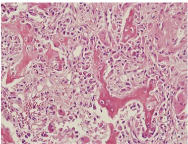
Based on clinical and radiological findings, the lesion was considered a benign primary tumor. An excisional biopsy was per-formed through a lateral approach and the resulting gap was filled with polymethylmethacrylate (PMMA). Macroscopic biopsy find-ings included a cavity containing spongy bone and reddish soft mesh. Histopathological appearance of the osteoblastoma con-sisted of a well-vascularized connective tissue stroma actively producing osteoid and primitive woven bone (Fig. 2).
Then the digit was immobilized by a cast, which was opened 10 days later to start physical therapy. At that time, the patient was pain-free; he recovered full function of his finger and remained without pain at one month post-surgery. The finger was moni-tored closely for two years; sequential films showed a radiopaque interface and no evidence of local recurrence (Fig. 3).
3. Discussion
Osteoblastoma is an uncommon primary bone tumor with clin-ical and histologclin-ical manifestations similar to those of osteoid osteoma. The most helpful single imaging technique for diagnos-ing osteoblastoma is radiography, on which the tumor appears as
Fig. 2. The hematoxylin eosin (H-E) stained histological sample of osteoblastoma
(with 40× magnification) consists of a well-vascularized connective tissue stroma in which there is active production of osteoid and primitive woven bone.
an expansile radiolucent lesion with an intact surrounding shell of bone and may contain varying degrees of calcification[13,14]. Osteoblastoma may have features similar to those of malignancy, such as cortical destruction and extra-osseous soft tissue expansion [13,19].
Other relevant imaging methods for this tumor type include computed tomography (CT), bone scan, and magnetic resonance imaging (MRI). CT can detect small mineralization in the tumor, which can help with diagnosis. CT also provides information about the size and extent of the lesion in the cortical bone, and aids preoperative evaluation and planning for surgery. Bone scans are also useful for localizing lesions, which appear as increased activity areas, but the scintigraphic appearance of the tumors is nonspecific. MRI findings of osteoblastoma are not specific, with a low signal in T1-weighted images and a high signal in T2-weighted images. However, MRI can reveal the extent of intra-osseous and soft tissue extension more accurately than other methods[4,13].
Although typical for an osteoblastoma, cortical attenuation may also be seen in other benign lesions such as fibrous dysplasia, aneurysmal bone cysts, and giant cell tumors[19].
The pathological features of osteoblastoma are similar to osteoid osteoma; both are bone-producing lesions arising from osteoblasts. Usually, the lesion is reddish-brown, even pinkish; friable with a gritty consistency; and hemorrhagic in appearance[9].
Osteoblastoma can be aggressive if associated with large epithelioid osteoblasts, and carries risk of local recurrence or malignant transformation into osteosarcoma[5]. These
“aggres-Fig. 3. Follow-up radiographs (a) and physical examination (b) at two years after
surgery. Radiographs showed radiopaque interface and no evidence of local recur-rence.
CASE REPORT – OPEN ACCESS
130 S.A. Gumustas et al. / International Journal of Surgery Case Reports 12 (2015) 128–131Table 1
Summary of treatment options and outcome for osteoablastoma in literature and in present case. Reference Number of cases Location of
osteoblastoma
Treatment Follow-up duration Recurrence
Arkader and Dormans[2] 17 Lower extremity (n = 7) Spine (n = 5)
Upper extremity (4) Sternum (n = 1)
4-step approach (extended curettage, high-speed burring, electrocauterization of cavity wall, and phenol 5% solution)
2 years (range, 2–5 years)
6%
Miszczyk et al.[16] 5 Long bones (n = 4)
pelvis (n = 1)
Curettages with or without postoperative irradiation
28.3 months None
Saglik et al.[20] 20 Spine (n = 8)
Extremities (n = 8) Talus (n = 3) Metatarsal (n = 1) Intralesional curettage (n = 11) Wide resection (n = 3) Marginal excision (n = 6) 50.6 months (range, 20–192 months) 13%
Present case 1 Distal phalanx Curettage and
polymethylmethacrylate filling
2 years None
sive osteoblastomas” are more mitotically active than conventional osteoblastoma[14].
Benign osteoblastoma has a reported recurrence of approxi-mately 10–20%[9,12,19]; therefore, close follow-up monitoring of patients is required. The size, location, and ability to resect the entire lesion are important factors influencing the risk of recur-rence, morbidity, and mortality, although metastasis has rarely been documented[14]. The patient in this case study was followed-up closely for two years and sequential films revealed a radiopaque interface and no evidence of local recurrence.
Simple curettage with or without bone grafting results in less local morbidity but a higher rate of recurrence[2,15,20]; total removal is required to eliminate recurrence. Complete excision may be possible by intra-lesional curettage aided by high-speed burr and adjuvant cryotherapy[2], or in expendable bones such as ribs, fibula, or distal ulna, by en bloc resection[8]. Cementation in conjunction with excision may be helpful for extending the surgical margin. Postoperative radiation is usually not recommended; how-ever, it may be considered for tumors that have recurred multiple times, particularly around the spine[11,16].
The majority of patients are cured by initial therapy. Recur-rences usually occur within two years of initial surgery because of growth of remaining tumor cells; recurrences after two years are very rare. In the present case, we successfully applied curettage (excisional biopsy) and filled the resulting gap with polymethyl-methacrylate filling without any complication or recurrence for two years postoperatively. Depending on the size of tumor or pref-erence of surgeon, the curettage cavity is filled with bone graft or polymethylmethacrylate[20]. The treatment approaches and out-come in previous reports[2,16,20]and for the present case were summarized inTable 1.
Case reports of osteoblastoma in the small bones of the hands describe swelling as the main feature of metacarpal and phalangeal lesions and curettage and bone grafting as sufficient treatment[18]. Mosher and Peckham[17] described a case of osteoblastoma of the fifth metacarpal in a 10-year-old boy that was treated with en bloc excision and fibular grafting. Although usually a solitary bone tumor, Adler[1] described a case with multiple osteoblastomas occurring in multiple bones of the right hand in a nine-year-old child. All lesions identified radiologically were treated by curettage. The phalanx is involved in less than 5% of cases, making this localization exceptional [3,14]. At least one case of benign osteoblastoma, localized on the proximal phalanx of the fifth digit of the left hand, was treated by en bloc surgical resection of the lesion and curettage[10]. In a summary of six cases of osteoblas-toma of the hand by Farzan and Mortazavi[7], half were on the metacarpals and the other half were on phalanges; recurrence was observed in one case.
In conclusion, to the best of our knowledge, this is the first report of osteoblastoma on the distal phalanx. Although rare,
osteoblastoma can be localized on phalanx. Unless the lesion is not very large, excisional biopsy followed by cementation with poly-methylmethacrylate may effectively treat osteablastoma without causing recurrence for up to two years.
Conflict of interest
There is no conflict of interest.
Funding
None.
Consent
In our study informed full consent was taken from the patient, and we did not need any detailed information about the patient.
Author contribution
Gumustas, Seyit Ali: surgeon and one of the author. Cagırmaz, Talat: study designer, data collector. Guler, Olcay: study designer, one of the author. Ofluoglu, Onder: surgeon, supervisor.
Kayahan, Sibel: pathologist.
References
[1] C.P. Adler, Multifocal osteoblastoma of the hand, Skeletal Radiol. 29 (2000) 601–604.
[2] A. Arkader, J.P. Dormans, Osteoblastoma in the skeletally immature, J. Pediatr. Orthop. 28 (2008) 555–560.
[3] M. Berry, H. Mankin, M. Gebhardt, A. Rosenberg, F. Hornicek, Osteoblastoma: a 30-year study of 99 cases, J. Surg. Oncol. 98 (2008) 179–183.
[4] S.D. Chakrapani, K. Grim, V. Kaimaktchiev, J.C. Anderson, Osteoblastoma of the spine with discordant magnetic resonance imaging and computed tomography imaging features in a child, Spine 33 (2008) E968–E970. [5] C.R. de Oliveira, B.B. Mendonc¸a, O.P. de Camargo, et al., Classical
osteoblastoma, a typical osteoblastoma, and osteosarcoma: a comparative study based on clinical, histological, and biological parameters, Clinics 62 (2007) 167–174.
[6] H.D. Dorfman, B. Czerniak, Benign osteoblastic tumors, in: Bone Tumors, 1st ed., Mosby, St Louis, 1998, pp. 85–127.
[7] M. Farzan, S.M.J. Mortazavi, Osseous tumors of the hand, Acta Med. Iran. 40 (2002) 207–211.
[8] R. Ferracini, E.L. Masterson, R.S. Bell, J.S. Wunder, Distal ulnar tumours. Results of management by en bloc resection in nine patients and review of the literature, J. Hand Surg. Br. 23 (1998) 517–521.
[9] F.S. Frassica, R.L. Waltrip, P.D. Sponseller, et al., Clinicopathologic features and treatment of osteoid osteoma and osteoblastoma in children and adolescents, Orthop. Clin. North Am. 27 (1996) 559–574.
[10] J.G. Herrera Tenorio, A.I. Núnuez Fernández, J.J. Mendoza Quiroga, R.A. Sesma Villalpando, Benign osteoblastoma in the proximal phalanx. A case report, Acta Ortop Mex. 23 (2009) 298–301.
[11] A.G. Huvos, Osteoblastoma, in: Bone Tumors: Diagnosis, Treatment and Prognosis, 2nd ed., WB Saunders, Philadelphia, 1991, pp. 67–83.
CASE REPORT – OPEN ACCESS
S.A. Gumustas et al. / International Journal of Surgery Case Reports 12 (2015) 128–131 131 [12] R.P. Jackson, Recurrent osteoblastoma: a review, Clin. Orthop. 131 (1978)
229–233.
[13] H.M. Kroon, J. Schurmans, Osteoblastoma: clinical and radiologic findings in 98 new cases, Radiology 175 (1990) 783–790.
[14] D.R. Lucas, K.K. Unni, R.A. McLeod, M.I. O’Connor, F.H. Sim, Osteoblastoma: clinicopathologic study of 306 cases, Hum. Pathol. 25 (1994) 117–134. [15] E. Marechal, Recurrent osteoblastoma of the hamate bone, J. Hand Surg. Br. 24
(1999) 757–758.
[16] L. Miszczyk, G. Woeˆniak, J. Spindel, P. Walichiewicz, Radiotherapy in the treatment of osteoblastoma – a report of five consecutive cases, Nowotwory J. Oncol. 54 (2004) 31–33.
[17] J.F. Mosher, A.C. Peckham, Osteoblastoma of the metacarpal. A case report, J. Hand Surg. 3 (1978) 358–360.
[18] C.A. Peimer, O.J. Moy, M.H. Dick, Tumors of bone and soft tissue, in: Operative Hand Surgery, Chirchill Livingstone, New York, 1998.
[19] D. Resnick, Tumors and tumor-like lesions of bone: imaging and pathology of specific lesions, in: Diagnosis of Bone and Joint Disorders, 3rd ed., WB Saunders, Philadelphia, 1995, pp. 3786–3796.
[20] Y. Saglik, H. Atalar, Y. Yildiz, K. Basarir, C. Gunay, Surgical treatment of osteoblastoma: a report of 20 cases, Acta Orthop Belg 73 (2007) 747–753.
Open Access
This article is published Open Access atsciencedirect.com. It is distributed under theIJSCR Supplemental terms and conditions, which permits unrestricted non commercial use, distribution, and reproduction in any medium, provided the original authors and source are credited.