Address for Correspondence: Dr. Uğur Abbas Bal, Başkent Üniversitesi Tıp Fakültesi, Kardiyoloji Anabilim Dalı, Fevzi Çakmak Cad. 10. Sokak, No: 45, 06490, Bahçelievler, Ankara-Türkiye Phone: +90 312 212 68 68 Fax: +90 312 223 86 97 E-mail: [email protected]
Accepted Date: 05.11.2013 Available Online Date: 14.02.2014
©Copyright 2014 by Turkish Society of Cardiology - Available online at www.anakarder.com DOI:10.5152/akd.2014.4922
A
BSTRACTObjective: The aim of this study was to investigate the factors associated with coronary stent restenosis and if there is an association between plasma asymmetric dimethylarginine (ADMA) levels and stent restenosis.
Methods: Ninety-one patients, who had a history of coronary bare metal stent implantation due to any cause in the last one year period, were admitted to this observational cross-sectional study. Coronary angiography was performed to all patients and quantitative angiography was used to determine the presence of stent restenosis. Laboratory parameters and angiographic features that contribute to stent restenosis were evaluated. Plasma ADMA levels were measured by using high performance liquid chromatography. Logistic regression analysis was used to determine the independent factors of stent restenosis.
Results: Angiographic restenosis was found in 35 patients (38.5%). Stent diameter (p=0.038) and left ventricular ejection fraction (p=0.023) were lower and stent implantation history due to acute coronary syndrome (p=0.029), plasma ADMA level (5.0±1.8x10⁻⁴ mmol/L vs. 3.9±1.0x10⁻⁴
mmol/L, p=0.001), C-reactive protein concentration (p=0.016), white blood cell count (p=0.044) and stent length (p=0.005) were higher in patients with restenosis. Plasma ADMA level (β=0.536; OR: 1.710; CI: 1.022-2.861; p=0.041), C-reactive protein concentration (β=0.062; OR: 1.064; CI: 1.003-1.129; p=0.041), stent diameter (β=-3.047; OR: 0.048; CI: 0.007-0.313; p=0.002) and length (β=0.165; OR: 1.179; CI: 1.036-1.343; p=0.013) were found to be the independent predictors of stent restenosis in logistic regression analysis.
Conclusion: We conclude that plasma ADMA levels may be used as a novel marker for stent restenosis beyond the classic stent restenosis markers. (Anadolu Kardiyol Derg 2014; 14: 491-7)
Key words: stent, restenosis, plasma asymmetric dimethylarginine
Uğur Abbas Bal, Aylin Yıldırır, Alp Aydınalp, Gamze Kaynar, Süleyman Kanyılmaz,
Koza Murat*, İbrahim Haldun Müderrisoğlu
Department of Cardiology and *Biochemistry, Faculty of Medicine, Başkent University; Ankara-Turkey
Could plasma asymmetric dimethylarginine level be a novel predictor
beyond the classic predictors of stent restenosis?
Introduction
In recent years, stents are widely used for the treatment of coronary artery disease. During the wide spread usage of coro-nary stent implantation, stent restenosis was found to be the major problem related to this intervention. Neointimal hyperpla-sia composed of vascular smooth muscle cells and the matrix is the mainstay of stent restenosis (1). Nitric oxide (NO) was con-verted from L-arginine by nitric oxide synthase (NOS) and released from intact endothelium (2, 3). Nitric oxide inhibits vascular smooth muscle cell proliferation and prevents neointi-mal hyperplasia. Thanyasiri et al. (4) showed that endothelium-dependent coronary artery dilatation was reduced in subjects with restenosis after percutaneous coronary intervention. It has also been shown that the NO prevents the development of
reste-nosis after percutaneous transluminal coronary angioplasty (PTCA) by inhibiting the formation of mitogenic substances resulted from injured endothelial tissue and the adhesion of leukocyte and thrombocytes (5, 6). Asymmetric dimethylarginine (ADMA) is a potent competitive inhibitor of NOS and occurs dur-ing protein degradation of arginine residues (7, 8). As a result of NOS inhibition by ADMA, NO formation and therefore its protec-tive effects against the stent restenosis diminishes.
Many factors contribute to stent restenosis. Stent diameter and length were well known predictors for stent restenosis but there is not enough knowledge about ADMA and its role on stent restenosis. In recent years few studies reveal a link between ADMA and stent restenosis due to its role on NO formation (9-11). Derkacz et al. (9) concluded that pre-procedural elevated plasma ADMA levels increases the risk of restenosis in patients
who underwent coronary angioplasty and stenting with bare metal stents. Khalifa et al. (10) found that the patients who devel-oped stent restenosis had an increase in ADMA levels following coronary stenting. Arı et al. (11) found that the plasma levels of ADMA obtained before the procedure predict the development of restenosis and major adverse cardiac events in patients who underwent elective percutaneous transluminal coronary angio-plasty and bare metal stent procedures. All of these studies attempted to evaluate the predictive value of ADMA on stent restenosis but more data is still needed in this regard. The aim of this study was to investigate the factors associated with coro-nary stent restenosis and also decide to evaluate the associa-tion between ADMA levels and stent restenosis on the patients with coronary stent and who need a diagnostic coronary angi-ography due to symptoms or high risk positive stress tests or laboratory parameters.
Methods
This study was designed as an observational cross-section-al study and has been approved by the Başkent University Ethical Committee. The patients who were admitted to our emer-gency service or outpatient clinics with a history of coronary bare metal stent implantation in the last one year period were evaluated for the study participation. Exclusion criteria were defined as chronic renal failure, chronic liver disease, cerebro-vascular event in the last year, severe peripheral arterial dis-ease, uncontrolled diabetes mellitus (HbA1c >%7), uncontrolled hypertension (systolic blood pressure >140 mm Hg or diastolic blood pressure >90 mm Hg regardless of hypertension status or preexisting antihypertensive medication use) (12), clinical hyper-thyroidism, erectile dysfunction and pulmonary hypertension (mean pulmonary arterial pressure >30 mm Hg). Patients with drug eluting stents (DES) were also excluded because DES have different mechanisms of restenosis and might be seen after a long time period (delayed restenosis). Ninety-one patients with recurrent angina pectoris under optimal medication or high risk positive stress tests or acute coronary syndrome were enrolled to the study. All patients were informed about the study protocol and written informed consent was obtained.
The demographic characteristics, cardiovascular risk fac-tors and the medication history of the patients were recorded and all underwent an extensive physical examination. Patients with two consecutive fasting serum glucose measurements > 126 mg/dL (13) or those on oral antidiabetic drug and/or insulin were diagnosed as diabetic. Hypertensive patients with the sys-tolic blood pressure <140 mm Hg and diassys-tolic blood pressure <90 mm Hg on physical examination under drug therapy were diagnosed as controlled hypertensive. Hyperlipidemia was defined as LDL cholesterol >100 mg/dL, HDL cholesterol <40 mg/ dL or triglyceride >150 mg/dL or usage of lipid lowering drug therapy (14). Body mass index (BMI) was calculated by division of weight to height’s square (kg/m2) and patients with BMI >30
kg/m2 were diagnosed as obese (15).
Serum levels of total cholesterol, low density lipoprotein (LDL) cholesterol, high density lipoprotein (HDL) cholesterol, tri-glyceride, glucose, creatinine, C-reactive protein and hemogram were determined by using commercial tests (Belliver Industrial Estate, Plymouth, UK) after 8 hours of fasting period.
Percutaneous coronary intervention views and reports of the patients were analyzed cautiously by one experienced cardiolo-gist and the factors (predilatation, stent diameter, stent length, oversizing, undersizing, residue stenosis) that might contribute to stent restenosis were noted.
Electrocardiography and echocardiography were performed to all patients. Especially left ventricle ejection fraction, left ven-tricle hypertrophy and pulmonary blood pressures were calcu-lated cautiously by one experienced echocardiographer.
After these steps, an elective coronary angiography was assessed for the patients. Blood samples (10 mL) were collected into the ethylenediaminetetraacetic acid containing tubes from femoral arterial sheath before coronary angiography was per-formed. Whole blood samples were centrifuged immediately in biochemistry laboratory at 3000 g for 10 minutes. Each plasma samples were put into two Eppendorf tubes (Labor Teknik, İstanbul, Turkey) by using a plastic pipe and stored in a deep freezer at -80 degree (Celsius) until analysis. One of the plasma samples was analyzed in biochemistry laboratory and the other sample was stored in the freezer for the backup.
Coronary angiographic examination was performed after local anesthesia by employing the modified Seldinger technique through the femoral artery. All coronary arteries were visualized at right and left anterior oblique projections with caudal and cranial angulations and left lateral projection (Philips, Artis zee, Munich, Germany). Left ventriculography was also performed at right and left anterior oblique projections. Iohexol was used in coronary angiography and left ventriculography as the contrast agent. Coronary angiography views were assessed by two expe-rienced cardiologists who had no knowledge about the patients’ clinical demographics and laboratory parameters. The degree of coronary stenosis was determined at the projection which shows the stenosis more severe. Quantitative coronary angiog-raphy was used to determine the severity of stenosis and angio-graphic restenosis is defined as ≥50% luminal narrowing (16).
Plasma ADMA levels were analyzed by using high perfor-mance liquid chromatography (HPLC). The method includes reversed-phase HPLC analysis by using fluorescence detection (Shimadzu LC 10A fluorescent detector, Japan) in a high-perfor-mance liquid chromatography system (Shimadzu RF 10XL, Japan). The plasma concentrations of ADMA were measured by HPLC with precolumn derivatization with o-phthaldialdehyde (OPA) and 3-mercaptopropionic acid. Samples and standards were incubated with OPA reagent for exactly 30 s before injec-tion into the HPLC system (17).
Statistical analysis
Statistical analyses were performed by using the statistical program for the social sciences (SPSS) version 15.0 (Chicago, IL,
USA). Data were submitted to a frequency distribution analysis by Kolmogorov-Smirnov’s test. Values displaying normal distri-bution were expressed as the mean (standard deviation; SD) and values with skewed distribution were expressed as median (interquartile range). While comparing parametric variables ‘Independent Samples T’ test and in nonparametric variables ‘Mann-Whitney U’ test were used. Categorical variables were compared using chi-square tests, or if small expected cell fre-quencies, exact tests. To determine the independent factors of stent restenosis, “Logistic Regression Analysis” method was used for significant variables which were found in binary com-parisons. The p values less than 0.05 were accepted as statisti-cally significant.
Results
The patients who had a history of coronary stent implanta-tion within one year (9.0±1.7 months) and planned to undergo an elective coronary angiography in our center were evaluated for study participation. 91 patients, 72 (79.1%) men, 19 women (20.9%) and mean age 58.96±8.72 years were included in the study. 32 patients were diabetic (35.2%), 62 (68.1%) had hyper-tension and 77 (84.6%) had hyperlipidemia. Out of 91 patients, 35 had an established stent restenosis after coronary angiography. The time period after stent implantation to coronary angiography had a mean 8.7±1.8 months in patients with stent restenosis.
1. Clinical characteristics/ Laboratory parameters and stent restenosis:
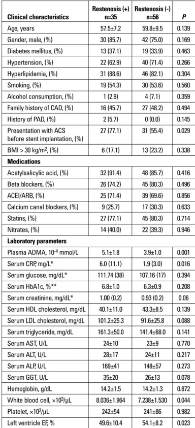
When we compared the clinical characteristics of the patients, only the incidence of stent implantation due to acute coronary syndrome was significantly higher in the patients with restenosis than the patients without restenosis (77.1% vs. 55.4%; p=0.029, respectively). There were no association between age and plasma ADMA levels (p=0.524 for women and p=0.260 for men). There was no significant difference between two groups according to the usage of medications and also between plasma ADMA levels and the type of medication (all p values >0.05).
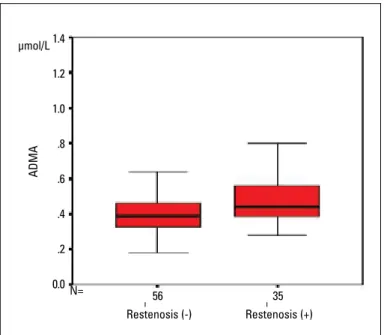
Among the laboratory parameters studied, patients with restenosis has significantly higher plasma ADMA levels than the patients without restenosis (5.1±1.8x10⁻⁴ mmol/L vs. 3.9±1.0x 10⁻⁴
mmol/L; p=0.001) (Fig. 1). Also serum C-reactive protein levels [6.0 (11.1) mg/L vs. 1.9 (3.0) mg/L; p=0.016] and white blood cell count (8.036±1.964×10³/μL vs. 7.238±1.530×10³/μL; p=0.044) were
higher and left ventricle ejection fraction was lower (49.6±10.4% vs. 54.1±8.2%; p=0.023) in the patients with restenosis than the patients without restenosis. All the clinical characteristics and laboratory parameters of patients with and without stent reste-nosis were summarized in Table 1.
2. Relationship between plasma ADMA levels and clinical characteristics/Laboratory parameters:
There was no significant relationship between plasma ADMA levels and clinical characteristics. But plasma ADMA levels tended to be higher in diabetic patients (n=32) compared
Restenosis (+) Restenosis (-) Clinical characteristics n=35 n=56 P Age, years 57.5±7.2 59.8±9.5 0.139 Gender, male, (%) 30 (85.7) 42 (75.0) 0.169 Diabetes mellitus, (%) 13 (37.1) 19 (33.9) 0.463 Hypertension, (%) 22 (62.9) 40 (71.4) 0.266 Hyperlipidemia, (%) 31 (88.6) 46 (82.1) 0.304 Smoking, (%) 19 (54.3) 30 (53.6) 0.560 Alcohol consumption, (%) 1 (2.9) 4 (7.1) 0.359 Family history of CAD, (%) 16 (45.7) 27 (48.2) 0.494 History of PAD, (%) 2 (5.7) 0 (0.0) 0.145 Presentation with ACS 27 (77.1) 31 (55.4) 0.029 before stent implantation, (%)
BMI > 30 kg/m2, (%) 6 (17.1) 13 (23.2) 0.338
Medications
Acetylsalicylic acid, (%) 32 (91.4) 48 (85.7) 0.416 Beta blockers, (%) 26 (74.2) 45 (80.3) 0.496 ACEI/ARB, (%) 25 (71.4) 39 (69.6) 0.856 Calcium canal blockers, (%) 9 (25.7) 17 (30.3) 0.633
Statins, (%) 27 (77.1) 45 (80.3) 0.714
Nitrates, (%) 14 (40.0) 22 (39.3) 0.946 Laboratory parameters
Plasma ADMA, 10-4 mmol/L 5.1±1.8 3.9±1.0 0.001
Serum CRP, mg/L* 6.0 (11.1) 1.9 (3.0) 0.016 Serum glucose, mg/dL* 111.74 (38) 107.16 (17) 0.394 Serum HbA1c, %** 6.8±1.0 6.3±0.9 0.208 Serum creatinine, mg/dL* 1.00 (0.2) 0.93 (0.2) 0.06 Serum HDL cholesterol, mg/dL 40.1±11.0 43.3±8.5 0.139 Serum LDL cholesterol, mg/dL 101.2±25.3 91.6±25.8 0.088 Serum triglyceride, mg/dL 161.3±50.0 141.4±68.0 0.141
Serum AST, U/L 24±10 23±9 0.770
Serum ALT, U/L 28±17 24±11 0.217
Serum ALP, U/L 169±41 148±57 0.273
Serum GGT, U/L 35±20 26±13 0.078
Hemoglobin, g/dL 14.2±1.5 14.2±1.3 0.872 White blood cell, ×103/μL 8.036±1.964 7.238±1.530 0.044
Platelet, ×103/μL 242±54 241±86 0.982
Left ventricle EF, % 49.6±10.4 54.1±8.2 0.023
ACS - acute coronary syndrome; ADMA - asymmetric dimethylarginine; ALP - alkaline phosphatase; ALT - alanine aminotransferase; AST - aspartate aminotransferase; BMI - body mass index; BUN - blood urea nitrogen; CAD - coronary artery disease; CRP - C-reactive protein; EF - ejection fraction; GGT - gamma-glutamyl transferase; HDL - high density lipoprotein; LDL - low density lipoprotein; PAD - peripheral artery disease Parametric; value given as mean±standard deviation
*Non-parametric; value given as median (interquartile range) **Data for diabetic patients
Table 1. Clinical characteristics and laboratory parameters of patients with and without stent restenosis
to nondiabetics (p=0.068) (Fig. 2). Also in hyperlipidemic patients a tendency for higher plasma ADMA levels were observed com-pared to patients with normal lipid levels (p=0.053).
3. Stent /implantation procedure properties and stent reste-nosis:
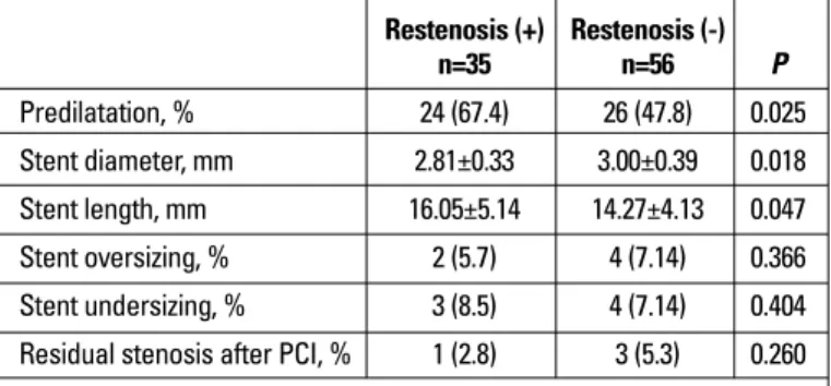
When we consider the properties of stent and the implanta-tion procedure, we found that the stent length (16.05±5.14 mm vs. 14.27±4.13 mm; p=0.047) was significantly higher and the stent diameter (2.81±0.33 mm vs. 3.00±0.39 mm; p=0.018) was signifi-cantly lower in patients with stent restenosis. Predilatation his-tory before stent implantation (67.4% vs. 47.8%; p=0.025) was also significantly higher in the patients with stent restenosis. The other properties of stent and implantation procedure were not significantly different between the patients with or without stent restenosis (Table 2).
4. Independent predictors of stent restenosis
Logistic regression analysis was used to determine the inde-pendent predictors for the development of stent restenosis. The variables (plasma ADMA levels, CRP, white blood cell count, left ventricle ejection fraction, acute coronary syndrome clinic before stent implantation, stent diameter, stent length and predilatation before stent implantation) which were significantly (p<0.05) differ-ent in patidiffer-ents with or without stdiffer-ent restenosis were evaluated by using backward elimination method. Plasma ADMA levels (β=0.536; OR: 1.710; CI: 1.022-2.861; p=0.041), CRP (β=0.062; OR: 1.064; CI: 1.003-1.129; p=0.041), stent diameter (β=-3.047; OR: 0.048;
CI: 0.007-0.313; p=0.002) and length (β=0.165; OR: 1.179; CI: 1.036-1.343; p=0.013) were found to be the independent predictors of stent restenosis in logistic regression analysis (Table 3).
Discussion
In our study we found higher plasma ADMA levels in patients with stent restenosis than without stent restenosis and stent diam-eter, stent length, CRP and ADMA were the independent predictor of stent restenosis. Our results indicate that plasma ADMA levels may be used to predict the development of stent restenosis.
Restenosis is the most important problem that limits the suc-cess of percutaneous coronary revascularization interventions in the long term. In 1980s, the high ratio of restenosis after bal-loon angioplasty declined to 20-30% with the usage of bare metal stents and to 8-15% with the drug eluting stents in 2000s (18, 19). Very complex molecular and cellular mechanisms play role in stent restenosis. Different growth factors/receptors, cyto-kines, secondary messengers and proto-oncogenes mediate this process (20). Most important mechanism in the development of stent restenosis is neointimal hyperplasia. Since stent implan-tation inhibits elastic recoil and negative remodeling by its mechanical effect, stent restenosis develops mainly by neointi-mal hyperplasia (1, 21). The development rate of neointineointi-mal hyperplasia is higher in the first six months and continues in a lower rate for 3 years after stent implantation. Neointima basi-cally contains proliferated smooth muscle cells and extracellu-lar matrix (22, 23).
Nitric oxide is a potent vasodilator which converted from its precursor L-arginine by NOS and it released by intact endothe-lium. Behind the potent vasodilator effect, NO also protects the healthy vessels against atherosclerosis and restenosis. This effect achieved by inhibition of smooth muscle cells’ migration and proliferation. Nitric oxide also inhibits leukocytes and plate-lets’ adhesion, aggregation and reconstruction attend to throm-bosis (2, 5, 24, 25).
After an injury on the vessel wall NO synthesis and its pro-tecting effect against the restenosis decreases because of endothelial dysfunction. Lee et al. (26) showed that NO inhala-tion decreases neointimal hyperplasia by 50% after angioplasty in rat vessels. Do et al. (27) reported that intima-media ratio was 32-46% less with NO releasing stent implantation than bare metal stent on rabbit aorta. In a human study Suziki et al. (28) found that intramural L-arginine implementation after stent implantation decreases neointimal volume by 35% at the end of six months. As a result, all these studies show the effects of NO against the development of restenosis.
At 1992, ADMA was discovered as an endogenous inhibitor of NOS and was thought to have an effect on the dysfunction of L-arginine/nitric oxide pathway (29). Asymmetric dimethylargi-nine inhibits NO production and high levels of ADMA causes superoxide radicals’ production instead of NO by endothelial NOS (30). Restenosis (+) Restenosis (-) n=35 n=56 P Predilatation, % 24 (67.4) 26 (47.8) 0.025 Stent diameter, mm 2.81±0.33 3.00±0.39 0.018 Stent length, mm 16.05±5.14 14.27±4.13 0.047 Stent oversizing, % 2 (5.7) 4 (7.14) 0.366 Stent undersizing, % 3 (8.5) 4 (7.14) 0.404 Residual stenosis after PCI, % 1 (2.8) 3 (5.3) 0.260
PCI - percutaneous coronary intervention Parametric; value given as mean±standard deviation
Table 2. Properties of stent and stent implantation procedure in patients with and without stent restenosis
Odds ratio Confidence
β (OR) P interval (CI)
Plasma ADMA 0.536 1.710 0.041 1.022-2.861
CRP 0.062 1.064 0.041 1.003-1.129
EF -0.064 0.938 0.065 0.877-1.004
Stent diameter -3.047 0.048 0.002 0.007-0.313 Stent lenght 0.165 1.179 0.013 1.036-1.343
ADMA - asymmetric dimethylarginine; CRP - C-reactive protein; EF - ejection fraction Binary logistic regression analysis was used and Odd’s ratios and 95% confidence intervals were calculated for the risk factors of stent restenosis
In our study we want to search ‘if there is any role of ADMA on stent restenosis beyond the classic stent restenosis mark-ers’. We thought that if ADMA inhibits NO synthesis, also it decreases the inhibiting effects of NO on the development of stent restenosis. So we admitted the patients who had a history of stent implantation in the past year and had a clinical indica-tion for coronary angiography to our study. We measured plasma ADMA levels in those patients and compared them with the clinical characteristics and the angiographic features.
When the clinical characteristics are considered, the key point for the development of restenosis is whether the patient’s clinic at the time of stent implantation was acute coronary syn-drome or not. Cutlip et al. (18) studied 6.186 patients (6.219 lesions) pooled from several recently completed coronary stent trials and unstable angina was marked as an independent predictor of stent restenosis. Similar to these findings we found significantly higher rates of stent restenosis in patients with acute coronary syn-drome presentation at the time of stent implantation.
Controversial data are displayed in literature on the relation-ship between ADMA and diabetes mellitus. Most of the studies reported that plasma ADMA levels were higher in patients with type II diabetes mellitus but not higher in patients with type I diabetes mellitus (31). In our study, all diabetic patients had type II diabetes mellitus. Similar to literature we found a tendency for higher plasma ADMA levels in patients with diabetes mellitus compared to nondiabetic patients.
In literature there are many studies which support the pres-ence of an inflammatory response against the stents after stent implantation. Almagor et al. (32) showed an inflammatory response with high CRP levels after stent implantation to patients who have stable angina pectoris. Xu et al. (33) also found that the CRP levels at both pre-percutaneous coronary intervention and follow-up were significantly correlated with stent restenosis. In our study, when we evaluated the laboratory parameters of the patients, we found significantly higher serum CRP levels and white blood cell count in the patients with restenosis. In a prospective study Kozinski et al. (34) highlighted that inflammatory response and elevated CRP levels were more prominent in the patients with stent restenosis and it is similar to our findings.
In a prospective study done by Derkacz et al. (9) it has been demonstrated that pre-procedural elevated plasma ADMA lev-els increased the risk of restenosis in patients who underwent coronary angioplasty and stenting with bare metal stents. In our study we did not analyzed the plasma ADMA levels before stent-ing. We analyzed the plasma ADMA levels at the time of control angiography and found higher plasma ADMA levels in patients with stent restenosis after restenosis developed. Khalifa et al. (10) found that the patients who developed stent restenosis had a 35% increase in ADMA levels following coronary stenting. This finding is similar to our study but they have enrolled 37 patients in that study. In our study we have enrolled 91 patients and this is statistically more powerful. Also Arı et al. (11) found that the plasma levels of ADMA obtained before the procedure predict the development of restenosis and major adverse cardiac events in patients who underwent elective percutaneous trans-luminal coronary angioplasty and bare metal stent procedures. The results of this study were also in parallel with our findings.
Study limitations
Coronary angiography was not routinely performed to every patient after stent implantation as control angiography. We
per-Figure 1. Plasma asymmetric dimethylarginine in restenotic and non-restenotic patients
ADMA - asymmetric dimethylarginine
Restenosis (+) Restenosis (-) ADMA μmol/L 35 56 N= 1.4 1.2 1.0 .8 .6 .4 .2 0.0
Figure 2. Plasma asymmetric dimethylarginine in diabetic and non-diabetic patients
ADMA - asymmetric dimethylarginine; DM - diabetes mellitus
Restenosis (-) ADMA Restenosis (+) N = 37 19 22 13 DM (-) DM (+) 14.0 12.0 10.0 8.0 6.0 4.0 2.0 0.0 10-4 mmol/L
formed control angiography to patients presenting with recur-rent angina pectoris under optimal medication or high risk posi-tive stress tests or acute coronary syndrome. So the rate of stent restenosis was found to be higher in the present study than general population.
Measurement of NO and arginine levels might improve the results of this study but we were not able to measure these parameters.
Fractional flow reserve might be better to define the stent restenosis than quantitative angiography. But it was not feasible to every restenotic patient in our country.
Conclusion
We conclude that plasma ADMA levels can be used as a novel marker for stent restenosis beyond the classic stent reste-nosis markers. By reducing plasma ADMA levels or by increas-ing NO synthesis, the development of stent restenosis may be prevented. Nevertheless, large randomized and controlled trials are needed to further clarify the effect of ADMA on the develop-ment of stent restenosis.
Acknowledgement: The authors thank M.A. Tekindal, A.C. Yazıcı (statisticians) for the contribution in statistical analysis.
Conflict of interest: None declared. Peer-review: Externally peer-reviewed.
Authorship contributions: Concept - U.A.B., A.Y., İ.H.M.; Design - U.A.B., A.Y., A.A., İ.H.M.; Supervision - A.Y., İ.H.M.; Resource - İ.H.M.; Material - A.Y., İ.H.M., A.A., G.K., S.K.; Data collection &/or processing - A.A., G.K., S.K., K.M.; Analysis &/or interpretation - A.Y., A.A., G.K., S.K., K.M.; Literature search - U.A.B., A.Y.; Writing - U.A.B., A.Y., K.M.; Critical review - A.Y., A.A., İ.H.M.; Other - A.Y.
References
1. Rajagopal V, Rockson SG. Coronary restenosis: a review of mecha-nisms and management. Am J Med 2003; 115: 547-53. [CrossRef] 2. Schulz R, Triggle CR. Role of NO in vascular smooth muscle and
car-diac muscle function. Trends Pharmacol Sci 1994; 15: 255-9. [CrossRef] 3. Marletta MA, Hurshman AR, Rusche KM. Catalysis by nitric oxide
synthase. Curr Opin Chem Biol 1998; 2: 656-63. [CrossRef] 4. Thanyasiri P, Kathir K, Celermajer DS, Adams MR. Endothelial
dys-function and restenosis following percutaneous coronary inter-vention. Int J Cardiol 2007; 119: 362-7. [CrossRef]
5. Garg UC, Hassid A. Nitric oxide-generating vasodilators and 8-bromo-cyclic guanosine monophosphate inhibit mitogenesis and proliferation of cultured rat vascular smooth muscle cells. J Clin Invest 1989; 83: 1974-7. [CrossRef]
6. De Caterina R, Libby P, Peng HB, Thannickal VJ, Rajavashisth TB, Gimbrone MA Jr, et al. Nitric oxide decreases cytokine-induced endothelial activation. Nitric oxide selectively reduces endothelial
expression of adhesion molecules and proinflammatory cytokines. J Clin Invest 1995; 96: 60-8. [CrossRef]
7. Gary JD, Clarke S. RNA and protein interactions modulated by protein arginine methylation. Prog Nucleic Acid Res Mol Biol 1998; 61: 65-131. [CrossRef]
8. Jin JS, D'Alecy LG. Central and peripheral effects of asymmetric dimethylarginine, an endogenous nitric oxide synthetase inhibitor. J Cardiovasc Pharmacol 1996; 28: 439-46. [CrossRef]
9. Derkacz A, Protasiewicz M, Poreba R, Doroszko A, Poreba M, Antonowicz-Juchniewicz J, et al. Plasma asymmetric dimethylargi-nine predicts restenosis after coronary angioplasty. Arch Med Sci 2011; 7: 444-8. [CrossRef]
10. Khalifa NM, Gad MZ, Hataba AA, Mahran LG. Changes in ADMA and TAFI levels after stenting in coronary artery disease patients. Mol Med Rep 2012; 6: 855-9.
11. Arı H, Arı S, Erdoğan E, Tiryakioğlu O, Üstündağ Y, Huysal K, et al. A novel predictor of restenosis and adverse cardiac events: asym-metric dimethylarginine. Heart Vessels 2010; 25: 19-26. [CrossRef] 12. Egan BM, Zhao Y, Axon RN. US trends in prevalence, awareness,
treatment, and control of hypertension, 1988-2008. JAMA 2010; 303: 2043-50. [CrossRef]
13. American Diabetes Association. Diagnosis and classification of diabetes mellitus. Diabetes Care 2011; 34: 62-9. [CrossRef]
14. Expert Panel on Detection, Evaluation, and Treatment of High Blood Cholesterol in Adults. Executive Summary of the Third Report of the National Cholesterol Education Program (NCEP) Expert Panel on Detection, Evaluation and Treatment of High Blood Cholesterol in Adults (Adult Treatment Panel III). JAMA 2001; 285: 2486-97. [CrossRef] 15. Clinical guidelines on the identification, evaluation, and treatment
of overweight and obesity in adults: executive summary. Expert Panel on the Identification, Evaluation and Treatment of Overweight in Adults. Am J Clin Nutr 1998; 68: 899-917.
16. Morice MC, Serruys PW, Sousa JE, Fajadet J, Ban Hayashi E, Perin M, et al; RAVEL Study Group. A randomized comparison of a siroli-mus-eluting stent with a standard stent for coronary revasculariza-tion. N Engl J Med 2002; 346: 1773-80. [CrossRef]
17. Teerlink T. HPLC analysis of ADMA and other methylated L-arginine analogs in biological fluids. J Chromatogr B Analyt Technol Biomed Life Sci 2007; 851: 21-9. [CrossRef]
18. Cutlip DE, Chauhan MS, Baim DS, Ho KK, Popma JJ, Carrozza JP, et al. Clinical restenosis after coronary stenting: perspectives from multi-center clinical trials. J Am Coll Cardiol 2002; 40: 2082-9. [CrossRef] 19. Stone GW, Ellis SG, Cox DA, Hermiller J, O'Shaughnessy C, Mann
JT, et al. A polymer-based, paclitaxel-eluting stent in patients with coronary artery disease. N Engl J Med 2004; 350: 221-31. [CrossRef] 20. Farb A, Sangiorgi G, Carter AJ, Walley VM, Edwards WD, Schwartz
RS, et al. Pathology of acute and chronic coronary stenting in humans. Circulation 1999; 99: 44-52. [CrossRef]
21. Lowe HC, Oesterle SN, Khachigian LM. Coronary in-stent resteno-sis: current status and future strategies. J Am Coll Cardiol 2002; 39: 183-93. [CrossRef]
22. Kearney M, Pieczek A, Haley L, Losordo DW, Andres V, Schainfeld R, et al. Histopathology of in-stent restenosis in patients with periph-eral artery disease. Circulation 1997; 95: 1998-2002. [CrossRef] 23. Virmani R, Farb A. Pathology of in-stent restenosis. Curr Opin
Lipidol 1999; 10: 499-506. [CrossRef]
24. Radomski MW, Palmer RM, Moncada S. An L-arginine/nitric oxide pathway present in human platelets regulates aggregation. Proc Natl Acad Sci USA 1990; 87: 5193-7. [CrossRef]
25. Armstead VE, Minchenko AG, Schuhl RA, Haywert R, Nossuli TO, Lefer AM. Regulation of P-selectin expression in human endothe-lial cells by nitric oxide. Am J Physiol 1997; 273: 740-6.
26. Lee JS, Adrie C, Jacob HJ, Roberts JD JR, Zapol WM, Bloch KD. Chronic inhalation of nitric oxide inhibits neointimal formation after balloon-induced arterial injury. Circ Res 1996; 78: 337-42. [CrossRef] 27. Do YS, Kao EY, Ganaha F, Minamiguchi H, Sugimoto K, Lee J, et al.
In-stent restenosis limitation with stent-based controlled-release nitric oxide: Initial results in rabbits. Radiology 2004; 230: 377-82. [CrossRef] 28. Suzuki T, Hayase M, Hibi K, Hosokawa H, Yokoya K, Fitzgerald PJ,
et al. Effect of local delivery of L-arginine on in-stent restenosis in humans. Am J Cardiol 2002; 89: 363-7. [CrossRef]
29. Vallance P, Leone A, Calver A, Collier J, Moncada S. Accumulation of an endogenous inhibitor of NO synthesis in chronic renal failure. Lancet 1992; 339: 572-5. [CrossRef]
30. Sydow K, Munzel T. ADMA and oxidative stress. Atherosclerosis 2003; 4: 41-51. [CrossRef]
31. Tousoulis D, Kampoli AM, Stefanadis C. Diabetes mellitus and vas-cular endothelial dysfunction: current perspectives. Curr Vasc Pharmacol 2012; 10: 19-32. [CrossRef]
32. Almagor M, Keren A, Banai S. Increased C-reactive protein level after coronary stent implantation in patients with stable coronary artery disease. Am Heart J 2003; 145: 248-53. [CrossRef]
33. Xu HY, Qiao SB, Zhang JF, Dong QT, Li JJ. Different impacts of C-reactive protein and lipid profile on coronary lesions following a percutaneous coronary intervention. Coron Artery Dis 2012; 23: 181-7. [CrossRef] 34. Kozinski M, Krzewina-Kowalska A, Kubica J, Zbikowska-Gotz M,
Dymek G, Piasecki R, et al. Percutaneous coronary intervention triggers a systemic inflammatory response in patients treated for in-stent restenosis -comparison with stable and unstable angina. Inflamm Res 2005; 54: 187-93. [CrossRef]