Near-Infrared Dyes and Their Use in Medical Science
Yakın Kızıl Ötesi (Near-IR) Boyalar ve Bu Boyaların Tıp Alanında Kullanımları
S. Sibel Erdem
Department of Medical Biochemistry, İstanbul Medipol University International School of Medicine, İstanbul, Turkey
Correspondence Author/Sorumlu Yazar: S. Sibel Erdem E-mail/E-posta: [email protected] Received/Geliş Tarihi: 17.02.2016 Accepted/Kabul Tarihi: 18.04.2016 DOI: 10.5152/clinexphealthsci.2016.011
©Copyright by 2016 Journal of Marmara University Institute of Health Sciences - Available online at www.clinexphealthsci.com ©Telif Hakkı 2016 Marmara Üniversitesi Sağlık Bilimleri Enstitüsü - Makale metnine www.clinexphealthsci.com web sayfasından ulaşılabilir
Öz
Abstract
Near-IR (NIR) (yakın kızıl ötesi) floresan boyalarla hedefe yönelik görüntüle-me (tanı) ve tedavi imkânı, görüntülerin renk yansıması ve floresan emisyo-nundan alınan datalar yardımıyla gerçekleştirilir ve özellikle derin yüzeyde bulunan dokuların görüntülenmesinde önemli rol oynar. Bu moleküllerin so-ğurma ve floresans yaptıkları bölge NIR spektroskopisinin (NIRS) moleküler görüntülemelerdeki seçici alanı olarak tanımlanır (650-850 nm). NIR floresan boyalar genel olarak NIR görüntüleme içeren çalışmalarda kullanılmasına rağmen günümüzde fotodinamik tedavi de kendine yer bulmaktadırlar. Kli-nik öncesi araştırmalarda, ftalosiyanin, klorin, porfirin, bakterioklorin, siyanin, alexfluore ve çeşitli bodipy serileri vb. NIR floresan boyalar/ajanlar kullanıl-maktadır. Bu boyalardan özellikle görüntüleme çalışmalarında en öne çıkanı, diğer boyalara kıyasla ileri fotofiziksel ve kimyasal özelliklere sahip ftalosiya-ninlerdir. NIR boyaların kullanılmasının mevcut ve potansiyel avantajlarının yanında bu boyaların toksisite sorunu boyaların klinikte kullanılmasını kısıt-lamaktadır. Klinikte kullanılan FDA onaylı tek boya indosiyanin yeşilidir ve ihmal edilebilir yan etkileriyle, kardiyak fonksiyonların kontrolü, karaciğer çık-tıları ve retinal anjiyografi gibi klinik alanlarda kullanılmaktadır. Sonuç olarak, birçok önemli sorunlar taşımakla birlikte günümüzde hala güvenli kullanıma uygun olmayan NIR boyaların yakın gelecekte bahsi geçen uygulamalarda uygun olarak kullanılabilecek kimyasal, fotokimyasal ve fotofiziksel özellikleri geliştirilmiş şekilde üretilmesi ihtiyacı kaçınılmazdır. Bununla beraber gelişti-rilmiş ve/veya geliştirilecek olan NIR floroforların nanopartiküler sistemlerle birleştirilerek ve/veya ajan moleküller ile hedef belirlenerek NIRS ve terapi yönünden daha avantajlı hale getirilmesi gereklidir.
Anahtar Kelimeler: Yakın kızıl ötesi boyalar, fotodinamik terapi,
ftalosiya-nin, teranostik, hedefe yönelik görüntüleme, floresan sistemler INTRODUCTION
Near-IR (NIR) Dyes
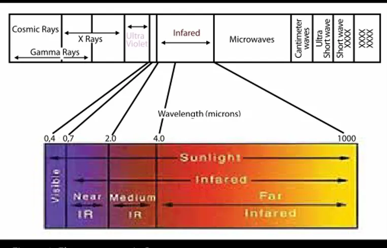
Fluorescence-based detection provides support to a number of biological assays including but not limited to immunoassays, flow cy-tometer, DNA sequencing, various proteomics assays, and several clinical chemistry applications. Because of recent advances in the field, the efficiency of these assays has to be amplified. In recent years, when minimal background fluorescence and low scattering in the 650–850 nm region of the electromagnetic spectrum (Figure 1) were coupled with photophysically and chemically improved fluoro-phores, near-infrared (NIR) dye-based fluorescence imaging gained importance (1, 2). Although the primary purpose of the NIR dyes is aiding imaging, some of the NIR dyes are also used as therapeutic agents for photodynamic therapy (PDT) of cancer and other diseases. NIR dyes show variety based on their solubility, molar absorptivity, photostability, and fluorescence efficiency. Phthalocyanines (Pcs), cy-anines, BODIPY dye series porphyrins, squarins, benzo [c] heterocyclic, and xanthine derivatives are the most commonly used NIR dyes (3-5). Among these dyes, cyanine dyes receive attention because of their high molar absorptivity, strong fluorescence emission, and good photostability. However, their excitation and emission spectra interference as a result of intrinsic small stokes shift is an obstacle for their use as NIR imaging agents (6). Porphyrins are tetrapyrolic compounds. Depending on the substituent pattern on the macrocycle, their chemical
Targeted imaging (diagnosis) and therapy using near-infrared (NIR) dyes can be accomplished with the help of the data obtained from fluorescence emission of the fluorophores and play an important role particularly in deep tissue imaging. The area NIR dyes absorb and emit light is defined as NIR spectroscopy (NIRS, 650–850 nm). Although NIR dyes are widely used for imagining purposes, they also find application in photodynamic ther-apy. In preclinical studies, phthalocyanine (Pc), chlorine, porphyrin, bac-teriochlorin, cyanine, Alexa-fluor, and various BODIPY dye series are used as NIR fluorescent dyes/agents. When compared to other dyes, one of the most promising NIR dye is Pc because of their photophysical and chemi-cal properties particularly for the imaging applications. Although NIR dyes have several advantages, their toxicity limits their usage in clinics. Indocy-anine green, having negligible side effects, is the only FDA approved NIR dye used in clinics. It is used for controlling of cardiac function, liver output, and retinal angiography. In conclusion, the development of new genera-tion NIR dyes with improved chemical, photophysical, and photochemical properties that are more appropriate for the aforementioned applications is inevitable. Nevertheless, the NIR dyes that have been developed and will be developed should be combined with the nanoparticular systems and/or targeting moieties to make them more advantageous for NIRS and therapy.
Keywords: Near-infrared dyes, photodynamic therapy, phthalocyanine,
theranostic, targeted imaging, fluorescent probes
and photochemical properties can be easily tuned. Instead of being used as NIR imaging dyes, porphyrins are predominantly used for PDT applications because of their ability to form triplet state complexes via intersystem crossing and to generate reactive oxygen species. A deriv-ative of porphyrin, chlorin, is a two electron reduced from of porphyrin and is widely used as a photosensitizer (PS) for PDT applications, which will be further elaborated in the following sections of this review. Compared to porphyrin derivatives and cyanines, BODIPY series have lower molecular weight (Figure 2) and last within the cells for a lon-ger period of time, which provides a better time scale for imaging. BODIPY dyes have many derivatives having absorption and emis-sion profile ranging from 500 nm to 660 nm in the electromagnetic spectrum (Figure 3). However, typically they have a relatively short-er wavelength emission maxima and smallshort-er extinction coefficients compared to the other NIR-active fluorophores (e.g., Pcs) (7, 8). Pcs, known systematically as tetraaza tetra benz porphyrins, are a member of porphyrinoid derivative aromatic compounds (Figure 4). Pcs are planar macrocycles with 18 p electrons. In the Pc structure, aza bridges connect four isoindole units linked together through their 1, 3 positions (Figure 4). Pcs have a number of characteristic proper-ties such as high thermal stability, unique photophysical properproper-ties, intense color, non-toxicity, and high phototoxicity upon irradiation with light, contributing to their effectiveness in different research areas. Their easily tunable photochemical and photophysical prop-erties such as narrow absorbance and emission band and resistance to photobleaching, by changing the substitution pattern around the Pc aromatic core and/or the central metal atom, makes these com-pounds highly appropriate for applications such as PCR, single gene mutation detection, and resonance energy transfer-based assays (9).
CLINICAL AND RESEARH CONSEQUENCES
For most of the applications, it is a “must” to have Pcs with high wa-ter solubility along with a functional group that can be used for bio-conjugation. However, difficulties in their synthesis limit their usage; therefore, a new synthetic method has been developed by Erdem et al. (1, 2) that has been published in two different articles in 2008 and 2009 in the Journal of Organic Chemistry Article (1, 2). In these stud-ies, using a new solid phase synthesis method, Erdem et al. (2) were able to synthesize monoamine and mono-hydroxyl functionalized highly water-soluble Pcs to be used as NIR imaging agents. Further expansion of the study included the conjugation of monoamine
functionalized Pcs to various length oligonucleotides. The authors successfully showed that the synthesis and isolation of the Pc–oligo-nucleotide conjugate takes just few hours with the help of a three-step specific bioconjugation method.
For many bioanalytical applications, Pcs are used as fluorophores to tag biomolecules because of their structural properties. They are specifically useful fluorescence on-off systems offering high target to background ratio due to Pcs high extinction coefficients and strong and narrow emission profiles. In the light of this information, Nest-erova et al. (9) have published another article in the Journal of Amer-ican Chemical Society in 2009 as a follow up study (9). In this study, authors, for the first time, have designed, prepared, and evaluated the use of double-labeled dimerization-based molecular beacons
Figure 2. General structure of BODIPY dyes (6)
Figure 1. Electromagnetic Spectrum
Cosmic Rays Gamma Rays
X Rays Infared Microwaves
Can timet er w ave s Ultr a Shor t w av e Shor t w av e XXXX XXXX XXXX 0,4 0,7 2.0 4.0 1000 Ultra Violet Wavelength (microns)
Figure 3. Normalized fluorescence emission spectra of BODIPY series
Fluor esc enc e emission 500 550 600 650 700 750 1 2 3 4 5 6 7 Wavelength (nm)
Figure 4. General structure of porphyrin and chlorin
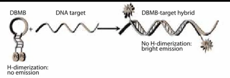
using Pcs as markers for single nucleotide base pair detection to be used in vivo cell imaging to trace cancer cells. For this purpose, the authors have designed “off/on probe” in a way that two identical Pc molecules in the molecular beacon’s closed state form a non-fluo-rescent H-dimer (off state). However, in the presence of a comple-mentary DNA, the molecular beacon’s loop hybridizes to its target forcing the molecular beacons to open, disrupting the Pc dimer and restoring fluorescence emission (on state) (Figure 5) (9). On the other hand, in the presence of a single base-pair mismatch, the loop does not open due to mismatch between the loop and the complimentary DNA, the probe stays in the off state and consequently fluorescence read-out does not get recorded.
A different perspective of Pc aggregation-based system (on/off probe) has been described in another study published in the Analyst in 2010. Nesterova et al. (10) developed a Pc aggregation-based NIR fluorescence quenching system employing only one type of a fluo-rophore. With the aim of discovering inhibitors for long interspersed Element 1 endonuclease, the authors successfully demonstrated that the probe could effectively distinguish differences in enzyme activity via H-aggregation of single Pc-labeled oligonucleotide. The probe designed in a way that Pc bearing oligonucleotide is used as a fluorescence quenching system. In the generated probe-based system, in the absence of the enzyme, two fluorophores are located far away from each other (on the either end of the double
strand-ed oligonucleotide) so that the fluorescence read out is recordstrand-ed (on state). As the enzyme is introduced in the system, it cleaves the double stranded oligonucleotide. Consequently, fluorophores (Pc) get close to each other in the solution resulting in quenched fluores-cence (off state) (Figure 6). The advantage of using only one type of a fluorophore is reduced cost and time for substrate preparation. The signal read-out format overcomes some disadvantages of the “off/on system.” In the signal read-out format, amplification of the signal is monitored against a low background (10). It is concluded that such a system would be particularly beneficial in high throughput screen-ing applications, in which enzyme activity is closely monitored. Besides on/off or off/on systems, NIR imaging can be coupled with nanoparticles to increase detection efficacy by measuring the num-ber of fluorophores in the tissue of interest. Nanoparticles have many advantages such as tunable pharmacokinetics, increased number of fluorophores per area, and the ability to carry more than one type of a molecule (therapeutic, diagnostic, and biomarker targeted). All these features make nanoparticles an attractive choice as drug carriers for theranostic studies in recent years (in vivo and in vitro). McCarthy et al. (11) study of thrombus targeted fibrinolytic nanoparticles is one of the best examples in the nanoparticles’ usage in NIR imaging (11). Thrombus, blood clot, is the final product of the blood coagulation step in hemostasis and is formed by aggregated platelets and a mesh of cross-linked fibrin protein (12-15). Although currents effective-ly effective-lyse clots and prevent tissue and organ death using exogenous plasminogen activators (PAs), these PAs may also damage normal hemostasis, which may lead to life-threatening bleeding such as in-tra-cerebral hemorrhage. McCarthy et al. (16) aimed to develop new thrombus-targeted fibrinolytic agents that employ the multifunc-tional theranostic nanomaterial to generate efficacious thrombolytic effects, while minimizing deleterious side effects (16). In this study, the authors developed thrombus targeting theranostic nanoparti-cles using cross-linked dextran coated iron-oxide (FeO) nanopartinanoparti-cles carrying imaging, therapeutic, and targeting agents. The surface of the FeO nanoparticles were functionalized with free amine groups so that imaging and targeting agents with amine reactive functional groups can be covalently attached to the surface of the nanoparticle. To accomplish thrombus targeting, the nanoparticle was decorat-ed with an activatdecorat-ed factor XIII (FXIIIa)-sensitive peptide. Therapeu-tic ability is provided using tPA, which is covalently conjugated to nanoparticles via PEG linker (Figure 7). Commercially available car-boxylic acid bearing VT680 dye is used as florescence reporter (16). Following the synthesis of nanoparticles, the applicability of the FXII-Ia-targeted thrombolytic nanoagent in the treatment of thromboem-bolism was demonstrated in vitro and in vivo in a murine model of arterial and venous thrombosis. Investigation of the safety profile of the nanoagent was planned to performed in another study (16). Although it is beneficial to use fluorescence imaging, NIR imaging can be coupled with other imaging technologies such as magnetic resonance imaging and optical coherence tomography (OCT) to im-prove detection/imaging efficiency. In one of the most recent papers published in European Heart Journal in 2015 by Hara et al. (16), the dual imaging (NIRF-OCT) system for intravascular fibrin after stent implantation, which has a potentially clinically translatable technolo-gy, has been studied. In this study, the authors reported about
diag-Figure 5. Dimerization-based molecular beacons assay using Pc dyes (9)
DBMB DBMB-target hybrid Pc Pc Pc Pc DNA target H-dimerization: no emission No H-dimerization: bright emission
Figure 6. Schematic of DNase action on substrate and emission (10)
non aggregated Pc: bright emission non aggregated Pc: bright emission aggregated Pc's: no emission L1-EN consensus sequence
Figure 7. Step-wise synthesis of thrombolytic nano particles synthesis (11)
NH2 NH2 NH2 NH2 NH2 NH2 NH2 NH2 NH2 NH2 NH2 NH2 NH2 NH2 NH2 NH2 NH2 NH2 NH2 NH2 NH2 NH2 NH2 NH2 NH2 NH2 NH2 NH2 NH2 NH2 CLIO Fluorophore Peptide
Nanomedicine © Future Science Group (2012) tPA PEG
NH2
NH2
nosis and prevention of stent thrombosis, which is a life-threatening complication of coronary artery stent implantation that occurs when a blood clot forms acutely within a stent in both bare metal stents (BMS) and drug-eluting stents (DES). OCT is a non-invasive imaging test that uses light waves to take cross-section pictures of your reti-na and is increasingly used for assessing stent tissue coverage as a measure of healed stents (17-21). Although OCT is increasingly used for assessing stent tissue coverage as a measure of healed stents, it cannot accurately identify whether an overlying tissue represents physiological neointima. In this study, the authors evaluated and compared fibrin deposition and persistence on BMS and DES using NIRF molecular imaging in vivo, in combination with simultaneous OCT (22).
The identification of overlying tissue represents physiological neoin-tima visualized by near-infrared fluorescence (NIRF) molecular imag-ing in vivo, in combination with simultaneous OCT stent coverage (23, 24). The authors employed CyAm7 as an NIR reporter dye. Fi-brin targeted imaging agent consisting of a fiFi-brin targeting peptide and CyAm7 was prepared and validated in murine thrombosis. For in vivo studies, rabbits underwent an implantation of one BMS and DES without overlap. At Days 7 and 28, intravascular NIRF-OCT was performed following the injection of fibrin-targeted NIRF molecu-lar imaging agent FTP11-CyAm7. Compared with BMS, DES showed greater fibrin deposition and fibrin persistence at Days 7 and 28. The results showed that the detection efficiency of unhealed stents is improved by intravascular NIRF fibrin molecular imaging techniques. A significant percentage of stents evaluated using OCT are found to be covered by fibrin, specifically in DES. Thus, there is a great possi-bility that such stents might remain prothrombotic. These findings indicate the specificity of clinical OCT for the evaluation and follow up of stent healing.
In another thrombosis-based study, Stein-Merlob et al. (22) NIR im-aging is coupled with different imim-aging technologies. In this study, published in 2015 in the authors utilized high-resolution in vivo op-tical molecular imaging with FTP11, a NIRF fibrin-specific reporter, to investigate the in vivo inter relationships of blood accessibility to fi-brin, thrombus age, thrombus neoendothelialization, and fibrinolysis in murine venous thrombosis (VT). Theranostic IVM fibrin molecular imaging strategy was developed to predict the fibrinolytic response based on fibrin accessibility of FTP11via imaging signal. NIRF micros-copy showed that FTP11 fibrin binding was thrombus age-depen-dent. FTP11 localized to the luminal surface of early-stage VT, but
only minimally to subacute VT. The authors concluded that VT fibri-nolysis diminishes with thrombus age and relates to the accessibility of fibrin to blood-based fibrinolytic enzymes. Also the in vivo FTP11 fibrin accessibility signal predicts the efficacy of exogenous fibrino-lysis (25).
Fibrin is the major proteinaceous component of the initial thrombus scaffold and provides a surface for thrombus propagation and even-tual vessel occlusion.
As discussed above, current imaging techniques provide accurate in-formation about the presence of thrombus and the blood flow within the vessel. However, important aspects, such as the age and anato-my, of the thrombus cannot be detected using the present method-ologies. Therefore, new techniques, having huge potential, are being explored (25).
NIR dyes are valuable not only for imaging but also for the detec-tion of enzyme activity (26-29). Konishi et al. (30) have published one recent example of this in 2015 in Circulation Journal. The au-thors proved that NIR imaging can be also used for the detection of enzymatic activity. This study validates a novel molecular imaging tool that enables the in vivo visualization of granzyme B activity, a major effector of cytotoxic CD8+ T lymphocytes. The authors syn-thesized and optimized a fluorogenic substrate capable of reporting on granzyme B activity. The substrate composed of polylysine graft copolymer and granzyme B specific peptide. The peptide having the sequence GIEFDSGGC is modified with Cy5.5-analogous (CyAl5.5B) fluorescent dye on the N terminus (Figure 8). The probe’s specifici-ty examined ex vivo in mice hearts with experimental cytotoxic CD8+-mediated myocarditis using fluorescence reflectance imaging. Granzyme B, released from CD8+ T cells, induces apoptotic death of target cells by caspase-dependent mechanisms, and a major effector of cytotoxic CD8+ T is lymphocytes. In the continuation of the study, in vivo experiments were carried for the detection of localized gran-zyme B activity in murine model with acute myocarditis. Fluorescent molecular tomography in conjunction with co-registered computed tomography imaging was employed for in imaging purposes. The authors confirmed that molecular imaging of granzyme B activity can visualize T cell-mediated myocardial injury and monitor the re-sponse to an anti-inflammatory intervention. Although for clinical translation there are still limitations such as the depth dependence of fluorescence imaging that prevents this method to be used on large animal or human hearts, this will be a pilot study for the future development of further novel tools that can investigate the mech-anisms of immune-mediated cardiac processes, including acute cardiac transplant rejection, and evaluate the effects of therapeutic interventions (30).
A recent study published in the Journal of Biomedical Optics by Er-dem et al. (31) in 2014 is another example of the detection of en-zymatic activity using fluorescence as a tool (31). Authors designed and synthesized a β-lactam specific probe (off/on probe) to be able to detect one of the most commonly prescribed antibiotic β-lact-am’s antibiotic susceptibility and resistance in less than 30 min. For this purpose, β-lactam core of the antibiotic was modified with two identical BODIPY dyes to serve as fluorophores. When the enzyme is not introduced, two fluorophore are in close proximity and quench each other’s fluorescence (off state). When the β-lactamase enzyme is introduced to the system, the β-lactam core is cleaved and
fluoro-Figure 8. Modification of the polylysine graft copolymer and fluorescence activation (30)
CGGSDFEIG CGGS DFEIG
phores are separated and get way from each other. Consequently, the system gains fluorescence (on state) and fluorophores dequench. According to the quenching-dequenching, antibiotic resistance of an individual can be clarified in an easy and quick way (Figure 9).
Therapy
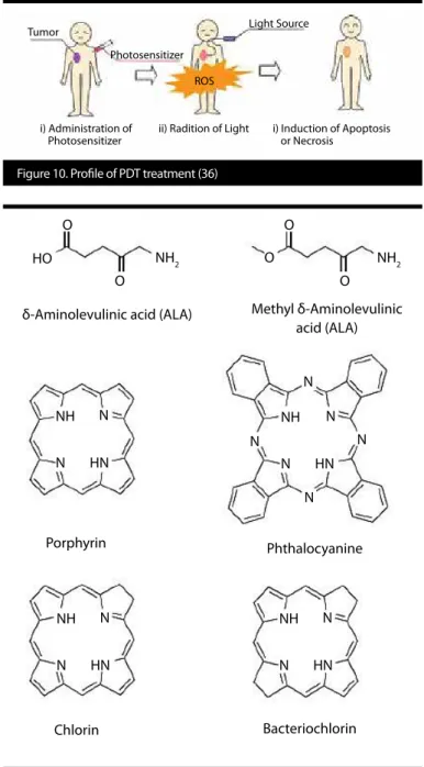
Besides their usage in various imaging platforms, some of the NIR im-aging agents are also used as a PS in PDT applications (32-35). PDT is a promising non-invasive localized treatment modality for a diverse range of diseases, including various types of cancers, infections, and inflammatory conditions. It involves combination of light and a PS. PS is activated after treatment with light, and it causes local tissue dam-age via the generation of reactive oxygen species (ROS) (Figure 10). PSs that are being used in PDT are usually highly hydrophobic mole-cules with very low water solubility. As a result of low solubility, these molecules tend to aggregate and cause reduced ROS generation. The aforementioned cascade of events causes low PDT efficiency. Some of the PSs’ general structures are shown in Figure 11 (36, 37). In a recent review paper published by Avcı et al. (37) the overall goal of PDT is described as destroying the target tissue, while leaving healthy tissues undamaged. However, while target tissues take up PS with some selectivity, some PS also accumulates in surrounding healthy tissues. Thus, another limiting factor of PDT is that light di-rected at the targeted tissue site may damage adjacent healthy tis-sues. This prevents the use of higher PS amounts, which potential-ly leads to incomplete treatment responses. To solve this problem, many laboratories have been working on the development of new methods for site-specific delivery of PS such as the encapsulation of PS into drug delivery vehicles (37).
The authors also elaborated the PSs’ self-assembly to form nanopar-ticles for PDT. To improve PS delivery to the specific site, they are of-ten encapsulated or conjugated in/on nano-drug delivery vehicles including but not limited to liposomes, fullerosomes, and nanocells. Taken together, based on their conclusion, PDT-based activation incorporated with nanotechnology can further enhance effective drug-delivery while minimizing side-effects and is expected to be clinically applicable in the near future.
Last but not the least, in 2014, another article has been published by Spring et al. (38) to show that it is possible to selectively treat mi-croscopic tumors using an activatable immunoconjugate utilizing
a NIR dye (38). The team chose to treat micro metastases because in standard therapies, they are not detected until the late stage of the disease. The detection is proceeded by immunoconjugate com-posed of self-quenching, near infrared chromophores loaded onto a cancer cell-targeting antibody. In the mentioned study, EGFR mono-clonal antibody (mAb) was used as to target cancer cells overex-pressing EGFR. EGFR is an important molecule for targeting cancer cells that displays elevated expression in up to 70% of EOCs and in many other carcinomas (39, 40). Benzoporphyrin derivative is a clin-ically-approved NIR photoactivable and cytotoxic chromophore. After conjugation with an antibody it undergoes electronic excited singlet state quenching and although fluorescence is activated as a result of lysosomal proteolysis, chromophore phototoxicity is gained after light treatment following cancer cell internalization enabling tumor-confined photocytotoxicity and the resolution of individual micrometastases. Although EGFR-targeting is the focus here,
immu-Figure 9. On/off state scheme of β-lactamase enzyme-activated fluorophore (β-LEAF) (31)
B-LEAF Probe
Quenching System does not produce fluorescence HN HN ß-lactamase (Drug Resistant) Enzyme Enzyme Fluorescence System produces high fluorescence S S S N N NH NH SH OH OH O O O
Figure 11. General structures of photosensitizers (6)
δ-Aminolevulinic acid (ALA)
NH NH NH NH N N N N HN HN HN HN N N N N N N N N NH2 NH2 HO O O O O O Porphyrin Chlorin Bacteriochlorin Phthalocyanine Methyl δ-Aminolevulinic acid (ALA)
Figure 10. Profile of PDT treatment (36)
Tumor
Photosensitizer ROS
Light Source
ii) Radition of Light i) Administration of
Photosensitizer i) Induction of Apoptosisor Necrosis
noconjugate synthesis, imaging, and taPIT can be applied to many other targets. This unique antibody-based system that couple both NIR imaging and PDT in the same platform does not only introduce a therapeutic strategy to help destroy residual drug-resistant cells but also provides a sensitive imaging method to monitor micromet-astatic disease in common sites of recurrence and provides effacing the residual micro metastases limits ability to cure many cancers in standard therapies. Fluorescence microendoscopy was used to monitor immunoconjugate activation and micrometastatic disease. Tumor-targeted, activatable photoimmunotherapy’ (taPIT) has been demonstrated in a mouse model of peritoneal carcinomatosis.
CONCLUSION
In conclusion, outstanding, rapid progress of great scope has put NIR dyes in the center of attention. NIR dyes’ tunable chemical, photo-chemical, and photophysical properties with their minimal toxicity make these dyes powerful tools not only for NIR imaging in divergent fields but also for PDT of various diseases. Although NIR dyes’ utility is widely accepted, the only clinically approved (FDA approved) mate-rial is indocyanine green (ICG). ICG has a quantum yield of only 0.01 in aqueous solution, which is very poor compared to other NIR dyes. Moreover, there have been reports of poor stability, rapid clearance from the liver, and cytotoxicity. It is very clear that there is a great need for new NIR dyes having high fluorescence quantum yield, im-proved molar absorptivity, and low cytotoxicity that could be used in clinics and/or in pre-clinical studies.
Peer-review: Externally peer-reviewed.
Acknowledgements: The author thanks to Prof. Dr. Nesrin Emekli for her
support and encouragement and Mrs. Vildan Akgül Obeidn for her assistance.
Conflict of Interest: No conflict of interest was declared by the author. Financial Disclosure: The author declared that this study has received no
fi-nancial support.
Hakem Değerlendirmesi: Dış Bağımsız.
Teşekkür: Yazar Prof. Dr. Nesrin Emekli’ye verdiği destekten, Vildan Akgül
Obeidin’e de sağlamış olduğu teknik destekten ötürü teşekkür eder.
Çıkar Çatışması: Yazar çıkar çatışması bildirmemiştir.
Finansal Destek: Yazar bu çalışma için finansal destek almadığını beyan
et-miştir.
REFERENCES
1. Erdem SS, Nesterova IV, Soper SA, Hammer RP. Solid-phase synthesis of asymmetrically substituted “AB3-Type” Phthalocyanines. J Org Chem 2008; 73: 5003-7. [CrossRef]
2. Erdem SS, Nesterova IV, Soper SA, Hammer RP. Mono-amine function-alized phthalocyanines: microwave-assisted solid-phase synthesis and bioconjugation strategies. J Org Chem 2009; 74: 9280-6. [CrossRef]
3. Umezawa K, Citterio D, and Suzuki K. Water-soluble NIR fluorescent probes based on squaraine and their application for protein labeling. Anal Sci 2008; 24: 213-7. [CrossRef]
4. Meek ST, Nesterov EE, and Swager TM. Near-infrared fluorophores containing benzo [c] heterocycle subunits. Org Lett 2008; 10: 2991-3. [CrossRef]
5. Yang Y, Lowry M, Xu X, Escobedo JO, Sibrian-Vazquez M, Wong L, et al. Seminaphthofluorones are a family of water-soluble, low molecular weight, NIR-emitting fluorophores. Proc Natl Acad Sci USA 2008; 105: 8829-34. [CrossRef]
6. Escobedo JO, Rusin O, Lim S, Strongin SM. NIR dyes for bioimaging appli-cations. Curr Opin Chem Biol 2010: 14: 64-70. [CrossRef]
7. Gallgher WM, O’Shea DF. Synthesis of BF2 Chelates of Tetraarylazadipyr-romethenes and Evidence for Their Photodynamic Therapeutic Behavior. ChemInform 2002; 33: 177-7.
8. Hilderbrand SA, Weissleder R. Near-infrared fluorescence: application to in vivo molecular imaging. Curr Opin Chem Biol 2010; 14: 71-9. [CrossRef]
9. Nesterova IV, Erdem SS, Pakhomov S, Hamer RP, Soper SA. Phthalocya-nine dimerization-based molecular beacons using near-IR fluorescence. J Am Chem Soc 2009; 131: 2432-3. [CrossRef]
10. Nesterova IV, Bennett CA, Erdem SS, Hammer RP, Deininger PL, Sopper SA. Near-IR single fluorophore quenching system based on phthalo-cyanine (Pc) aggregation and its application for monitoring inhibitor/ activator action on a therapeutic target: L1-EN. Analyst 2011; 136: 1103-5. [CrossRef]
11. McCarthy JR, Sazonova IY, Erdem SS, Hara T, Thompson BD, Patel P, et al. Multifunctional nanoagent for thrombus-targeted fibrinolytic therapy. Nanomedicine 2012; 7: 1017-28. [CrossRef]
12. Fruie B, Furie BC. Mechanisms of Thrombus Formation. N Engl J Med 2008; 359: 938-49. [CrossRef]
13. Ruggeri ZM. Thrombosis and Haemostasis. Thromb Haemost 1997; 78: 611-6. 14. Rauch U, Osende JI, Fuster V, Badimon JJ, Fayad Z, Chesebro JH. Throm-bus Formation on Atherosclerotic Plaques: Pathogenesis and Clinical Consequences. Ann Intern Med 2001; 134: 224-38. [CrossRef]
15. Svilaas T, Vlaar PJ, van der Horst IC, Diercks GFH, de Smet BJGL, van den Heuvel AFM, et al. Thrombus aspiration during primary percutaneous coronary intervention. N Engl J Med 2008; 358: 557-67. [CrossRef]
16. Hara T, Ughi GJ, McCarthy JR, Erdem SS, Mauskapf A, Lyon SC. Intravascu-lar fibrin molecuIntravascu-lar imaging improves the detection of unhealed stents assessed by optical coherence tomography in vivo. Eur Heart J 2015; pii: ehv677 [Epub ahead of print]. [CrossRef]
17. Huang D, Swanson EA, Lin CP, Schuman JS, Stinson WG, Chang W, et al. Optical coherence tomography. Science 1991; 254: 1178-81. [CrossRef]
18. Tamburino C, Manna AL, Geraci S. Optical coherence tomography for coronary imaging. ESC Council for Cardiology Practice 2010: 9.
19. Bouma BE, Tearney GJ, Yabushita H, Shishkov M, Kauffman CR, DeJoseph GD, et al. Evaluation of intracoronary stenting by intravascular optical coherence tomography. Heart 2003; 89: 317-20. [CrossRef]
20. Gonzalo N, Serruys PW, Okamura T, van Beusekom HM, Garcia-Garcia HM, van Soest G, et al. Optical coherence tomography patterns of stent restenosis. Am Heart J 209: 158: 284-93. [CrossRef]
21. Cook S, Wenaweser P, Togni M, Billinger M, Morger C, Seiler C, et al. Incom-plete stent apposition and very late stent thrombosis after drug-eluting stent implantation. Circulation 2007; 115: 2426-34. [CrossRef]
22. Stein-Merlob AF, Kessinger CW, Erdem SS, Zelada H, Hilderbrand SA, Lin CP. Blood Accessibility to fibrin in venous thrombosis is thrombus age-dependent and predicts fibrinolytic efficacy: An in vivo fibrin mo-lecular imaging study. Theranostics 2015; 5: 1317-27. [CrossRef]
23. Ughi GJ, Verjans J, Fard AM, Wang H, Osborn E, Hara T, et al. Dual mo-dality intravascular optical coherence tomography (OCT) and near-in-frared fluorescence (NIRF) imaging: a fully automated algorithm for the distance-calibration of NIRF signal intensity for quantitative molecular imaging. Int J Cardiovasc Imaging. 2015; 31: 259-68. [CrossRef]
24. Mehanna EA, Attizzani GF, Kyono H, Hake M, Bezerra HG. Assessment of coronary stent by optical coherence tomography, methodology and definitions. Int J Cardiovasc Imaging. 2011 Feb; 27: 259-69. [CrossRef]
25. Erdem SS, McCarthy JR. Multifunctional Nanoagents for the Detection and Treatment of Thromboses. In: Hunter RJ, Preedy VR, eds. Nanomedicine and the Cardiovascular System. New York: CRC Press; 2011.p. 324-44. [CrossRef]
26. Tung CH, Mahmood U, Bredow S, Weissleder R. In Vivo Imaging of Pro-teolytic Enzyme Activity Using a Novel Molecular Reporter. Cancer Re-search 2000; 60: 4953-58.
27. Kiyose K, Kojima H, Urano Y, Nagano T. Development of a Ratiometric Fluorescent Zinc Ion Probe in Near-Infrared Region, Based on Tricarbocy-anine Chromophore. J Am Chem Soc 2006; 128: 6548-9. [CrossRef]
28. Xing B, Khanamiryan A, Rao J. Cell-Permeable Near-Infrared Fluorogenic Substrates for Imaging β-Lactamase Activity. J Am Chem Soc 2005; 127:
29. Josephson L, Kircher MF, Mahmood U, Tang Y, Weissleder R. Near-Infra-red Fluorescent Nanoparticles as Combined MR/Optical Imaging Probes. Bioconjugate Chem 2002; 13: 554-60. [CrossRef]
30. Konishi M, Erdem SS, Weissleder R, Lichtman AH, McCarthy JR, Libby P. Imaging Granzyme B Activity Assesses Immune-Mediated Myocarditis. Circ Res 2015; 117: 502-12. [CrossRef]
31. Erdem SS, Khan S, Palanisami A, Hasan T. Rapid, low-cost fluorescent as-say of β-lactamase-derived antibiotic resistance and related antibiotic susceptibility. J Biomed Opt 2014; 19: 105007. [CrossRef]
32. Basu U, Khan I, Hussain A, Kondaiah P, Chakravarty AR. Photodynamic Ef-fect in Near-IR Light by a Photocytotoxic Iron(III) Cellular Imaging Agent. Angew Chem Int Ed Engl 2012; 51: 2658-61. [CrossRef]
33. Kostenich G, Orenstein A, Roitman L, Malik Z, Ehrenberg B. In vivo dynamic therapy with the new near-IR absorbing water soluble photo-sensitizer lutetium texaphyrin and a high intensity pulsed light delivery system. J Photochem Photobiol B 1997; 39: 36-42. [CrossRef]
34. Yates NC, Moan J, Western A. Water-soluble metal naphthalocyanines— near-IR photosensitizers: Cellular uptake, toxicity and photosensitizing properties in nhik 3025 human cancer cells. J Photochem Photobiol B 1990; 4: 379-90. [CrossRef]
35. Sasmal PK, Saha S, Majumdar R, Dighe RR, Chakravarty AR. Oxovanadi-um (IV)-based near-IR PDT agents: design to biological evaluation. Chem Commun (Camb) 2009; 13: 1703-5. [CrossRef]
36. Yano S, Hirohara S, Obata M, Hagiya Y, Ogura SI, Ikeda A, et al. Current states and future views in photodynamic therapy. J Photochem Photobi-ol C: Photochemistry Reviews 2011; 12: 46-7. [CrossRef]
37. Avci P, Erdem SS, Hamblin MR. Photodynamic Therapy: One Step Ahead with Self-Assembled Nanoparticles. J Biomed Nanotechnol 2014; 10: 1937-52. [CrossRef]
38. Springa BQ, Abu-Yousifa AO, Palanisamia A, Zhenga X, Rizvia I, Maia Z, et al. Selective treatment and monitoring of disseminated cancer microme-tastases in vivo using dual-function, activatable immunoconjugates. Proc Natl Acad Sci USA 2013; 111: 933-42. [CrossRef]
39. Psyrri A, Kassar M, Yu Z, Bamias A, Weinberger PM, Markakis S, et al. Effect of epidermal growth factor receptor expression level on survivalin pa-tients with epithelial ovarian cancer. Clin Cancer Res 2005; 11: 8637-43.
[CrossRef]
40. Mendelsohn J, Baselga J. Status of epidermal growth factor receptor an-tagonists in the biology and treatment of cancer. J Clin Oncol 2003; 21: 2787-99. [CrossRef]