208
M
M
y
y
e
e
l
l
o
o
m
m
e
e
n
n
i
i
n
n
g
g
o
o
s
s
e
e
l
l
O
O
n
n
a
a
r
r
ı
ı
m
m
ı
ı
S
S
ı
ı
r
r
a
a
s
s
ı
ı
n
n
d
d
a
a
K
K
ö
ö
k
k
v
v
e
e
P
P
l
l
a
a
k
k
o
o
d
d
u
u
n
n
İ
İ
n
n
t
t
r
r
a
a
o
o
p
p
e
e
r
r
a
a
t
t
i
i
f
f
D
D
i
i
r
r
e
e
k
k
t
t
E
E
l
l
e
e
k
k
t
t
r
r
i
i
k
k
S
S
t
t
i
i
m
m
ü
ü
l
l
a
a
s
s
y
y
o
o
n
n
S
S
o
o
n
n
u
u
ç
ç
l
l
a
a
r
r
ı
ı
T
T
h
h
e
e
R
R
e
e
s
s
u
u
l
l
t
t
s
s
o
o
f
f
I
I
n
n
t
t
r
r
a
a
o
o
p
p
e
e
r
r
a
a
t
t
i
i
v
v
e
e
D
D
i
i
r
r
e
e
c
c
t
t
E
E
l
l
e
e
c
c
t
t
r
r
i
i
c
c
a
a
l
l
S
S
t
t
i
i
m
m
u
u
l
l
a
a
t
t
i
i
o
o
n
n
o
o
f
f
R
R
o
o
o
o
t
t
s
s
a
a
n
n
d
d
P
P
l
l
a
a
c
c
o
o
d
d
e
e
D
D
u
u
r
r
i
i
n
n
g
g
M
M
y
y
e
e
l
l
o
o
m
m
e
e
n
n
i
i
n
n
g
g
o
o
c
c
e
e
l
l
e
e
R
R
e
e
p
p
a
a
i
i
r
r
Hüseyin Canaz1, Ezgi Tuna Erdoğan2, İbrahim Alataş1
1 İstanbul Bilim Üniversitesi, Nöroşirurji Anabilim Dalı, İstanbul, Türkiye 2 İstinye Üniversitesi Tıp Fakültesi, Fizyoloji Anabilim Dalı, İstanbul, Türkiye
Kocaeli Med J 2018; 7; 3:208-213 ARAŞTIRMA MAKALESİ/ ORIGINAL ARTICLE
ÖZ
GİRİŞ ve AMAÇ: Transkraniyal yüksek akım elektriksel
uyarımın güvenliği yenidoğanlarda tartışmalıdır. Ancak, intraoperatif direkt stimülasyon teknikleri yenidoğanlarda da fonksiyonel sinir dokusunu belirlemek ve daha güvenli bir cerrahi sağlamak için güvenli ve faydalı
yöntemlerdendir. Hastanın total paraplejisi olmadıkça, enstitümüzde myelomeningosel onarım operasyonlarında direkt sinir stimülasyon yöntemini rutin olarak
kullanmaktayız. Bu yazıda, myelomeningoseli olan 20 bebekte sinir ve nöral plakodun intraoperatif direkt stimülasyonunun sonuçlarını analiz ettik ve sunduk.
YÖNTEM ve GEREÇLER: İntraoperatif direkt stimülasyon
uygulanan 20 myelomeningoselli hastanın sonuçları incelendi. Hastaların alt ekstremite kaslarındaki
elektromiyografi aktivitesi hem tetiklenmiş hem de spontan aktivite açısından takip edildi.
BULGULAR: Elde edilen bileşik kas aksiyon potansiyeli
yanıtları alt ekstremitelerin preoperatif motor fonksiyonu ile ilişkilidir. Motor yollardaki iletim bloğu seviyesi bazı durumlarda sinir köklerini içerse de, stimüle edilen köklerin çoğunun işlevsel olması motor iletim bloğunun spinal kordun üst seviyelerinde olduğunu göstermiştir. Bir olguda, dismorfik görünen ince kökler fonksiyonel bulunmuş ve ameliyat boyunca korunmuştur.
TARTIŞMA ve SONUÇ: İntraoperatif direkt stimülasyon,
myelomeningosel operasyonlarında nöral tüpün diseksiyonu ve onarımı sırasında fonksiyonel sinir dokusunun tespit edilip korunması için yararlı ve güvenilir bir yöntemdir.
Anahtar Kelimeler: myelomeningosel, intraoperatif
nörofizyoloji, direkt sinir stimülasyonu
ABSTRACT
INTRODUCTION: The safety of transcranial high current
stimulation is controversial in newborns. However, intraoperative direct stimulation techniques are safe and useful methods even for newborns to determine the functional neural tissue and to provide a safer surgery. We routinely use direct nerve stimulation techniques during myelomeningocele closure in our institution unless patient has total paraplegia. In this paper, we analyzed and presented the results of intraoperative direct stimulation of nerves and neural placode in 20 infants with
myelomeningocele.
METHODS: Intraoperative direct stimulation was performed
and electromyography was followed from lower extremity muscles both for triggered and spontaneous activity during myelomeningocele repair.
RESULTS: The compound muscle action potentials were
correlated with motor examination of lower extremities. While, the level of conduction block in motor pathways involved nerve roots in some cases, most of the stimulated roots were
functional which indicates the motor conduction block was upper in spinal cord. In one case dysmorphic appearing rootlet was found functional and preserved throughout the surgery.
DISCUSSION and CONCLUSION: In our practice,
intraoperative direct stimulation is a useful and reliable method to check the functional neural tissue and spare it to preserve during releasing and closure of neural tube in
myelomeningocele operations.
Keywords: myelomeningocele, intraoperative
neurophysiology, direct nerve stimulation
İletişim / Correspondence: Dr. Hüseyin Canaz
İstanbul Bilim Üniversitesi, Nöroşirurji Anabilim Dalı, İstanbul, Türkiye E-mail: [email protected]
Başvuru Tarihi: 03.05.2018 Kabul Tarihi: 03.06.2018
INTRODUCTION
Today, intraoperative neurophysiological monitoring is an essential part of surgeries in which nerve roots and spinal cord are under a high risk of being damaged by surgical manipulations. Monitoring modalities including transcranial motor evoked potentials (MEP) and direct nerve stimulation are routinely used and well described techniques. However, monitoring in newborns is questionable due to the safety concerns and impractical because of the immature neural development. Since, the safety of transcranial high current stimulation is controversial in newborns, we do not use motor evoked potentials for monitoring motor functions in newborns. On the other hand, direct nerve stimulation is a very practical and safe method to provide information about functionality of nerves inside surgical area and it helps to prevent them. A significant part of our knowledge about direct stimulation of nerve roots are obtained from tethered cord surgeries in which sacral roots and conus are under risk [1, 2]. Despite the young ages of those patients, there are few data in the literature about intraoperative mapping in newborns [3, 4]. In this paper, we present the results of intraoperative direct stimulations in 20 newborns with myelomeningocele (MMC). Myelomeningocele is the most common (1per /1000 births) form of neural tube defects which requires an early repair operation to close the spine for preventing infection or direct trauma to open neural tissue [5]. Because of the failure of neural tube development, both open tube which is known as neural placode and the nerve roots can be formed in anatomically abnormal positions [6, 7]. This abnormal anatomy cause a challenge for neurosurgeons during closing the neural tube. Furthermore, since open spinal cord is exposed to toxicity of amniotic fluid and direct trauma, axons are damaged and lost their functions in utero [8, 9]. Therefore, we are routinely use intraoperative direct nerve stimulation and free-run electromyography (EMG) techniques during closure operations to check the functionality of nerve roots and to spare and preserve functional neural elements. In this paper, we retrospectively analyze and present the results of intraoperative direct nerve stimulation of 20 infants during MMC closure.
METHODS
We did not use neurophysiological monitoring in the operations of newborns who had no movement in lower extremities. We monitored twenty newborn (9 male, 11 female) who underwent surgery for myelomeningocele repair in Spina Bifida Center during 6-month period. Eighteen of 20 operations were performed in 72 hours after birth while 2 of 20 newborn were taken in a late closure operation (more than 72 hours) due to out clinic reasons. Two newborns had kyphosis and hydrocephalus together with myelomeningocele.
Intraoperative neurophysiological monitoring method
Free-run EMG and triggered EMG were used to detect neural functions during MMC closure operations. All intraoperative neurophysiological recordings and stimulations were done with Cadwell Cascade Elite System (Cascade Laboratories, WA, USA). Anesthesia was maintained with fentanyl infusion, sevoflurane and %50 nitrous oxide. Short acting muscle reluctant was used for intubation but not after. After induction of anesthesia, subdermal needle electrodes were placed for recording electromyography. Recording electrodes were inserted into M.quadriceps femoris (L3-L4), M.tibialis anterior (L4-S1), M.gastrocnemius (S1-S2) and external anal sphincter muscle (S2-S4) (Figure 1). We preferred to use one pair of needle electrodes -one for each side- for anal sphincter EMG recording. Patient was grounded with a needle electrode from lateral gluteal region. Impedances of all electrodes were kept below 2 kOhm. Free-run EMG window parameters set to 100 ms/div and 30 µV/div (1000 ms total window length). Free-run EMG were followed from first incision to the closure of the lesion. Surgeons were informed about any irregular EMG bursts repeating with same surgical manipulations and prolonged high frequency train activity (>1sn) in free-run EMG monitoring which is related to direct stretching of the roots.
For mapping, we used a bipolar probe with 2 mm distance between tips and a monopolar probe together in the first two surgeries to make a comparison of usefulness. In the rest of the operations (18 operation) bipolar probe was not preferred for direct stimulations because of the difficulty of contacting both tips on tiny roots. So,
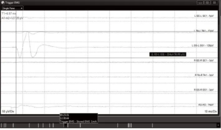
210 monopolar probe was used in all operations (Figure 1). The tip of the probe was cathode and a needle electrode inserted in a muscle close to surgical incision was anode. Constant current was delivered with a 2.82 Hz stimulation frequency and 200 µs pulse width. The current intensity was increased in 0.1 mA steps started from 0 to a maximum of 5 mA until a triggered compound muscle action potential (CMAP) response was seen on EMG. Triggered EMG window parameters were set to 10 ms/div and 100 µV/div (100 ms total window length) (Figure 2).
Figure 1. Left: Needle elecrodes placed after induction of anesthesia; Right: Stimulation of sacral root with a monopolar probe
Figure 2. The response of left gastrocnemius muscle during mapping of roots with direct nerve stimulation
RESULTS
Results of root stimulation
Four operations were finished without any direct nerve stimulation since surgeons did not need it due to relatively simple closure of lesion was sufficient. In those operations only free-run EMG was monitored during the closure and no critical discharge was observed. In remaining 16 operations, compound muscle action potentials were elicited from lower extremity muscles with
direct stimulation of nerve roots. The CMAP responses were obtained with stimulation intensities between 0.05 – 2 mA. When CMAP responses were compared with clinical deficits of newborns, it was obvious that muscle responses were easily obtained from all newborns with normal movements in lower extremities. In 3 newborns with low distal movements, we elicited CMAPs from lower extremity down to gastrocnemius muscle. In 4 newborns with low proximal movements with no movement in distal muscles, CMAP responses were obtained from proximal muscles in all cases (with a very low amplitude in one case). Furthermore, we could elicit bilateral distal responses in one case and unilateral distal responses in another one of those newborns whose proximal weakness was asymmetric. In 2 newborns with distal muscle plegia, we could not take any CMAP responses from distal muscles with direct root stimulation.
During releasing of spinal cord, some dysmorphic appearing rootlet was thought to sacrifice in two cases. Before dissection, the dysmorphic appearing rootlet was stimulated to check the functionality and in one case it was found nonfunctional. However, in the second case the rootlet was found functional, so surgical manipulations were changed with the purpose of preserving functional rootlet.
We did not use external anal sphincter muscle data in our interpretations about roots because in most cases lower sacral roots were not inside the critical surgical area and were in some cases they were not visible to stimulate. We could stimulate lower sacral roots only in 6 cases. In 4 of them external anal sphincter responses elicited while in 2 patients there was no response who had preoperative neurological dysfunction in lower extremities.
Results of placode stimulation
We stimulated neural placode in 12 cases and in 10 of them CMAP responses were elicited. We needed higher stimulation intensity according to root stimulation. The CMAP responses were obtained with stimulation intensities between 0.4 – 3 mA. The CMAP responses were compatible with the side and level of the stimulation in 9 infants.
Two newborns had no CMAP responses with placode stimulation. Those infants had preoperative distal muscle plegia in lower extremities and had also no response with root stimulation. In one case, we started to stimulate neural placode from caudal part and continued stimulating by moving the probe towards cranially. First, we took left side lower limb responses with left side stimulation placode. Secondly, we started to stimulate right side of the placode, CMAP responses from right gastrocnemius were obtained as we expected. When we moved the probe more cranially on the right side we obtained left tibialis responses unexpectedly. Then, we stimulated the left side again from the same level with same stimulation intensity and we took left tibialis responses. On that level, we stimulated two side with same intensity and in every try, we took only left tibialis response without any other muscle responses. In conclusion, we took CMAP responses from left lower extremity with stimulation of both side of neural placode at a particular upper level.
DISCUSSION
There are few studies about intraoperative neurophysiological evaluations in newborns with myelomeningocele. Sala and his colleagues mentioned the feasibility of neurophysiological mapping in four myelomeningocele newborns to spare and preserve neural tissue from intraoperative damage [3]. Two studies are about intraoperative evaluation of ascending somatosensory pathways with direct stimulation of neural placode [10, 11]. To our knowledge, there is only one study in the literature with direct electrical stimulation of placode to induce motor evoked activity [4]. We did not monitor roots at thoracic level because of the technical difficulty to take reliable evoked responses from thoracic myotomes with the stimulation of roots. The same muscle groups with adults are used successfully in newborns for monitoring sacral and lumbar roots with direct stimulation (quadriceps, tibialis anterior, gastrocnemius and external anal sphincter muscles). Today, free-run EMG is routinely used to monitor motor functions of the roots intraoperatively [12, 13]. It is suggested to set the amplitude scale to 50-100 µV/division during monitoring of
electromyography in adults. However, we preferred to follow both triggered and free-run electromyography potentials with a lower (30 µV/division) scale not to miss any response due to low amplitude of responses in newborns, especially if patient had muscle weakness. We did not observe any critical discharges in free-run EMG during closure of the neural tube which supports that roots were not being stretched and injured due to dissecting and closure manipulations.
We used single pulse currents to stimulate placode directly. Under general anesthesia, we could obtain muscle responses with the activation of anterior horn cells in the cord with single pulse technique. When we increase the current intensity, the underlying axons of long motor tracts can also be activated. Even tough the electrical signal can travel down to lower levels in the spinal cord, it cannot pass the synapse to activate alpha-motor neuron and cannot induce muscle responses due to the inhibition effect of general anesthesia. It has been shown in studies that motor evoked potentials of single pulse transcranial electrical stimulation can be abolished by halogenated inhalational gases since they produce an inhibition of synaptic transmission on alpha motoneurons in spinal cord [14, 15]. So, we could elicit responses only from the muscles matching with stimulation level on neural placode due to direct activation of alpha motor neurons on same level which pathway does not include synapse, but not elicited muscle responses from lower levels of stimulation which involve synapses on the way and thus need synaptic transmission to generate a response in alpha motor neurons. A possible limitation of the study is the type of stimulation probe used on neural placode. We preferred monopolar probe to bipolar probe for root stimulation due to unpractical usage of two tips on tiny roots. Hence, we did not change the probe in the surgery to stimulate the neural placode and continue to use it for both stimulation. Since, monopolar probe has a risk of spreading current to close neural tissue, we may have induced activation on peripheral roots in course of placode stimulation. Bipolar concentric probe is more suitable to elicit a focal current flow during stimulation of tiny roots. Unfortunately, we have to choose one of the two type: monopolar probe or bipolar probe with two tips, due to problems in purchasing procedures in our
212 institution.
One of the substantial factor of direct stimulation is the distance between the tip of the probe and motor neurons. The maximum intensity needed to stimulate neural placode was higher than the maximum intensity needed to stimulate a root. The probable reason of this was a covering tissue on neural placode blocking the current in some patients, so we needed to increase the current intensity to activate motor neurons. It was emphasized in researches that somatosensory evoked potentials were present with stimulation of neural placode and distal nerves, which is an evidence of functional sensory pathway in open neural tube [10, 11]. Furthermore, some patients can gain motor functions after closure of MMC even presents a motor deficit in examination preoperatively [16]. Those results supports the functionality of neural placode in MMC patients. The results of placode stimulations in our study are also compatible with Pugh and colleagues’ research [4]. Both of the research found more proximal muscle responses with the stimulation of more proximal in the cord. Moreover, we took an unexpected response on neural placode in one case. When we stimulated right side of the neural placode from a more proximal level, we observed evoked responses from contralateral lower limb without any response at ipsilateral muscles. Technical reasons were ruled out, cerebrospinal fluid/saline were sucked from surgical area to prevent a possible current jump. Same stimulation was repeated at both side of the placode, yet the observed left tibialis responses (without any other muscle response) remained. If this responses resulted from a current spread from monopolar probe, we would expect to see responses at ipsilateral muscles together with contralateral responses. This result brings to mind a question: Is there a possible failure in intra-spinal neuronal (motor neuron) organization beside neural tube closure or is it an outcome of a spinal cord plasticity due to injury [17, 18]?
Kumar and his colleagues used only external anal sphincter for monitoring sacral roots during tumor excision and dissection of lipomyelomeningocele [19]. Some of the newborns had weakness in lumbar segments of lower extremities in our patient group, hence we added
lower extremity muscles to external anal sphincter to monitor wider myotomes which includes all lumbar and sacral roots. We monitored newborns who had weakness in lower extremities and took responses from those muscle groups with root stimulation. In 2 cases with distal plegia, we could elicit distal muscle responses in very low amplitude with root stimulation which shows that the roots were functional. The result of direct stimulation supports that the level of conduction block in motor pathway may be in spinal cord, not in roots. The levels of MMC lesion in those patients were thoracolumbar (1) and lumbosacral (1). However, in other 2 cases with distal plegia, we could not take evoked response from distal muscles which shows roots were not functional. The level of conduction block in motor pathway could be in roots or both in spinal cord and roots together. Some roots may lose their function due to abnormal development of neural tissue. Both of the patients had MMC at thoracolumbar level. Lastly, of 6 newborns with MMC at thoracolumbar level, 4 (66%) had neurological deficit at lower extremities while, of 9 newborns with MMC at lumbosacral level, 3 (33%) had neurological deficit in lower extremities. This is compatible with the common knowledge that higher the level of the MMC lesion, higher the severity of neurological dysfunction [20].
In conclusion, according to our practice, neurophysiological mapping with direct root and placode stimulation is a useful and reliable method to check the functional neural tissue and spare it to preserve during releasing and closure of neural tube in myelomeningocele operations. It is crucial for cases in particular, which involve dysmorphic appearing roots in surgical area. In addition, we elicited motor responses arising from anterior horn cells which contribute to literature about the integrity of grey matter-anterior horn functions in neural placode.
REFERENCES
1. Pouratian N, Elias WJ, Jane JA Jr, Phillips LH, Jane JA: Electrophysiologically guided untethering of secondary tethered spinal cord syndrome. Sr.Neurosurg Focus 2010; 29(1):E3. doi:10.3171/2010.3.FOCUS09299.
2. Hoving EW, Haitsma E, Oude Ophuis CM,
Journée HL: The value of intraoperative neurophysiological monitoring in tethered cord surgery. Childs Nerv Syst 2011; 27(9):1445-52. 3. Sala F, Tramontano V, Squintani G, Arcaro C,
Tot E, Pinna G, Meglio M: Neurophysiology of complex spinal cord untethering. J Clin Neurophysiol 2014; 31(4):326-36.
4. Pugh JA, Aronyk KE, Norton JA.: Neural activity generated in the neural placode and nerve roots in the neonate with spina bifida. J Neurosurg Pediatr 2012;9(4):452-6.
5. Copp AJ, Adzick NS, Chitty LS, Fletcher JM, Holmbeck GM, Shaw GM: “Spina Bifida”Nature Reviews Disease Primers 2015 Published online:30 April 2015 doi: 10.1038/nrdp.2015.7 6. Hendelman WJ: Atlas of Functional
Neuroanatomy, ed 2. Boca Raton, FL: CRC Press, 2006
7. Sanes DH, Reh TA, Harris WA: Development of the Nervous System, ed 2. London: Academic Press, 2006
8. Stiefel D, Copp A J, Meuli M: Fetal spina bifida: loss of neural function in utero. J Neurosurg 2007;106, 213–221.
9. Meuli M, Meuli-Simmen C, Hutchins GM, Seller MJ, Harrison MR, Adzick NS: The spinal cord lesion in human fetuses with myelomeningocele: Implications for fetal surgery. J Pediatr Surg 1997; 32, 448–452. 10. McLone DG: Technique for closure of
myelomeningocele Childs Brain 1980; 6:65-73 11. Reigel DH, Dallmann DE, Scarff TB, Woodford
J: Intra-operative evoked potential studies of newborn infants with myelomeningocele. Dev Med Child Neurol Suppl 1976 ;(37):42-9. 12. Kothbauer K, Schmid UD, Seiler RW, Eisner W
: Intraoperative motor and sensory monitoring of the cauda equina. Neurosurgery 1994;34(4):702-7; discussion 707.
13. Krassioukov AV, Sarjeant R, Arkia H, Fehlings MG: Multimodality intraoperative monitoring during complex lumbosacral procedures: indications, techniques, and long-term follow-up review of 61 consecutive cases. J Neurosurg Spine 2004;1:243–53
14. Kalkman, C, Drummond J, Ribberink A : Low concentrations of isoflurane abolish motor evoked responses to transcranial electrical stimulation during nitrous oxide/opioid
anesthesia in humans. Anesth Analg 1991;73, 410–5.
15. Yamada H, Transfeldt EE, Tamaki T, Torres F, Iaizzo PA : The effects of volatile anesthetics on the relative amplitudes and latencies of spinal and muscle potentials evoked by transcranial magnetic stimulation. Spine 1994; 19, 1512–7. 16. McLone DG : Results of treatment of children
born with a myelomeningocele. Clin Neurosurg 1983;30:407-12
17. Ramer LM, Ramer MS, Bradbury EJ : Restoring function after spinal cord injury: towards clinical translation of experimental strategies. Lancet Neurol 2014;13(12):1241-56.
18. ChédotalA:Development and plasticity of commis sural circuits:from locomotion to brain repair Trends Neurosci 2014;37(10):551-62. 19. Kumar GS, Rajshekhar V, Babu KS:
Intraoperative mapping of sacral nervous system (S2-4). Br J Neurosurg 2006;20(6):396-402.
20. Oakeshott P, Hunt GM, Poulton A, Reid F : Open spina bifida: birth findings predict long-term outcome. Arch Dis Child 2012;97, 474–6.