Concomitant inactivation of p53 and Chk2 in breast cancer
Alexandra Sullivan
1, Martin Yuille
2, Claire Repellin
1, Archana Reddy
1, Olivier Reelfs
1,
Alexandra Bell
3, Barbara Dunne
3, Barry A Gusterson
3, Peter Osin
4, Paul J Farrell
1, Isik Yulug
5,
Abigail Evans
6, Tayfun Ozcelik
5, Milena Gasco
7and Tim Crook*
,11Ludwig Institute for Cancer Research, Imperial College Faculty of Medicine, St Mary's Campus, Norfolk Place, London W2 1PG,
UK;2HGMC, Hinxton, CB;3Department of Pathology, University of Glasgow, Western In®rmary, Glasgow, UK;4Department of
Histopathology, University College and Middlesex Hospital Medical School, University Street, London WC1, UK;5Department of
Molecular Genetics, Bilkent University, Ankara, Turkey; 6Department of Surgery, Poole General Hospital, Poole, Dorset, UK; 7UO Oncologia Medica, Azienda Ospedaliera S Croce e Carle, Via Coppino 26, 12100, Cuneo, Italy
The structure and expression of the human Rad53 homologue Chk2 was analysed in breast cancer. The previously described silent polymorphism at nucleotide 252 in codon 84 (GAA4GAG) was observed in 5/141 cases. Somatic Chk2 coding mutations were detected in 7/141 cases, these occurring in 4/18 BRCA1-associated breast cancers, 1/78 sporadic breast cancers and 2/25 typical medullary carcinomas. Each of the BRCA1-associated cancers with Chk2 mutations also contained mutations in p53, whereas the single sporadic cancer with Chk2 mutation was wild-type for p53. Expression of Chk2 was ubiquitously detected in normal ductal epithelium of the breast, but there was loss of expression in a signi®cant proportion of breast carcinomas, and this occurred in cancers both with and without p53 mutation. A CpG island was identi®ed 5' of the Chk2 transcrip-tional start site, but there was no evidence of cytosine methylation in any of the cancers with down-regulated Chk2 expression. Analysis of the germ-line of 45 individuals with hereditary or early onset breast cancer revealed wild-type Chk2 sequence in all cases. Thus, despite the rarity of somatic mutations in Chk2 in sporadic breast carcinomas, our results nevertheless reveal that concomitant loss of function in Chk2 (via down-regulation of expression) and p53 (via mutation) occurs in a proportion of sporadic cases. However, consistent with other studies, we show that germ-line mutations in Chk2 are unlikely to account for a signi®cant proportion of non BRCA1-, non BRCA2-associated hereditary breast cancers.
Oncogene (2002) 21, 1316 ± 1324. DOI: 10.1038/sj/ onc/1205207
Keywords: p53; Chk2; breast cancer
Introduction
Breast cancer is one of the most common neoplasms to aect women. Inherited mutations may account for up to 10% of breast cancers, of which BRCA1 and BRCA2 may be mutated in the germ-line of half of such cases. Mutation in p53 occurs in 10 ± 30% of sporadic adenocarcinomas of the breast with higher frequencies with increasing histopathological grade. The proportion of cancers with p53 mutation is, however, increased in some forms of breast cancer. In BRCA1-associated cancers, several studies have re-ported an increased frequency of mutations compared to grade-matched sporadic cases (Crook et al., 1998; Phillips et al., 1999). Furthermore, mutations in p53 occur at extremely high frequencies in typical medul-lary breast cancers (de Cremoux et al., 1999). The latter observation is of particular interest in view of the recognized pathobiological similarities between medul-lary and BRCA1-associated breast cancers (Eisinger et al., 1998).
Chk2 (hcds 1) is the human homologue of the Saccharomyces cerevisiae RAD53 and Schizosacchar-omyces pombe CDS1 kinases. The protein contains a 60 amino acid fork head-associated (FHA) domain (residues 115 ± 175) and a kinase domain (residues 226 ± 486). Chk2 functions downstream of ATM to directly phosphorylate p53 at serine 20. A second substrate for Chk2 is BRCA1 (Chehab et al., 2000; Hirao et al., 2000; Lee et al., 2000). The FHA domain is essential for phosphorylation of Chk2 by ATM (Lee and Chung, 2001). Heterozygous germ-line mutations in Chk2 were reported in individuals with the cancer predisposition Li-Fraumeni syndrome who lacked p53 mutations (Bell et al., 1999), implying that loss of function in Chk2 might be functionally equivalent to p53 mutation. Analysis of Chk2 in breast cancer families has revealed that germ-line mutations are unlikely to account for a signi®cant proportion of non-BRCA1, non-BRCA2, non-p53 hereditary cancers. In one study, only a single mis-sense mutation was identi®ed in 79 families, but this change also occurred in healthy controls (Allinen et al., 2001), whereas a second study found Chk2 mutation in two of 44
www.nature.com/onc
*Correspondence: T Crook, E-mail: [email protected].
Received 30 October 2001; revised 21 November 2001; accepted 28 November 2001
families (Vahteristo et al., 2001). Studies of somatic mutations in sporadic and familial breast cancers have not been described. In this report, we have analysed the structure and expression of Chk2 in breast cancer. Results
Novel somatic Chk2 mutations are detected in breast cancer
Using SSCP and RT ± SSCP, the sequence of Chk2 was analysed in 141 primary breast cancers. Primers for SSCP analysis were designed from the intron ± exon structure of the human Chk2 gene (Accession numbers AL117330 and AL121825). SSCP mobility shifts were observed in 13/141 cases (Figure 1). Sequencing of multiple plasmid clones of each case with SSCP shifts revealed that ®ve were due to the recognized non-coding polymorphism at nucleotide 252 A4G (codon
84). Of the remaining eight cancers, coding mutations were present in seven (Table 1). Four of the coding mutations occurred in cancers arising in carriers of BRCA1 germ-line mutations (from 18 cases analysed). A further non-coding mutation was detected in a BRCA1 case.
No Chk2 mutations were detected in cancers occurring in BRCA2 mutation carriers (from 20 analysed). One of the mutations was in a sporadic case (from 78 analysed) and two were from typical medullary breast carcinomas (from 25 analysed). One of the medullary breast carcinomas occurred in an individual with a germ-line mutation in BRCA1 (185 del Ag) (Table 1), this change also being present in three further medullary breast cancers, whereas in the other case with Chk2 mutation, there was methylation of the BRCA1 promoter (data not shown). Normal tissue was available for two of the cases with mutations and sequencing of DNA from this revealed that the mutations were somatically acquired rather than
germ-Figure 1 Novel mutations in Chk2 are detected in BRCA1-associated breast cancers. germ-Figure 1a: SSCP analysis of the 5' portion of exon 1 of Chk2. Mobility shifts are seen in lanes 4, 6 and 9. Sequencing revealed mutations in each of these cases. Figure 1b: sequence analysis of exon 1 of Chk2 in BRCA1-associated breast cancers. The upper sequence shows the mutation TCC4CCC (Ser4Pro) at codon 49 of Chk2. The lower sequence shows the non-coding mutation TCC4TCA (Ser4Ser) at codon 62
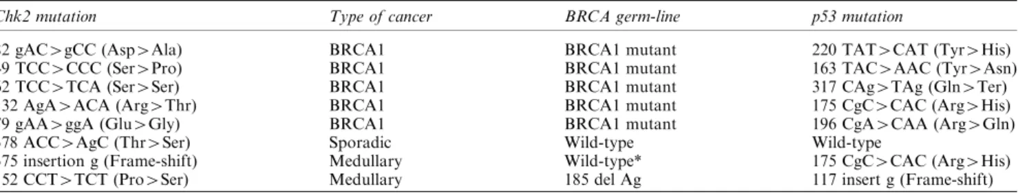
Table 1 Somatic mutations inChk2 in breast cancer
Chk2 mutation Type of cancer BRCA germ-line p53 mutation
82 gAC4gCC (Asp4Ala) BRCA1 BRCA1 mutant 220 TAT4CAT (Tyr4His)
49 TCC4CCC (Ser4Pro) BRCA1 BRCA1 mutant 163 TAC4AAC (Tyr4Asn)
62 TCC4TCA (Ser4Ser) BRCA1 BRCA1 mutant 317 CAg4TAg (Gln4Ter)
132 AgA4ACA (Arg4Thr) BRCA1 BRCA1 mutant 175 CgC4CAC (Arg4His)
79 gAA4ggA (Glu4Gly) BRCA1 BRCA1 mutant 196 CgA4CAA (Arg4Gln)
378 ACC4AgC (Thr4Ser) Sporadic Wild-type Wild-type
375 insertion g (Frame-shift) Medullary Wild-type* 175 CgC4CAC (Arg4His) 152 CCT4TCT (Pro4Ser) Medullary 185 del Ag 117 insert g (Frame-shift) *BRCA1 promoter hypermethylation
line (378 ACC4AgC (sporadic) case and 79 gAA4g-gA (BRCA1-associated) case. In an attempt to determine whether the remaining Chk2 mutations were somatic rather than germ-line, we analysed DNA from cancers occurring in individuals in the same families. In none of these cases did we detect Chk2 mutations (either the same or dierent from those originally identi®ed). These results strongly suggest that the mutations are somatically acquired rather than gem-line. Direct sequencing of the PCR products revealed that the mutated allele was the predominant one present in two of the seven cases (378 ACC4AgC (sporadic) and 49 TCC4CCC (BRCA1-associated)), consistent with loss of the wild-type allele. In the remaining ®ve cancers, there was no evidence of loss of heterozygosity (LOH). It should be noted, however, that the presence of normal tissue may mask LOH. As such, we cannot be certain that these are heterozygous mutations.
Absence of germ-line mutations in hereditary, familial, early onset and male breast cancer
Germ-line mutations in Chk2 have been described in individuals with the Li-Fraumeni syndrome (Bell et al., 1999) and in a very small number of BRCA1, non-BRCA2 hereditary breast cancers (Allinen et al., 2001; Vahteristo et al., 2001). We therefore analysed the germ-line structure of Chk2 in 45 individuals with various forms of breast cancer, in whom the BRCA1 and BRCA2 status has been previously described (Ozdag et al., 2000). In these cases, direct sequencing of genomic DNA isolated from peripheral blood lymphocytes detected the codon 84 polymorphism in three of the 45 cases, but no germ-line mutations in any of the cases.
Mutations in Chk2 and p53 co-exist in breast cancers The frequency of p53 mutations is higher in BRCA-associated breast carcinomas than in grade-matched sporadic cases (Crook et al., 1997, 1998; Phillips et al., 1999). We therefore determined the p53 sequence in the cancers with Chk2 mutation. Each of the BRCA1-associated cases with Chk2 mutation also contained mutation in p53 (Table 1). No p53 mutation was detected in the single sporadic cancer with a Chk2 mutation despite both SSCP and sequencing of independent clones of each p53 coding exon. We also determined the status of p53 in the medullary breast cancers with Chk2 mutations. In both cases, the p53 gene was mutated (Table 1). Our data show, therefore, that p53 mutations co-exist in the majority of cases with Chk2 mutation.
Down-regulation of Chk2 mRNA in sporadic breast cancer
The presence of Chk2 mutations in breast cancers prompted us to examine other possible mechanisms by which the function of Chk2 might be abrogated in
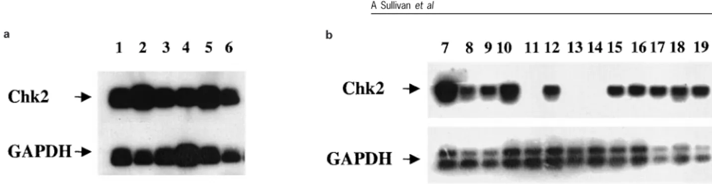
cancer. We therefore performed analysis of Chk2 expression in normal and malignant breast tissue by RT ± PCR. Frozen sections of the cancers selected for this analysis were initially checked by a breast pathologist to verify that at least 50% of the cells in the tissue were malignant. Analysis of normal breast tissue revealed that Chk2 mRNA was abundantly expressed in all cases analysed (Figure 2a), but steady-state levels of Chk2 mRNA were down-regulated in 16/ 34 cases of sporadic breast cancer (Figure 2b). In some cases, Chk2 mRNA was undetectable under the semi-quantitative conditions of our assay (Figure 2b). Of the 16 cancers with down-regulated Chk2 mRNA, seven cases had mutations in p53 while nine were wild-type for p53. Of the 18 cancers which did not exhibit down-regulated Chk2, p53 mutations were present in four, whereas 14 were wild-type. To verify the results of the RT ± PCR analysis, we performed a detailed immuno-cytochemical study of Chk2 expression in breast cancer. In preliminary characterization, the speci®city of the polyclonal reagent used for immunocytochem-istry was veri®ed by immunoblotting of cell lysates prepared from g-irradiated MCF-7 cells. The antibody clearly recognized a single protein of the appropriate molecular weight in MCF-7 cells (Figure 3a, top panel). A small but reproducible mobility shift, representing the active form of the protein, was detected in irradiated cells. This shift was more readily detectable when the lysates were subjected to extended electrophoresis to increase resolution (Figure 3a, lower panel).
In initial immunocytochemical analysis, it was clear that there was considerable variability in the percen-tage of normal cells that was positive in the normal glands and in the intensity of staining of individual nuclei between cases, although the intensity was similar within the same case. When the staining was strong in the normal tissue, this was always associated with a high proportion of positive cells. Thus variation in intensity between cases was probably a feature of dierences in epitope access to the primary antibody. As the objective was to assess the staining in the associated carcinomas, it was decided to use staining of the normal glandular elements present in the sections as an internal control for the associated carcinoma and to directly compare normal and tumour within the same section. Comparative staining intensity and percentage of nuclei positive in the normal tissues of the 81 cases that had normal tissue present are shown in Table 2. Table 3 shows the same data for the carcinomas in those 75 cases that had tumour and normal in the same slide. From these analyses, it was clear that the intensity of staining and the proportion of positive cells was lower in the carcinomas compared with the normal tissues. Comparing the normal and the tumour in the same sections, there were 32 cases with the same intensity of staining and 41 cases where the staining intensity was at least one point less in the tumour compared with the normal. There were two cases where the tumour was greater than the normal. If a cut o was taken for dierence of two levels of
Table 2 Immunocytochemical analysis of Chk2 expression in normal breast tissue (a) Proportion of cells positive (b) Intensity of staining
Score % of Cases Score % of Cases
1 1.2% 1 9.9% 2 2.5% 2 26% 3 11.0% 3 64% 4 12.0% 5 23.5% 6 49.4% a b
Figure 2 RT ± PCR analysis of Chk2 mRNA in breast tissue. Lanes 1 ± 6: RT ± PCR analysis of mRNA from reduction mammoplasties. Chk2 is invariably expressed in normal breast tissue. Lanes 7 ± 19: Expression in sporadic breast carcinomas. Note the loss of expression in lanes 11, 13 and 14. Also shown is the GAPDH control RT ± PCR
Figure 3 (a) The polyclonal antibody speci®cally recognizes Chk2. Lysates prepared at the indicated times after g -irradiation of MCF-7 breast carcinoma cells were resolved on 10% polyacryamide gels, transferred to membranes and probed with the anti-Chk2 polyclonal antiserum used for subsequent immunocytochemical analysis. The antiserum recognizes only a single species of the appropriate molecular weight. The lower panel is Western blot analysis of the same lysates following extended electrophoresis. The active, shifted form of Chk2 is recognized by the antibody. (b) Immunocytochemical analysis of expression of Chk2 in breast cancer. The sections show the indicated tissues (normal, DCIS and invasive cancer). Immunocytochemistry was performed as described in Materials and methods. Note the abundant expression in glandular epithelium in the two normal breast sections and the complete absence of nuclear Chk2 immunoreactivity in the DCIS and carcinoma sections shown
intensity (e.g. three to one or two to zero), then there were 17 tumours that were lower than the associated normal. If the cut o was three levels of intensity then there were nine cases that had score of three in the normal and a score of zero in the tumour. Examples of staining are shown in Figure 3. Amongst cases analysed were the 18 BRCA1-associated cancers. Of these, there was reduced Chk2 expression in seven cases. These comprised two cases with Chk2 mutations (132 AgA4ACA, 82 Asp4Ala) and ®ve cases lacking mutations.
Down-regulation is not associated with CpG Methylation in Chk2
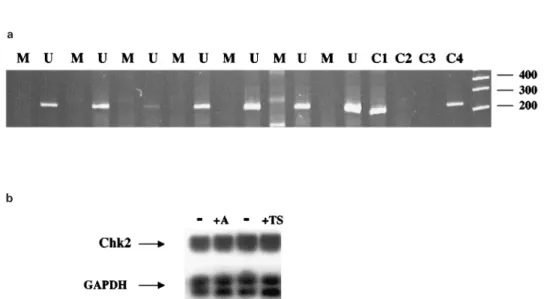
By 5'RACE and database searching, we identi®ed a CpG island 5' of the transcriptional start site of the Chk2 mRNA. We analysed both normal breast tissue and 24 cancers by bisulphite sequencing and methyla-tion speci®c PCR (MSP) to determine whether cytosine methylation was present. However, only unmethylated alleles were detected in all cases (Figure 4a). Similar analysis of breast cancer cell lines detected weak methylation only in MCF-7, but treatment with 5-azacytidine or trichostatin A did not aect the steady-state levels of Chk2 mRNA (Figure 4b). These results suggest that mechanisms other than aberrant methyla-tion in the CpG island 5' of Chk2 must underlie the down-regulation of Chk2 expression in breast cancer. Discussion
Germ-line mutations in Chk2 have been described in some Li-Fraumeni families lacking p53 mutations
(Bell et al., 1999). This observation suggests that loss of Chk2 might have an eect functionally equivalent to mutation of p53. Such a suggestion is compatible with Chk2-dependent phosphorylation of serine 20 of p53 in response to DNA damage. Because breast cancer is a recognized neoplasm in individuals with the Li-Fraumeni syndrome, we have addressed this possibility by examination of the structure and expression of Chk2 in breast cancers of varying p53 status and in the germ-line of individuals with hereditary breast cancer.
We have detected four previously unreported somatic mutations in BRCA1-associated breast cancers and medullary breast carcinomas. In contrast, analysis of sporadic breast cancers matched for grade revealed only a single Chk2 mutation in 78 cases analysed. One of the four BRCA1-associated cancers contained a mutation in the FHA domain of Chk2 and one of the mutations in the medullary breast cancers was also in the FHA domain. The remaining mutation in the medullary cancer was a frame-shift in the kinase domain. These mutations are, therefore, highly likely to result in inactivation of Chk2 (Wu et al., 2001).
a
b
Figure 4 Analysis of methylation in Chk2 in breast cancer. (a) MSP analysis of CpG island 5' of the Chk2 gene. Bisulphite-modi®ed genomic DNA from breast cancers with down-regulated Chk2 mRNA was prepared as described in Materials and methods and used as a substrate for MSP. The gel shows unmethylated (U) and methylated (M) reactions of cancers with down-regulated Chk2 expression. C1=control methylated DNA with M primers; C2=control methylated DNA with U primers; C3=control unmethylated DNA with M primers; 4=control unmethylated DNA with U primers. Molecular weight size markers are also shown. (b) Treatment with demethylating agents does not aect Chk2 mRNA levels. MCF7 cells were treated with 3 mM 5-aza-2'-deoxcytidine (A) is the 5 mMtrichostatin A (TS). RNA was harvested after 72 h, and Chk2 mRNA levels determined by RT ± PCR. Also shown is the GAPDH control RT ± PCR
Table 3 Immunocytochemical analysis of Chk2 expression in breast carcinoma
(a) Proportion of cells positive (b) Intensity of staining
Score % of Cases Score % of Cases
1 33.3% 0 23.5% 2 16.0% 1 16% 3 3.7% 2 33.3% 4 12.3% 3 18.5% 5 14.8% 6 11.1% 1320
Mutations outside the kinase domain or the FHA domain have not previously been reported in human cancer and, as such, the changes in the remaining 3 BRCA1-associated cases which are substitutions in exon 1, upstream of the FHA domain, represent novel mutations. The biological eect(s) of these mutations await detailed biochemical analysis. Interestingly, previous studies of p53 in BRCA1-associated breast cancers also identi®ed novel mutations not detected in a control series of cancers matched for pathobiological and patient characteristics (Crook et al., 1998; Phillips et al., 1999). This suggests that a distinct spectrum of genetic mutations may occur in such cases (Greenblatt et al., 2001). A further noteworthy observation from our work is the detection of somatic Chk2 mutations in typical medullary breast cancers. A number of studies have described similarities between medullary breast cancers and BRCA1-associated breast cancers (Eisinger et al., 1998). These include not only pathobiological factors such as pushing margins, high lymphocytic in®ltrate and low level of steroid hormone receptor expression (Lakhani et al., 2000), but also molecular genetic properties such as high mutation rate in p53 (de Cremoux et al., 1999). Moreover, methylation-depen-dent transcriptional silencing of BRCA1 occurs at high frequency in medullary cancers (Esteller et al., 2000). In the context of these similarities, it is of interest that the two medullary breast cancers with Chk2 mutations both had BRCA1 inactivation, in one case via germ-line BRCA1 mutation, the other via promoter silencing.
A major rationale for our study was to address the possibility that Chk2 mutation might compensate for the absence of p53 mutations. However, in the cases where Chk2 mutations were identi®ed, each of the BRCA-associated and medullary breast cancers also contained mutant p53. Only the single sporadic breast cancer with Chk2 mutation was wild-type for p53. Previous studies of human cancers have also reported the co-existence of p53 and Chk2 mutations in colon cancer (Bell et al., 1999) and in small cell lung cancer (Haruki et al., 2000). Thus, mutations in Chk2 and p53 appear to occur more frequently concomitantly than independently and this argues against the hypothesis that Chk2 mutations occur commonly as an alternative to p53 mutation. The ®nding of mutations in a subset of breast cancers prompted us to investigate whether abrogation of Chk2 function might occur via down-regulation of expression in breast cancer. Analysis by RT ± PCR revealed abun-dant expression of Chk2 mRNA in normal breast tissue, but loss of expression in a signi®cant proportion of carcinomas. Extensive, detailed analysis by immunocytochemistry veri®ed the results of RT ± PCR and demonstrated down-regulation or loss of expression of Chk2 in a proportion of carcinomas, including those with mutations in p53. Thus, in addition to the simultaneous mutation of Chk2 and p53 in familial and medullary cancers, there was down-regulation of Chk2 expression in a signi®cant number of breast cancers which also contained mutant
p53. Insight into the possible signi®cance of such concomitant defects in Chk2 and p53 was provided recently by Falck et al. (2001) who showed that concomitant loss of p53 and Chk2 results in a selective advantage over and above that resulting from loss of function in either protein alone. It will certainly be of interest in future studies to determine whether the Chk2 and p53 status in¯uences the clinico-pathological characteristics of breast cancers.
Down-regulation, but not mutation, of Chk2 in sporadic breast cancer in some respects resembles p16INK4a wherein loss of expression, rather than
mutation, occurs in a subset of cancers (Herman et al., 1995). It will clearly be of interest to evaluate Chk2 expression in other common cancers, and Chk2 has recently been shown to be down-regulated in a subset of testicular germ-cell tumours (Bartkova et al., 2001). The authors of this study emphasized the importance of studying promoter silencing in cancers with reduced Chk2 expression and in the present report we have addressed the possibility of methyla-tion-dependent silencing. Having identi®ed a CpG island 5' of the Chk2 transcriptional start site, we designed primers for MSP and bisulphite sequencing. We did not, however, detect evidence of cytosine methylation in any of the cancers with down-regulated expression, implying that mechanisms other than methylation, such as changes in pathways regulating expression and/or stability, regulate Chk2 expression in cancers. Alternatively, methylation in other regions of the Chk2 gene may mediate transcriptional silencing, but the absence of changes in mRNA levels following treatment with demethylating agents sug-gests that this is a less likely explanation. Methylation-independent silencing of other tumour suppressor genes has been described (Banelli et al., 2000; Acquati et al., 2001).
Germ-line mutations in BRCA1 and BRCA2 confer a signi®cant risk of breast cancer. However, the molecular genetic basis of inherited predisposition to breast cancer in non-BRCA1, non-BRCA2, non-Li-Fraumeni individuals is not known (Nathanson et al., 2001). We therefore sought germ-line mutations in Chk2 in a panel of breast cancers previously char-acterized for BRCA1 status (Ozdag et al., 2000). Other than three individuals heterozygous for a previously described non-coding polymorphism, no deviations from the published coding sequence of Chk2 were detected in the germ-line of these individuals. These results suggest that germ-line mutations in the coding sequence of Chk2 are unlikely to account for a signi®cant proportion of non-BRCA1, non-BRCA2, non-p53 hereditary breast carcinomas. Similar conclu-sions have been reached in other recent studies (Allinen et al., 2001).
In conclusion, our results are consistent with a role for Chk2, via loss of expression, in a signi®cant proportion of sporadic breast cancers, but support other studies suggesting that it is unlikely to represent a susceptibility gene for a high proportion of hereditary breast cancers.
Materials and methods Tumours
Sporadic breast cancers were collected, with ethical approval, at surgical resection. Diagnosis was con®rmed by histopatho-logical examination. Paran sections of medullary breast cancers were obtained from the pathology archives of three Israeli hospitals. Twenty-®ve cases were available for analysis. For analysis of germ-line Chk2 mutations, genomic DNA samples were analysed from 45 individuals, comprising hereditary (seven cases), familial (six cases), early onset (23 cases) and male breast cancer (nine cases) groups. The BRCA1 and BRCA2 status of these cases has been described previously (Ozdag et al., 2000). Each patient gave fully informed consent for participation in the study.
Gene analysis
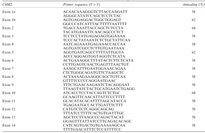
Fresh-frozen tumour tissue was available from six reduction mammoplasties and 34 sporadic breast carcinomas. Genomic DNA was isolated from frozen tissues (normal and tumour) by proteinase K digestion, and total RNA by RNAzo1 B. DNA was isolated from paran-embedded samples by extended digestion in proteinase K. Genomic DNA was isolated from the leukocytes of the samples analysed for germ-line changes in Chk2 by phenol/chloroform extraction. The p53 status of each cancer was determined by SCCP and DNA sequencing as described previously (Brooks et al., 2000). Mutations in Chk2 were sought in genomic DNA by SSCP and, for the sporadic cases where RNA was available, by RT ± PCR SSCP. Primers for SSCP from genomic DNA were designed from AL117330 and AL121825 (Table 4). Primers for RT ± PCR SSCP were as described (Haruki et al.,
2000). cDNA was synthesized in 50 ml reaction from 3 mg of total RNA using the Stratagene ProStar system. Two ml of this solution was used for each PCR. In all cases, PCR products were resolved on 6% native polyacrylamide gels with and without 10% glycerol. When SSCP amplimers exhibited aberrant mobility, the fragment was re-ampli®ed with Pfx DNA polymerase and multiple plasmid clones in the pCRBlunt vector were sequenced. During these studies a number of highly conserved 3' fragments of Chk2 were identi®ed which map to several autosomes and the X chromosome (Sodha et al., 2000). These fragments are homologous to sequences in exons 10 ± 14 of Chk2. We therefore performed additional analyses to con®rm that the sequence changes we had detected which occurred in these regions of Chk2 represent bona®de Chk2 mutations rather than rare polymorphisms within the homologous fragments. We used forward primers located within exon nine, which is unique to Chk2, together with reverse primers from exons 10 and 11 to amplify Chk2-speci®c genomic sequences and sequenced these. These con®rmed the presence of the mutations. Finally, we performed sequence analysis of matched normal tissue, where available, for each cancer with mutation for which matched normal tissue was available. The mutations were not detected in the normal tissue, con®rming their authenticity as somatic Chk2 mutations.
To seek potential sites of CpG methylation by which transcription might be silenced, we used the GRAIL CpG island program to search GI 6453388 which contains the Chk2 locus. This search identi®ed a CpG island on the -strand (in AL117330) upstream of the putative Chk2 transcriptional start site. We designed the following primer pairs to perform MSP (Herman et al., 1995). Unmethylated: TACAACAACCCATAAAACCCCAAACAAA-3' and 5'-TAGATTTTGATGTGTTTTTTGTTTGGGTTT-3', giving a
Table 4 Primers for SSCP of CHK2 from genomic DNA
CHK2 Primer sequence (5'43') Annealing (8C)
Exon 1a ACAACAAAGGGTCTTACCAAGATT 58 AGGGCATATCCAGCTCCTCTAC Exon 1b AGTGAGAGGACTGGCTGGAGT 62 GGCCCATCATTTACTTTTTAATTTT Exon 2 TGACCAAATTACCAGCTCTCCTA 64 TACATGAAATTCAACAGCCCTCT Exon 3 TCCTCCTATGAGAGAGTGGAAAA 58 TCCCACTATAAATCTCTGCTATTCAA Exon 4 AATCAGAAATGAGAAACCACCAA 58 AGTGATCGCCTCTTGTGAATAAA Exon 5 AGGTGATCAGCCTTTTATTGGTA 62 ACCCAGGAGTGGTAGGTCTCATA Exon 6 ACTGAAAGGCTTTATACTCTTCTCATA 58 CCTTGAGTCAACTGAGTTTAACTGT Exon 7 AAAGCATTTGAATGGAAACAGAA 58 CTCTGGGCAGATGTTCTAAGCTC Exon 8 ACTAAAAGAAAGGCAGCTGTCAA 58 GTTTTCCCCCAGGAATGAAC Exon 9 TTTCTGAACAAGAATCTACAGGAAT 58 TTAAGTATCTACTGCATGAATCTGAGG Exon 10 ATCACCTCCTACCAGTCTCTGC 64 GCAAGTTCAACATTATTCCCTTTT Exon 11 GCACATACACATTTTAGCATACCA 58 TGAGAATGCCACTTGATTTCTTT Exon 12 CATGTCTCTCAGGCAGCAG 58 TTTATCCTTTTCACTGTGATTTGC Exon 13 AGCTCCTTAAGCCCAGACTACAT 58 GGAGTTTATTATCCTTCAGACACAGC Exon 14 CATCAGTGACTGTGAAAAAGCAA 58 TTTTGAACATTTCTCCATTTTCC 1322
product of 216 bp. Methylated: 5'-GACGACCCATAAAA-CCCCGAACGAA-3' and 5'-TTTCGACGTGTTTTTCGTT-CGGGTTC-3', giving a product of 209 bp. Genomic DNA (1 mg) was modi®ed using the Intergen kit as recommended by the manufacturer. Following PCR, products were resolved on 10% polyacrylamide gels, stained in ethidium bromide and visualized under UV. Each PCR included control methylated and unmethylated DNA samples. Primers for bisulphite sequencing were as follows. Forward: GAT-TAATGAGGAGTAGTAGATGTGGT-3' and Reverse: 5'-CCTCTCAATTTCCTCCTAAAAAATCAA-3'. Following PCR, ampli®ed products were puri®ed with the Qiagen PCR clean up kit and sequenced directly.
To determine the eect of demethylating agents on Chk2 expression, MCF7 cells were treated with 5-aza-2'-deoxycy-tidine (1 mM) or trichostatin A (5 mM). Total RNA was
harvested after 72 h and RT ± PCR performed for Chk2 mRNA as described below.
RT ± PCR analysis of Chk2 expression
Chk2 mRNA expression in normal breast tissue and in breast cancers was analysed by RT ± PCR, using primers described previously (Haruki et al., 2000). Following PCR, reaction products were resolved on 2% agarose gels, transferred to nylon and probed with a Chk2-speci®c oligonucleotide, located internal to the PCR primers, end-labelled with g-32P
ATP using polynucleotide kinase. To ensure the presence of equal amounts of cDNA in each PCR, a GAPDH control PCR was performed under similar limiting conditions. Immunohistochemical analysis of CHK2 expression
The speci®city of the antibody reagent used for these studies was determined by Western blotting of MCF-7 cells. Cells were subjected to 5 Gy, then incubated for 0, 0.5 and 3 h after which time lysates were prepared in sample buer and resolved on 10% polyacrylamide gels. For immunocytochem-ical analysis, 3 m unstained sections were taken from anonymized breast cancer samples from the histology ®les. One hundred and twenty cases were used, of which one had no carcinoma in the blocks available. Ten breast carcinomas containing normal breast were used to optimize the working dilution of the ®rst antibody. Tissue sections were de-waxed in xylene, rinsed well in ethanol then washed in tap water. Antigen retrieval was performed by pressure cooking in
0.01M citric acid buer, pH 6.0. Sections were blocked in
1% hydrogen peroxide for 10 min, rinsed in tap water, then placed in a humid staining chamber and covered with TBS buer pH 7.6 for 5 min. The TBS buer was drained and the sections incubated with normal rabbit serum, NRS, (Dako) diluted 1 : 5 with TBS. Sections were covered in primary antibody, Chk2 polyclonal goat antibody (Santa Cruz) at 1 : 1000 dilution and incubated for 1 h at room temperature, then incubated for 30 min at room temperature with biotinylated rabbit anti-goat immunoglobulin (Dako) (1 : 200 in 5% NRS). Rinsed sections were incubated in HRP Strept AB Complex (Dako) then in DAB for 5 min. Finally sections were rinsed in running tap water, counter-stained as required, dehydrated and mounted. The antibody to Chk2 was used at a dilution which produced strong nuclear staining on the normal breast. It was noted that there was considerable variation in the intensity of staining so a control block was selected that had strong staining of the tumour and of the normal in the same section. This level of staining was scored as strong intensity and the intensity in all sections was relative to this. All batches of staining included this case as a control. Sections were read independently by two pathologists (BD and BAG). All cases of discordance of scoring were reviewed together and a joint decision reached. All normal epithelial elements and carcinomas were indepen-dently assessed using the quick score method (Detre et al., 1995). Brie¯y, nuclei for normal breast and invasive carcinoma were given scores of (a) nuclei staining positive as a proportion of the total number of nuclei and (b) the intensity of staining. The product of a and b was the ®nal score.
From this it can be seen that the scores can range from 0 ± 18. As there is no clinical correlate or biological activity to assess the cut o as a positive or negative, these data were analysed as a continuum and the objective was to use this semi-quantitive method to assess dierences between the normal tissue and the tumour in the same section.
References
Acquati F, Morelli C, Cinquetti R, Bianchi MG, Porrini D, Varesco L, Gismondi V, Rocchetti R, Talevi S, Possati L, Magnanini C, Tibilett MG, Bernasconi B, Daidone MG, Shridhar V, Smith DI, Negrini M, Barbanti-Brodano G and Taramelli R. (2001). Oncogene, 20, 980 ± 988. Allinen M, Huusko P, Mantyniemi S, Launonen V and
Winqvist R. (2001). Br. J. Cancer, 85, 209 ± 212.
Banelli B, Casciano I and Romani M. (2000). Oncogene, 19, 4553 ± 4556.
Bartkova J, Falck J, Rajpert-De Meyts E, Skakkebaek NE, Lukas J and Bartek J. (2001). Oncogene, 20, 5897 ± 5902. Bell DW, Varley JM, Szydlo TE, Kang DH, Wahrer DC,
Shannon KE, Lubratovich M, Verselis SJ, Isselbacher KJ, Fraumeni JF, Birch JM, Li FP, Garber JE and Haber DA. (1999). Science, 286, 2528 ± 2531.
Brooks LA, Tidy JA, Gusterson B, Hiller L, O'Nions J, Gasco M, Marin MC, Farrell PJ, Kaelin WG Jr and Crook T. (2000). Cancer Res., 60, 6875 ± 6877.
Chehab NH, Malikzay A, Appel M and Halazonetis TD. (2000). Genes Dev., 14, 278 ± 288.
de Cremoux P, Salomon AV, Liva S, Dendale R, Bouchind'homme B, Martin E, Sastre-Garau X, Magde-lenat H, Fourquet A and Soussi T. (1999). J. Natl. Cancer Inst., 91, 641 ± 643.
Crook T, Crossland S, Crompton MR, Osin P and Gusterson BA. (1997). Lancet, 350, 638 ± 639.
Crook T, Brooks LA, Crossland S, Osin P, Barker KT, Waller J, Philp E, Smith PD, Yulug I, Peto J, Parker G, Allday MJ, Crompton MR and Gusterson BA. (1998). Oncogene, 17, 1681 ± 1689.
Detre S, Saclani Jotti G and Dowsett M. (1995). J. Clin. Pathol., 48, 876 ± 878.
Eisinger F, Jacquemier J, Charpin C, Stoppa-Lyonnet D, Bressac-de Paillerets B, Peyrat JP, Longy M, Guinebre-tiere JM, Sauvan R, Noguchi T, Birnbaum D and Sobol H. (1998). Cancer Res., 58, 1588 ± 1592.
Esteller M, Silva JM, Dominguez G, Bonilla F, Matias-Guiu X, Lerma E, Bussaglia E, Prat J, Harkes IC, Repasky EA, Gabrielson E, Schutte M, Baylin SB and Herman JG. (2000). J. Natl. Cancer Inst., 92, 515 ± 517.
Falck J, Lukas C, Protopopova M, Lukas J, Selivanova G and Bartek J. (2001). Oncogene, 20, 5503 ± 5510.
Greenblatt MS, Chappuis PO, Bond JP, Hamel N and Foulkes WD. (2001). Cancer Res., 61, 4092 ± 4097. Haruki N, Saito H, Tatematsu Y, Konishi H, Harano T,
Masuda A, Osada H, Fujii Y and Takahashi T. (2000). Cancer Res., 60, 4689 ± 4692.
Herman JG, Merlo A, Mao L, Lapidus RG, Issa JP, Davidson NE, Sidransky D and Baylin SB. (1995). Cancer Res., 55, 4525 ± 4530.
Hirao A, Kong YY, Matsuoka S, Wakeham A, Ruland J, Yoshida H, Liu D, Elledge SJ and Mak TW. (2000). Science, 287, 1824 ± 1827.
Lakhani SR, Gusterson BA, Jacquemier J, Sloane JP, Anderson TJ, van de Vijver MJ, Venter D, Freeman A, Antoniou A, McGuog L, Smyth E, Steel CM, Haites N, Scott RJ, Goldgar D, Neuhausen S, Daly PA, Ormiston W, McManus R, Scherneck S, Ponder BA, Futreal PA, Peto J, Stoppa-Lyonnet D, Bignon YJ and Stratton M. (2000). Clin. Cancer Res., 6, 782 ± 789.
Lee CH and Chung JH. (2001). J. Biol. Chem., 276, 30537 ± 30541.
Lee JS, Collins KM, Brown AL, Lee CH and Chung JH. (2000). Nature, 404, 201 ± 204.
Nathanson KN, Wooster R and Weber BL. (2001). Nat. Med., 7, 552 ± 556.
Ozdag H, Tez M, Sayek I, Muslumanoglu M, Tarcan O, Icli F, Ozturk M and Ozcelik T. (2000). Eur. J. Cancer, 36, 2076 ± 2082.
Phillips KA, Nichol K, Ozcelik H, Knight J, Done SJ, Goodwin PJ and Andrulis IL. (1999). J. Natl. Cancer Inst., 91, 469 ± 473.
Sodha N, Williams R, Mangion J, Bullock SL, Yuille MR and Eeles RA. (2000). Science, 289, 359.
Vahteristo P, Tamminen A, Karvinen P, Eerola H, Eklund C, Aaltonen LA, Blomqvist C, Aittomaki K and Nevanlinna H. (2001). Cancer Res., 61, 5718 ± 5722. Wu X, Webster SR and Chen J. (2001). J. Biol. Chem., 276,
2971 ± 2974.