REVIEW
Management of cervical cancer during pregnancy
K. Gungorduk
1, A. Sahbaz
2, A. Ozdemir
1, M. Gokcu
1, M. Sancı
1& M. F. Köse
31Department of Gynecologic Oncology, Tepecik Education and Research Hospital, Izmir, Turkey, 2Department of Obstetrics and Gynecology, Bulent Ecevit University School of Medicine, Zonguldak, Turkey, and 3Department of Gynecologic Oncology, Medipol University School of Medicine, İstanbul, Turkey
Introduction
Cervical cancer (CC) is the most common gynaecological can-cer during pregnancy with an estimated incidence of 0.1–12 cases per 10,000 births (Al-Halal et al. 2013, Takushi et al. 2002, Han et al. 2013). The incidence of abnormal cervical cytol-ogy during the pregnancy period is approximately 5–8% (Creasman 2001, Van Calsteren et al. 2005). Approximately 1–3% of CC patients are diagnosed during or after pregnancy (Nguyen et al. 2000, Creasman 2001). Stage-I disease is three times more common in pregnant than non-pregnant patients, which may be explained by routine prenatal Pap screening (Morice et al. 2012, Guner and Taskıran 2007). Unfortunately, the incidence of CC diag-nosed during pregnancy is unclear due to a lack of organised data collection in Turkey. Although the management of CC diagnosed during pregnancy appears to be a significant dilemma for both the patients and the specialists, the prognosis of CC is not influenced by pregnancy (Guner and Taskıran 2007, Morice et al. 2012).
The rarity of the disease and lack of randomised control studies have resulted in a lack of established treatment guidelines. The management of CC mainly follows the guidelines for the non-pregnant disease state, expert opinions and limited case reports. Previously, CC diagnosed during pregnancy was treated radically, with termination of the pregnancy and immediate initiation of treatment (Morice et al. 2012). However, the disease is now man-aged more conservatively during pregnancy, and the treatment is tailored according to various factors. Mainly, the disease is man-aged according to disease stage, gestational age of pregnancy and the patient’s decision regarding the continuation of pregnancy
Correspondence: Kemal Gungorduk, MD., Department of Gynecologic Oncology, Tepecik Education and Research Hospital, Gaziler Street, 35120, Izmir, Turkey. Tel: 90 0505 492 17 66. Fax: 90 (0232) 433 07 56. E-mail: [email protected]
(Han et al. 2013; Morice et al. 2012; Yang 2012; Amant et al. 2014; Karam 2014).
In this review, we present current opinion and management protocols for pregnancies complicated by CC.
CC symptoms during pregnancy
Generally, the symptoms of CC may be confused as pregnancy-related complaints, and symptoms can change according to the size and character of the cervical lesion and disease stage. CC during pregnancy may present as an abnormal cervical cytology, post-coital bleeding, gross tumour found during vaginal exami-nation, or abnormal vaginal bleeding throughout pregnancy or the postpartum period (Kehoe S 2008). The most important factor for disease exclusion is suspicion and routine prenatal Pap smear screening. In one study, approximately 100% of pregnant women with stage-IA disease were asymptomatic and diagnosed based on an abnormal Pap smear (Duggan et al. 1993). The most common symptom is post-coital vaginal bleeding. Although rare, the disease might present with vaginal discharge (purulent, blood stained with offensive smell). In advanced stages, patients present with flank pain or pelvic pain, which easily may be confused with pregnancy complaints (Karam 2014, Kehoe S 2008).
Diagnosis during pregnancy
A Pap smear should be offered at the initial visit. Although physi-ological changes in the cervix during pregnancy complicate the Pap smear evaluation, the test accuracy is similar during preg-nancy and non-pregpreg-nancy (Karam 2014, Morimura et al. 2002). The evaluation Pap smears should be performed by experienced pathologists. The management of pregnant women with abnor-mal cervical cytology is similar to that of non-pregnant women (Yang 2012, Massad et al. 2013). The pregnancy does not affect the cervical lesions, and progression to invasive disease is very rare; thus, conservative management is possible during pregnancy. The evaluation and treatment of pre-invasive disease should be completed within six weeks after birth (Yang 2012, Karam 2014). Colposcopy and biopsy are regarded as safe procedures during pregnancy (Han et al. 2013). The sensitivity of colposcopy dur-ing pregnancy is 73–95% (Selleret and Mathevet 2008, Economos et al. 1993). The ablative and excisional therapies during preg-nancy are not indicated. Although conisation can be performed when microinvasive disease is highly suspected, insufficient
© 2015 Taylor & Francis Group, LLC ISSN 0144-3615 print/ISSN 1364-6893 online DOI: 10.3109/01443615.2015.1065235
Cervical cancer (CC) is the most common gynaecological cancer during pregnancy. The rarity of the disease and lack of
randomised control studies have prevented the establishment of treatment guidelines. The management of CC mainly follows the guidelines for the non-pregnant disease state, expert opinions and limited case reports. Although the management of CC diag-nosed during pregnancy appears to be a significant dilemma for the patients and specialists, the prognosis of CC is not influenced by pregnancy. The treatment decision should be made collabora-tively with a multidisciplinary team consisting of an obstetrician, gynaecologist, oncologist and paediatrician. The concerns of the patient should be taken into account.
Keywords: Cervical cancer, pregnancy, treatment Journal of Obstetrics and Gynaecology, April 2016; 36: 366–371
colposcopic evaluation and a discrepancy between colposcopy and cytology may increase the risk of the procedure (foetal loss, bleeding, cervical insufficiency) and should be discussed with the patient (Karam 2014, Palle et al. 2000).
Staging
CC during pregnancy is staged the same as CC in non-pregnant patients according to 2009 International Federation of Gynecol-ogy and Obstetrics (FIGO) staging guidelines (Pecorelli et al. 2009). Staging not only can be performed under ambulatory conditions, but also can be performed under anaesthesia in cases with suboptimal conditions.
Imaging modalities
Magnetic Resonance Imaging (MRI)
Magnetic resonance imaging (MRI) can provide detailed information via 3-dimensional evaluation of tumour size and stromal, vaginal, parametrial and lymph node involvement of the disease. Side effects of MRI have not been reported dur-ing any of the three trimesters of pregnancy (Han et al. 2013). Gadolinium as a contrast agent in MRI during pregnancy is classified as a category © drug. Gadolinium crosses the placenta and is filtered by the foetal kidneys and expelled into the amni-otic fluid (AF) (Kanal et al. 2004, Akata et al. 2005). Insufficient data exist regarding the length of time gadolinium remains in the AF. MRI can be used to evaluate local disease spread dur-ing pregnancy. MRI finddur-ings of CC were similar between non-pregnant and non-pregnant patients (Han et al. 2013, Balleyguier et al. 2013). However, poor imaging quality due to foetal move-ments, physiological changes in the cervix of pregnant women and imaging without a contrast agent cause difficulties when evaluating MRIs, although the overall accuracy is high (93%) (Reznek and Sahdev 2005).
Ultrasonography (USG)
Ultrasonography (USG) can be effective when evaluating the extent of local involvement preoperatively. The diagnostic accu-racy of transvaginal and transrectal ultrasonography is similar to that of MRI in experienced hands (Epstein et al. 2013, Fischerova et al. 2008).
Computerised Tomography (CT) and Nuclear Imaging
During pregnancy, the radiological and nuclear imaging exposure should be justified and used in lower doses. To prevent radiation side effects, the radiation dose and area of screening should be limited (Han et al 2013, Stabin et al. 2012). In high-risk patients with pulmonary metastasis, thorax CT can be performed by pro-tecting the foetus using abdominopelvic shields. In animal stud-ies, fluorodeoxyglucose or FDG was reported to remain in the foetal liver longer than in the maternal liver (Bartlett et al 2010). Although the reported foetal exposure dose from positron emis-sion tomography (PET)/CT is 1.1–2.43 mGy, which is below the deterministic effect (50–100 mGy), PET/CT during pregnancy is not recommended due to a lack of safety data (Han et al. 2013, Takalkar et al. 2011).
Disease management
Traditionally, the radical hysterectomy (Type II or III) combined with bilateral pelvic lymph node dissection (PLND) has been considered the standard surgical treatment for early-stage CC (FIGO stage IA1 LVAI , IIA2, IB1 and IIA1). Locally advanced
CC (FIGO stage IB2, IIA2, IIB, III, IVA) were treated with concurrent chemoradiation (DiSaia and Creasman 1997).
CC diagnosis during pregnancy is challenging ethically and psychologically for the physicians and families. The presence of a foetus impedes the decisions made by families and gynaecolo-gists; thus, a multidisciplinary team consisting of an obstetrician, gynaecologist, oncologist and paediatrician is needed for disease management. The lack of large randomised trials and evidence-based guidelines are additional issues for disease management.
The definitive treatment should be initiated in lymph-node-positive patients who opt to terminate the pregnancy and prevent disease progression (Han et al. 2013, Morice et al. 2012, Yang 2012, Amant et al. 2014, Karam 2014).
In CC patients who choose pregnancy termination and are past 20 weeks’ gestation, disease management is similar to that of non-pregnant CC patients (Han et al. 2013). During the earlier weeks of gestation, radical hysterectomy can be performed with the foetus in situ or after hysterotomy (Han et al. 2013, Sood et al. 1997). During the first trimester, pelvic radiation causes sponta-neous abortion, whereas during the second trimester, radiation causes foetal death approximately 1 month later (Han et al. 2013, Sood et al. 1997). Although pelvic radiation after hysterectomy may result in less maternal psychological stress and obstetrical complications, it may cause abdominopelvic adhesions, surgical-site infections and metastasis. The choice of delaying treatment should be discussed with those CC patients at less than 20 weeks’ gestation who have stage-IA1 disease. In previous studies, the treatment was delayed 3–32 weeks (Han et al. 2013, Morice et al. 2012, Yang 2012, Amant et al. 2014, Karam 2014). The overall mortality and recurrence rates were similar to those of pregnant patients treated immediately.
In those patients desiring to continue their pregnancy, dis-ease management depends on gestational age, stage, lymph node involvement and histological CC subtype (Han et al 2013, Morice et al. 2012, Yang 2012, Amant et al. 2014, Karam 2014). Accord-ing to the European Society of Gynaecological Oncology (ESGO) guidelines for management of CC during pregnancy, patients are divided into two groups according to gestational age: less than 22–25 weeks (Group I) and more than 22–25 weeks (Group II) (Amant et al. 2014). The management strategies of CC during pregnancy are summarised in Figure 1. Abdominopelvic radia-tion is contraindicated for the treatment of cancers in pregnant patients due to adverse effects on the foetus (foetal death, mental retardation, central nervous system and skeletal system anoma-lies) (Amant et al. 2014).
Group I (gestational age less than 22–25 weeks)
After evaluation of lymph node negativity, the management of the disease depends on its stage. ESGO recommends para-aortic lymph node dissection or PALND only for tumours 4 cm in size. For tumours 4 cm in size, only PLND is recommended (Amant et al. 2014).
Stage-IA1 disease is diagnosed using conisation, recommended at 12–20 weeks’ gestation. In patients with squamous histological subtype and negative surgical margins, the treatment is consid-ered to be complete (Amant et al. 2014).
Simple trachelectomy or large conisation is recommended for patients with stage-IA2 and stage-IB1 tumours 2 cm in size for whom the parametrial involvement risk is 1% (Rob et al. 2011; Schmeler et al. 2011). Trachelectomy can be performed vaginally or abdominally. In 2006, Ungar et al. first published the use of abdominal radical trachelectomy in five pregnant patients between 7 and 18 weeks’ gestation, and three of the cases resulted in miscarriage after surgery (Ungar et al. 2006). Van de
Does patient desire continuation of pre
gnanc
y?
No
Iniate treatment as in non- pre
gnant patients according to
disease stage
Ye
s
Gestational age less than 22 - 25 weeks at disease diagnosis : For stage IA1: conization is suf
ficieent
Fo
r stages IA2 to IB1:
Gestational age 22 - 25 weeks or higher at disease diagnosis
Lymph node (–)
Stage IA2 to IB1 (tumor size < 2 cm):
T
rachelectomy
Stage 1B1 (tumor size
≥
2 cm) and higher stages:
NA
C (up to 34
-35 weeks of gestation), stop
NA
C three weeks before the planned
deli very . Lymph node (+) Of fer termination of pre gnanc y and start
treatment, If insists on continuation of pre
gnanc y, administer NA C and deli ver earl y.
Stage IA to IB1 (tumor size < 2cm): postpone the treatment up to del
iv
ery
Stages greater than IB1 (tumor size
≥
2cm): If the
patient insists on continuation of pre
gnanc
y,
administer
NA
C, otherwise start def
init iv e treatment. Fig ur e 1. C er vi ca l c an ce r m an ag em en t d ur in g p re gn an cy . K. Gungorduk et al. 368
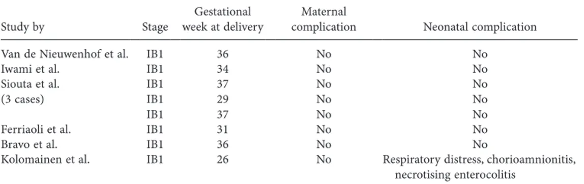
Nieuwenhoft et al. reported (2008) the first radical vaginal tra-chelectomy performed in a pregnant patient with CC. Currently, there are only eight cases in the literature of CC diagnosed during pregnancy managed with vaginal radical trachelectomy (Table I) (van de Nieuwenhof et al. 2008, Iwami et al. 2011, Siuotas et al. 2011, Ferriaoli et al. 2012, Bravo et al. 2012, Kolomainen et al. 2013). Of these eight cases, all the patients were in stage IB1. At the time of delivery, the gestational age ranged between 26 and 37 weeks, and the risk of preterm delivery was 20% (Kolomainen et al. 2013). Miscarriage rates were reported to be higher in the abdominal radical trachelectomy compared with radical vaginal trachelectomy. This is explained by higher uterine manipulation and reduced uterine vasculature due to severing of the uterine artery during abdominal trachelectomy and also explained by higher uterine artery preservation and less uterine manipulation involved in radical vaginal trachelectomy (Kolo-mainen et al. 2013).
Simple trachelectomy is a less complicated procedure, defined as an excision of the cervix 1 cm above the tumour border together with removal of the endocervical channel (Rob et al. 2011). Due to higher abortion rates with radical trachelectomy procedures, simple trachelectomy is recommended. The operation should be performed by experienced surgeons and the patient should be informed of the complications (foetal loss and bleeding) (Karam 2014, Kehoe 2008).
In pregnant patients with a tumour size 2 cm and with stage IB1 or higher disease, neo-adjuvant chemotherapy (NAC) is the treatment of choice. NAC is used to prevent cancer pro-gression and allow foetal development. The chemotherapeutic regimens used most commonly are cisplatin and paclitaxel regi-men (Karam 2014, Zagouri et al. 2013). The most effective NAC regimen in non-pregnant CC patients is the combined treatment of paclitaxel, ifosfamide and cisplatin; however, ifosfamide has foetal nephrotoxic effects (Karam 2014). Cisplatin is appropriate for first-trimester pregnancies and can be used at a dose of 25– 50 mg/m2 per week or 50–100 mg/m2 per three weeks for up to six cycles (Han et al. 2013, Karam 2014). The currently recom-mended NAC regimen is cisplatin with or without paclitaxel every three weeks. In a review evaluating 50 patients treated with NAC, the overall response rate was close to 90% (Amant et al. 2014). The median gestational age at diagnosis was 19.2 weeks, and platinum-based chemotherapy was used every three weeks up to the median gestational age of 33.2 weeks.
The estimated teratogenic risk with the use of single and mul-tiple chemotherapy agents during the first trimester is 7.5–17% and 25%, respectively (Weisz et al. 2001). Adverse effects due to NAC during the second and third trimesters include intrauterine growth retardation, in utero death, low birth weight and prema-turity (Islam et al. 2012). NAC should be terminated three weeks
before the planned delivery to decrease the possible maternal and foetal complications caused by haematopoietic suppression (Amant et al. 2014, Karam 2014, Peccatori et al. 2013).
In a systematic review by Zagouri et al. of platinum deriva-tive use during pregnancy in CC patients, 24 studies including 48 patients were evaluated (Zagouri et al. 2013). Early-stage disease was reported in 93.5% and advanced-stage disease was reported in 6.5% of the patients. The mean gestational age at diagnosis was 23.9 weeks. In 61.7% of the cases, cisplatin was used as monotherapy, and in the remaining cases, cisplatin com-bined with other regimens (cisplatin bleomycin, 2.1%; cispla-tin 5-fluorouracil, 2.1%; cisplacispla-tin paclitaxel, 12.8%; cisplacispla-tin vincristine, 17%; cisplatin vincristine bleomycin, 4.3%; cisplatin carboplatin paclitaxel in one patient) was used. At the time of birth, 67.4% of newborns were healthy, and serum creatinine elevation, anaemia, hypoglycaemia, supraventricular tachycardia, hypotension, intraventricular haemorrhage and respiratory syndrome disorder were reported in the remaining. After long-term follow-up, 100% were reported to be healthy. The reported health problems were the following: The platinum deriv-atives were well tolerated both by patients and foetuses. Complete and partial responses were 10% and 63.4%, respectively. Of the 48 pregnant patients, one gave birth to twins, and only Ayhan et al. (2012) have reported triplets born to a CC patient, who was successfully managed with cisplatin during pregnancy.
Lymph-node-positive pregnant patients who choose preg-nancy continuation should be informed of the poor prognosis with treatment. If the patient insists on continuing the pregnancy, NAC is the treatment of choice with a planned early delivery (Pec-catori et al. 2013, Karam 2014).
Group II (gestational age 22–25 weeks or later)
Evaluation of lymph node involvement with lymphadenectomy after 20 weeks’ gestation becomes less feasible due to surgery-related complications. Therefore, when CC is diagnosed at this stage of pregnancy, the disease management is determined mainly by the stage of the disease (Karam 2014, Amant et al. 2014).
In stage-IA and stage-IB1 patients with tumour sizes 2 cm, postponing definitive treatment after delivery is possible. Although large randomised trials have not been conducted, several studies evaluated the effect of delaying definitive treatment in non-pregnant and pregnant patients. The 6-week treatment interval between diagnostic conisation and definitive treatment in non-pregnant patients was associ-ated with adverse outcomes. In a study involving 98 pregnant patients with stage-I and stage-II CC, the definitive treatment was delayed to allow foetal maturation (Karam et al. 2007). This delay interval ranged from 3 to 40 weeks, and 96% of patients were alive without recurrence.
Table I. Radical vaginal trachelectomy during pregnancy.
Study by Stage week at deliveryGestational complicationMaternal Neonatal complication
Van de Nieuwenhof et al. IB1 36 No No
Iwami et al. IB1 34 No No
Siouta et al.
(3 cases) IB1IB1
IB1 37 29 37 No No No No No No
Ferriaoli et al. IB1 31 No No
Bravo et al. IB1 36 No No
Kolomainen et al. IB1 26 No Respiratory distress, chorioamnionitis, necrotising enterocolitis
If patients with disease stages higher than IB1 opt to continue their pregnancies, NAC is the treatment of choice and is used pri-marily for preventing disease progression rather than curing the disease. Patients should be given detailed information regarding the experimental CC treatment features during pregnancy.
Patients with stage-IA1 disease who choose continuation of pregnancy should be followed up with repeated colposcopy and clinical examination during each trimester up until the time of delivery. Patients who prefer delaying treatment after delivery should be followed clinically and by performing pelvic examina-tions every 3–4 weeks (Karam 2014). Imaging modalities such as USG and MRI can be used when disease progression is suspected (Karam 2014, Duggan et al. 1993).
Timing and mode of delivery
Although the optimal delivery time is 37 weeks’ gestation, the delivery time and the method should be determined individu-ally based on disease conditions. For patients with abnormal cervical cytology, the mode of delivery is determined based on obstetrical reasons (Karram 2014). Caesarean section (C/S) is not indicated in patients with an abnormal cervical cytology (Kathleen 2012). Pregnant women with stage-IA1 or -IA2 CC can deliver vaginally (Yang 2012). For stages IB1 and higher, C/S is the preferred mode of delivery, and vaginal delivery should be avoided (Yang 2012, Karam 2014, Amant et al. 2014). Vaginal delivery during advanced CC stages might increase the risk of lymphatic spread, infections, cervical lacerations and episiotomy-related metastasis. In a study by Sood et al. (2000) comparing vaginal delivery with C/S in CC patients, the recur-rence rate was higher in the patients who delivered vaginally. Based on multivariate analysis, vaginal delivery was deter-mined as the most important prognostic factor for CC recur-rence. In patients with long bone metastasis, the pushing that occurs during vaginal delivery may result in bone fractures; in patients with central nervous system metastasis, normal vagi-nal delivery may cause increased intra-cranial pressure, and therefore, vaginal delivery should be avoided in these patients (Amant et al. 2014).
There are only two reported cases of placental metastasis of CC in the literature. Although metastasis into the placenta is very rare, the placenta should be carefully examined macroscopically and histopathologically for any metastatic lesions (Can et al. 2013, Cailliez et al. 1980).
Definitive treatment for patients who want future
pregnancies
Definitive treatment can be performed at the time of deliv-ery or postpartum (Karam 2014). For patients with stage IA1 and negative margins who want to preserve their fertility, the conisation treatment is sufficient (Karam 2014). In patients with positive margins, a second conisation can be repeated 6– 8 weeks after delivery. In studies by Dunn et al. and Yakata et al., the margin positivities were 46% and 50%, respectively (Dunn et al. 2003, Yahata et al. 2008). They repeated the coni-sation procedure postpartum and reported no residual dis-ease. In patients with stage IA1 and no lymphovascular space involvement (LVSI) who are not concerned with preserving their fertility, a simple hysterectomy can be performed during the C/S or later (Karam 2014). In patients with stage-IA2 dis-ease and tumour sizes up to 4 cm who want to preserve their fertility, the definitive treatment is radical trachelectomy per-formed postpartum (Karam 2014). In patients with stage IA1
with LVSI, IA2 or IB1 disease and a tumour size 2 cm who do not want to preserve their fertility, the definitive treatment is radical hysterectomy during the C/S or later (Karam 2014). The treatment for advanced disease after delivery is the same as that for non-pregnant patients.
Lymphadenectomy
Lymph node involvement is an important prognostic factor in CC prognosis (Delgado et al. 1990). Although pelvic lymph node involvement can be evaluated surgically and by MRI, the histopathological evaluation of lymph nodes remains the most accurate method (Han et al. 2013, Morice et al. 2012). Several studies have reported the feasibility of lymphadenectomy dur-ing pregnancy (Han et al. 2013, Morice et al. 2012, Yang, 2012, Amant et al. 2014, Karam, 2014). In a study of 31 patients, the risk of maternal–foetal morbidity due to lymphadenectomy was negligible (Alouini et al. 2008, Siuotas et al. 2011). During the first and second trimesters, laparoscopic lymphadenectomy can be performed in patients with early-stage CC. Similar to cervical physiological changes due to pregnancy, physiologi-cal changes in the lymph nodes can be mistaken as metastatic disease; therefore, specimens should be evaluated by experi-enced pathologists (Morice et al 2012). Although there have been cases of laparoscopic lymphadenectomy performed after 20 weeks’ gestation, removal of the recommended number of lymph nodes ( 10) is difficult (Alouini et al. 2008, Stan et al. 2005, Chvatal et al. 2011). Laparoscopic lymphadenectomy should be performed by experienced surgeons. Lymph-node-positive pregnant patients should be informed of the need for immediate treatment.
Conclusion
In conclusion, the management of CC diagnosed during preg-nancy is a stressful event both for the caregivers and the families. The decision should be made collaboratively with a multidisci-plinary team. The desires of the patient should be addressed, and all options should be discussed carefully together with the limita-tions, as determined by evidence-based studies.
Disclosurestatement: None of the authors has any conflict of
interest relative to this work and this review did not receive phar-maceutical company support.
References
Al-Halal H, Kezouh A, Abenhaim HA. 2013. Incidence and obstetrical outcomes of cervical intraepithelial neoplasia and cervical cancer in pregnancy: a population-based study on 8.8 million births. The Archives of Gynecology and Obstetrics 287:245–250.
Amant F, Halaska MJ, Fumagalli M, Dahl Steffensen K, Lok C, Van Calsteren K, et al. 2014. ESGO task force ‘Cancer in Pregnancy’. Gynecologic cancers in pregnancy: guidelines of a second international consensus meeting. International Journal of Gynecological Cancer 24:394–403.
Alouini S, Rida K, Mathevet P. 2008. Cervical cancer complicating preg-nancy: implications of laparoscopic lymphadenectomy. Gynecologic Oncology 108:472–477.
Akata D, Kerimoglu U, Hazirolan T, Karcaaltincaba M, Köse F, Ozmen MN, et al. 2005. Efficacy of transvaginal contrast-enhanced MRI in the early staging of cervical carcinoma. European Radiology 15:1727–1733.
Ayhan A, Dursun P, Karakaya BK, Ozen O, Tarhan C. 2012. Neoadjuvant chemotherapy followed by cesarean radical hysterectomy in a triplet pregnancy complicated by clear cell carcinoma of the cervix: a case pre-sentation and literature review. International Journal of Gynecological Cancer 22:1198–1202.
K. Gungorduk et al.
Balleyguier C, Fournet C, Ben Hassen W, Zareski E, Morice P, Haie-Meder C, et al. 2013. Management of cervical cancer detected during pregnancy: role of magnetic resonance imaging. Clinical Imaging 37:70–76.
Bartlett RM, Nickles RJ, Barnhart TE, Christian BT, Holden JE, DeJesus OT. 2010. Fetal dose estimates for (18)F-fl uoro-L-thymidine using a pregnant monkey model. Journal of Nuclear Medicine 51:288–292.
Bravo E, Parry S, Alonso C, Rojas S. 2012. Radical vaginal trachelectomy and laparoscopic pelvic lymphadenectomy in IB1 cervical cancer during pregnancy. Gynecologic Oncology Case Reports 2:78–79.
Cailliez D, Moirot MH, Fessard C, Hémet J, Philippe E. 1980. Placental localisation of cancer of the cervix. The European Journal of Obstetrics & Gynecology and Reproductive Biology 9:461–463.
Can NT, Robertson P, Zaloudek CJ, Gill RM. 2013. Cervical squamous cell carcinoma metastatic to placenta. International Journal of Gynecological Pathology 32:516–519.
Chvatal R, Oppelt P, Koehler C, Habelsberger A, Yaman C. 2011. Simple tra-chelectomy of early invasive cervix carcinoma in the second trimester. Journal of the Turkish German Gynecological Association 12:121–123. Creasman WT. 2001. Cancer and pregnancy. Annals of the New York
Acad-emy of Sciences 943:281.
DiSaia PJ, Creasman WT. Clinical gynecologic oncology. 5th ed. St. Louis: Mosby, 1997.
Duggan B, Muderspach LI, Roman LD, Curtin JP, d’Ablaing G 3rd, Morrow CP. 1993 Cervical cancer in pregnancy: reporting on planned delay in therapy. Obstetrics & Gynecology 82:598–502.
Delgado G, Bundy B, Zaino R, Sevin B, Creasman W, Major F. 1990. Prospec-tive surgical-pathological study of disease-free interval in patients with stage IB squamous cell carcinoma of the cervix: a Gynecologic Oncology Group study. Gynecologic Oncology 38:352–357.
Dunn T, Ginsburg, V, Wolf D. 2003. Loop-cone cerclage in pregnancy: a 5-year review. Gynecologic Oncology 90:577–580.
Economos K, Perez Veridiano N, Delke I, Collado ML, Tancer ML. 1993.Abnormal cervical cytology in pregnancy: a 17-year experience. Obstetrics & Gynecology 81:915–918.
Epstein E, Testa A, Gaurilcikas A, Di Legge A, Ameye L, Atstupenaite V, et al. 2013. Early-stage cervical cancer: tumor delineation by magnetic reso-nance imaging and ultrasound - a European multicenter trial. Gyneco-logic Oncology 128:449–453.
Fischerova D, Cibula D, Stenhova H, Vondrichova H, Calda P, Zikan M, et al. 2008. Transrectal ultrasound and magnetic resonance imaging in staging of early cervical cancer. International Journal of Gynecological Cancer 18:766–772.
Ferriaoli D, Buenerd A, Marchiole P, Constantini S, Venturini PL, Mathevet P. 2012. Early invasive cervical cancer during pregnancy: different therapeutic options to preserve fertility. International Journal of Gynecological Cancer 22:842–849.
Guner H, Taskıran C. 2007. Serviks kanser epidemiyolojisi ve Human papil-loma virus. Turkish Journal of Obstetrics and Gynecology 2:11–19 Han SN, Mhallem Gziri M, Van Calsteren K, Amant F. 2013. Cervical cancer
in pregnant women: treat, wait or interrupt? Assessment of current clini-cal guidelines, innovations and controversies. Therapeutic Advances in Medical Oncology 5:211–219.
Islam S, Mukhopadhyay L, Howells R. 2012. Neo-adjuvant chemotherapy and radical surgery for stage 1B cervical cancer in pregnancy. Journal of Obstetrics & Gynaecology 32:191–192.
Iwami N, Ishioka S, Endo T, Baba T, Nagasawa K, Takahashi M. 2011. First case of vaginal radical trachelectomy in a pregnant Japanese woman. International Journal of Clinical Oncology 16:737–740.
Karam A, Feldman N, Holschneider CH. 2007. Neoadjuvant cisplatin and radical cesarean hysterectomy for cervical cancer in pregnancy. Nature Clinical Practice Oncology 4:375–380.
Kanal E, Borgstede JP, Barkovich AJ, Bell C, Bradley WG, Etheridge S, et al. 2004. American College of RadiologyAmerican College of Radiology White Paper on MR Safety: 2004 update and revisions. American Journal of Roentgenology 182:1111–1114.
Karam A. 2014. Cervical cancer in pregnancy. http://www.uptodate.com/ contents/cervical-cancer-in pregnancy
Kathleen Y. Yang. 2012. Abnormal Pap Smear and Cervical Cancer in Preg-nancy. Clinical Obstetrics and Gynecology 55:838–848.
Kehoe S. 2008. Cervical and Endometrial Cancer. During Pregnancy. Recent Results in Cancer Research, Vol. 178 © Springer-Verlag Berlin Heidel-berg, page: 70.
Kolomainen DF, Bradley RJ, Larsen-Disney P, Shepherd JH. 2013. Radical vaginal trachelectomy at 16 weeks’ gestation: A case report. Gynecologic Oncology Case Reports 14:28–30.
Massad LS, Einstein MH, Huh WK, Katki HA, Kinney WK, Schiffman M, et al. 2013. 2012 ASCCP Consensus Guidelines Conference 2012 updated consensus guidelines for the management of abnormal cervical cancer screening tests and cancer precursors. Obstetrics & Gynecology 121: 829–846.
Morice P, Uzan C, Gouy S, Verschraegen C, Haie-Meder C. 2012. Gynaeco-logical cancers in pregnancy. Lancet 379:558–569.
Morimura Y, Fujimori K, Soeda S, Hashimoto T, Takano Y, Yamada H, et al. 2002. Cervical cytology during pregnancy–comparison with non-preg-nant women and management of pregnon-preg-nant women with abnormal cytol-ogy. Fukushima Journal of Medical Science 48:27–37.
Nguyen C, Montz FJ, Bristow RE. 2000. Management of stage I cervical can-cer in pregnancy. Obstetrical & Gynecological Survey 55:633.
Palle C, Bangsbøll S, Andreasson B. 2000. Cervical intraepithelial neoplasia in pregnancy. Acta Obstetricia et Gynecologica Scandinavica 79:306. Pecorelli S, Zigliani L, Odicino F. 2009. Revised FIGO staging for carcinoma
of the cervix. International Journal of Gynecology & Obstetrics 105: 107–108.
Peccatori FA, Azim HA Jr, Orecchia R, Hoekstra HJ, Pavlidis N, Kesic V, et al. 2013. ESMO Guidelines Working GroupCancer, pregnancy and fertility: ESMO Clinical Practice Guidelines for diagnosis, treatment and follow-up. Annals of Oncology 24:160–170,
Reznek RH, Sahdev A. 2005. MR imaging in cervical cancer: seeing is believ-ing. 2004. Mackenzie Davidson Memorial Lecture. The British Journal of Radiology 2:73.
Rob L, Skapa P, Robova H. 2011 Fertility-sparing surgery in patients with cervical cancer. The Lancet Oncology 12:192–200.
Siuotas A, Schedvins K, Larson B, Gemzell-Danielsson K. 2011. Three cases of vaginal radical trachelectomy during pregnancy. Gynecologic Oncol-ogy 121:420–421.
Selleret L, Mathevet P.2008. Precancerous cervical lesions during pregnancy: diagnostic and treatment. Journal de Gynécologie Obstétrique et Biologie 37 (suppl 1):S131–138.
Stan C, Megevand E, Irion O, Wang C, Bruchim I, Petignat P. 2005. Cervical cancer in pregnant women: laparoscopic evaluation before delaying treatment European Journal of Gynaecological Oncology 26:649–650.
Sood AK, Sorosky JI, Mayr N, Anderson B, Buller RE, Niebyl J. 2000. Cervi-cal cancer diagnosed shortly after pregnancy: prognostic variables and delivery routes. Obstetrics & Gynecology 95:832–838.
Sood AK, Sorosky JI, Mayr N, Krogman S, Anderson B, Buller RE, Hussey DH. 1997. Radiotherapeutic management of cervical carcinoma that complicates pregnancy. Cancer 80:1073–1078.
Schmeler KM, Frumovitz M, Ramirez PT. 2011. Conservative management of early stage cervical cancer: is there a role for less radical surgery? Gynecologic Oncology 120:321–325.
Stabin M, Xu X, Emmons M, Segars W, Shi C, Fernald M. 2012. RADAR reference adult, pediatric, and pregnant female phantom series for internal and external dosimetry. Journal of Nuclear Medicine 53: 1807–1813.
Takalkar A, Khandelwal A, Lokitz S, Lilien D, Stabin, M. 2011. 18F-FDG PET in pregnancy and fetal radiation dose estimates. Journal of Nuclear Medicine 52:1035–1040.
Takushi M, Moromizato H, Sakumoto K, Kanazawa K. 2002. Management of invasive carcinoma of the uterine cervix associated with pregnancy: outcome of intentional delay in treatment. Gynecologic Oncology 87: 185–189.
Ungár L, Smith JR, Pálfalvi L, Del Priore G. 2006. Abdominal radical trachelectomy during pregnancy to preserve pregnancy and fertility. Obstetrics & Gynecology 108(3 Pt 2):811–814.
Van Calsteren K, Vergote I, Amant F. 2005.Cervical neoplasia during preg-nancy: diagnosis, management and prognosis. Best Practice & Research Clinical Obstetrics & Gynaecology 19:611.
van de Nieuwenhof HP, van Ham MA, Lotgering FK, Massuger LF. 2008. First case of vaginal radical trachelectomy in a pregnant patient. Interna-tional Journal of Gynecological Cancer 18:1381–1385.
Weisz B, Schiff E, Lishner M. 2001. Cancer in pregnancy: maternal and fetal implications. Human Reproduction Update 7:384–393.
Yang KY. 2012 Abnormal pap smear and cervical cancer in pregnancy. Clinical Obstetrics and Gynecology 55:838–848
Yahata T, Numata M, Kashima K, Sekine M, Fujita K, Yamamoto T, Tanaka K. 2008. Conservative treatment of stage IA1 adenocarcinoma of the cervix during pregnancy Gynecologic Oncology 109:49–52.
Zagouri F, Sergentanis TN, Chrysikos D, Bartsch R. 2013. Platinum derivatives during pregnancy in cervical cancer: a systematic review and meta-analysis. Obstetrics & Gynecology 121:337–343.