Is bioresorbable vascular scaffold acute recoil affected by baseline renal
function and scaffold selection?
Haci Murat Gunes
⁎
, Filiz Kizilirmak Y
ılmaz, Tayyar Gokdeniz, Gultekin Gunhan Demir, Ekrem Guler,
Gamze Babur Guler, O
ğuz Karaca, Beytullah Cakal, Ersin İbişoğlu, Bilal Boztosun
Medipol University, Faculty of Medicine, Cardiology Department, Istanbul, Turkey
a b s t r a c t
a r t i c l e i n f o
Article history: Received 20 June 2016
Received in revised form 8 September 2016 Accepted 12 September 2016
Available online 20 September 2016
Objective: The aim of the present study was to investigate the relationship between glomerularfiltration rate (GFR) and acute post-scaffold recoil (PSR) in patients undergoing bioresorbable scaffold (BVS) implantation. Methods: We included 130 patients who underwent everolimus-eluting BVS device (Absorb BVS; Abbott Vascular, Santa Clara, CA, USA) or the novolimus-eluting BVS device (Elixir Medical Corporation) implantations for single or multi-vessel disease. Clinical, angiographic variables and procedural characteristics were defined and pre-procedural GFR was calculated for each patient. Post-procedural angiographic parameters of each patient were analyzed. Primary objective of the study was to evaluate the effect of GFR on angiographic outcomes after BVS implantation while secondary objective was to compare post-procedural angiographic results between the two BVS device groups.
Results: Baseline clinical characteristics and angiographic parameters were similar between the two BVS groups. Post-procedural angiographic analysis revealed significantly lower PSR in the DESolve group than the Absorb group (0.10 ± 0.04 vs. 0.13 ± 0.05, p: 0.003). When PSR in the whole study population was evaluated, it was positively correlated with age, tortuosity , calcification and PBR as there was a negative correlation between GFR. Besides GFR were found to be independent predictors for PSR in all groups and the whole study population. Conclusion: In patients undergoing BVS implantation, pre-procedural low GFR is associated with increased post-procedural PSR. Calcification, age, PBR, dyslipidemia and tortuosity are other independent risk factors for PSR. DESolve has lower PSR when compared with Absorb.
© 2016 Elsevier Ireland Ltd. All rights reserved.
Keywords:
Bioresorbable vascular scaffold Glomerularfiltration rate Stent recoiling
1. Introduction
Percutaneous coronary intervention (PCI) has made substantial progress in the last decades with the introduction of bare metal stents (BMS), drug-eluting stents (DES) and a newcomer to thefield, biore-sorbable vascular scaffolds (BVS)[1]. The main benefits anticipated by the use of biocompatible materials include gradually reduced in flam-matory and fibrotic response through resorption mechanism and reconstruction of normal vessel wall structure[2]. BVSs have been de-veloped to overcome difficulties related with DES technology; including side-branch occlusion, late adverse events, inability of by-pass grafting to stented segments and limited image resolution with computed tomography or magnetic resonance imaging modalities, in addition to their capability of restoring normal vessel vasomotion[1]. BVSs have been developed to overcome these difficulties related with DES
technology. BVSs remain within the vessel long enough to provide pro-tection against subacute closure, wall recoil and restenosis. Besides it has the potential to reduce problems such as very late stent thrombosis and the need for prolonged dual antiplatelet therapy. More aggressive lesion preparation is required due to singular design and composition of BVSs[3]. Similarly, it was shown that plaque composition, morphol-ogy and burden were effective factors for BVS expansion and eccentric-ity[4]. Increasedfinal luminal diameter of target vessel is associated with improved long-term patency after PCI and lower recurrence rates[5–7]. Stent recoil is one of the major mechanisms responsible for suboptimal stent expansion and severe residual lesion post-PCI
[8,9]. Danzi et al. detected significantly greater acute recoil of the BVS in comparison with everolimus-eluting stents and also correlation between recoil percentage and residual stenosis after predilatation in patients treated with BVS[10]. The relationship between renal impair-ment (RI) and coronary artery disease is well established[11,12]. Com-plex coronary artery disease, left main coronary artery disease, ostial lesions, multi-vessel disease and heavily-calcified lesions are more common in patients with RI than those without RI[13,14]. Budoff et al. demonstrated a powerful and graded relationship between
⁎ Corresponding author at: Medipol University Hospital, Cardiology Department, TEM Avrupa Otoyolu Göztepe Çıkışı No: 1 Bağcılar, 34214 İstanbul, Turkey.
E-mail address:[email protected](H.M. Gunes).
http://dx.doi.org/10.1016/j.ijcard.2016.09.024
0167-5273/© 2016 Elsevier Ireland Ltd. All rights reserved.
Contents lists available atScienceDirect
International Journal of Cardiology
lower glomerularfiltration rate (GFR) and increased coronary artery calcifications (CAC)[15].
In this study we aimed to examine the relationship between pre-procedural GFR and acute post-scaffold recoil (PSR) in patients treated with BVS.
2. Methods
2.1. Study population
In this cross-sectional and single-center study, we included 135 patients with single or multi-vessel disease undergoing PCI with everolimus-eluting BVS device (Absorb BVS; Abbott Vascular, Santa Clara, CA, USA) or the novolimus-eluting BVS device (Elixir Medical Corporation) between January 2013 and December 2015 in Medipol University. PCI could not be performed due to inability to cross the lesion with stent in 5 patients with single lesion (2 in LAD, 2 in Cx and 1 in RCA). We included total 130 patients. Baseline clinical, angiographic variables and procedural characteristics were defined.
Eligible patients were those with reference vessel diameter (RVD) ≥2.25 mm, stentable lesions, stable coronary artery disease or unstable angina or non-ST segment elevation myocardial infarction. All treated lesions consisted of stenosisN50% but less than 100% vessel occlusion in native coronary arteries with thrombolysis in myocardial infarction (TIMI)flow grade ≥1. Major exclusion criteria were defined as acute ST-segment elevation myocardial infarction, hemodynamically unstable arrhythmias, left ventricular ejection fraction (LVEF)b30%, lesions in ar-terial or saphenous vein grafts and restenotic lesions. No restrictions were applied regarding the number of treated lesions and vessels, im-planted stents or lesion length. The patients were asked for their per-mission about implantation of one or more BVSs and in the case of DES or BMS.
2.2. Procedures
All interventions were performed according to current PCI standards including mandatory predilatation and pressures under rated burst pressure. Post-dilatation or additional stent implantation was left to the discretion of the operator. Predilatation was performed at the same diameter of RVD and post-dilatation balloon was selected with a diameter of 0.5 mm larger than implanted BVS diameter. Upstream 300 mg loading dose of oral aspirin was followed with 100 mg daily aspirin in patients not receiving chronic aspirin treatment. Upstream loading dose of clopidogrel 600 mg, prasugrel 60 mg or ticagrelor 180 mg was followed with daily maintenance dose of clopidogrel 75 mg, prasugrel 10 mg or ticagrelor 90 mg bid for 12 months in thienopyridine-naive patients. None of the patients received glycopro-tein IIb/IIIa inhibitors.
2.3. Study objectives and definitions
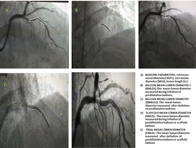
Primary objective of the study was to evaluate the effect of GFR on angiographic outcomes after BVS implantation. Secondary goal was to compare post-procedural angiographic results between the BVS device types (Absorb and Elixir Medical Corporation). Pre-procedural GFR was estimated by the use of the Modification of Diet in Renal Disease (MDRD) equation[16]. Quantitative coronary angiography (QCA) was performed using standard techniques with automated edge-detection algorithms (version 1.4) in the hospital's angiographic analysis center. Scaffold segment and peri-scaffold segment (5 mm proximal and distal to the scaffold edge) were analyzed for each patient. Minimal luminal diameter (MLD), lesion length, RVD and percent diameter stenosis (DS) were calculated. In addition to baseline images, four images of post-PCI cine frames were analyzed (Fig. 1). Two images were acquired while predilatation balloon catheter was at the highest pressure, fully expanded and after balloon deflation was followed with nitroglycerine
injection. The other two images were acquired while post-dilatation balloon (or scaffold balloon instead) was at the highest pressure, fully expanded and after balloon deflation was followed with nitroglycerine injection. These images were analyzed from the same angiographic projection in order to minimize foreshortening.
2.4. Angiographic parameters
2.4.1. Calcification
Calcification was identified as readily apparent radiopacity within the vascular wall at the site of the stenosis, and was classified as moder-ate (radiopacity noted only during the cardiac cycle before contrast in-jection), and severe (radiopacity noted without cardiac motion before contrast injection and generally compromising both sides of the arterial lumen)[17].
2.4.2. Tortuosity
Coronary lesion is defined as severely tortuous when it satisfies the following criteria: one or more bends of 90° or more, or three or more bends of 45–90° proximal to the diseased segment. Lesions out of these criteria were defined as moderate[18].
2.4.3. Predil. diameter
Predil. diameter is defined as the balloon diameter for predilatation. 2.4.4. Postdil. diameter
Postdil. diameter is defined as the balloon diameter for post-dilatation.
2.4.5. BMLD1(balloon mean lumen diameter1)
BMLD1(balloon mean lumen diameter1) is defined as the mean
lumen diameter measured during inflation of predilatation balloon. 2.4.6. BMLD2(balloon mean lumen diameter2)
BMLD2(balloon mean lumen diameter2) is defined as the mean
lumen diameter measured after deflation of predilatation balloon. 2.4.7. PBR (post-balloon recoiling)
PBR (post-balloon recoiling) is defined as recoiling ((BMLD1−
BMLD2) / BMLD1) after predilatation.
2.4.8. SMLD (scaffold mean lumen diameter)
SMLD (scaffold mean lumen diameter) is defined as the mean lumen diameter measured during inflation of post-dilatation balloon or scaffold balloon (when post-dilatation was not performed).
2.4.9. FMLD (final mean lumen diameter)
FMLD (final mean lumen diameter) is defined as the mean lumen diameter measured after deflation of post-dilatation balloon or scaffold balloon (when post-dilatation was not performed).
2.4.10. PSR (post-scaffold recoil)
PSR (post-scaffold recoil) is defined as recoiling (( SMLD − FMLD) / SMLD) after scaffold implantation.
2.4.11. Statistical analysis
SPSS 23.0 statistical software (SPSS Inc., Chicago, IL, USA) were used for statistical analysis. Continuous variables were expressed as mean ± standard deviation (SD) or median and interquartile range as appropriate. Categorical variables were expressed as percentages. The Kolmogorov–Smirnov test was used to test normality of distribution of continuous variables. Group means for continuous variables were compared with the use of Student's t-test or the Mann–Whitney U test, as appropriate. Pearson or Spearman correlation analysis was used for assessing correlation between PSR and clinical and procedural variables of patients in all BVS population and each BVS type (Absorb,
Elixir Medical Corporation) depending on Gaussian distributions. Stepwise multivariate linear regression analysis involving significant parameters in univariate analysis was performed tofind independent predictors of PSR in all BVS population and each BVS type. A p value of ≤0.05 was considered statistically significant.
3. Results
A total of 130 patients underwent 162 BVS implantations consisting of Absorb (n:131 80.9%) and DESolve (n:31, 19.1%). Femoral access was used in 111 (85.4%) patients and radial access was used in 19 (14.6%) patients. Baseline characteristics and clinical data of both groups are shown inTable 1. Mean age of the study population was 59 ± 11 and male gender was 82%. Patients with stable angina and unstable angina were 111 (85%) and 19 (14%), respectively. Mean LVEF of the patients was 56 ± 10% and mean GFR was 88 ± 23 ml/min/1.73 m2. There
was no difference between scaffold groups with regard to baseline clinical characteristics.
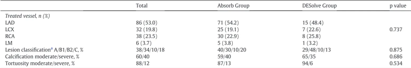
Similarly, there were no differences among two scaffold groups in terms of angiographic characteristics of the patients (Table 2). Pre-procedural and in-scaffold post-Pre-procedural QCA data of patients in each scaffold group were shown inTable 3. Pre-procedural QCA param-eters were not different among two groups. Scaffold diameter was significantly higher in the DESolve group than the Absorb group (3.1 ± 0.3 vs. 2.9 ± 0.3, p: 0.037). PBR in the DESolve group was lower than the Absorb group (0.17 ± 0.06 vs. 0.20 ± 0.07, p: 0.005). FMLD was significantly higher in the DESolve group than the Absorb group (3.08 ± 0.41 vs. 2.84 ± 0.4, p: 0.005). PSR was lower in the
DESolve group than the Absorb group (0.10 ± 0.04 vs. 0.13 ± 0.05, p: 0.003).
Table 4shows the correlation analysis between PSR and clinical and procedural variables. When the patients in the whole study population were taken into consideration, PSR was positively correlated with age (r: + 0.147, p: 0.006), tortuosity (r: + 0.146, p: 0.024), calcification (r: +0.364, pb 0.001) and PBR (r: +0.364, p b 0.001) while it was neg-atively correlated with GFR (r:−0.342, p b 0.001). In the Absorb group, there was a positive correlation between PSR and age (r: + 0.252, p: 0.001), calcification (r: +0.438, p: 0.001) and PBR (r: +0.475,
Fig. 1. A) Baseline parameters; reference vessel diameter (RVD), min lumen diameter (MLD), lesion length (LL). B) Balloon mean lumen diameter 1 (BMLD1): the mean lumen diameter
measured during inflation of predilatation balloon. C) Balloon mean lumen diameter 2 (BMLD2): the mean lumen diameter measured after deflation of predilatation balloon. D) Scaffold
mean lumen diameter (SMLD): the mean lumen diameter measured during inflation of post-dilatation balloon or scaffold balloon. E) Final mean lumen diameter (FMLD): the mean lumen diameter measured after deflation of post-dilatation balloon or scaffold balloon.
Table 1
Baseline clinical characteristics. Total n = 130 Absorb Group n = 107 DESolve Group n = 23 p value Age, years 59 ± 11 59.7 ± 11.3 59 ± 11.8 0.772 Male gender, n (%) 107 (82) 86 (80) 21 (91) 0.174 Hypertension, n (%) 81 (62) 66 (61) 15 (65) 0.473 Diabetes mellitus, n (%) 57 (43) 50 (46) 7 (30) 0.115 Current smoker, n (%) 60 (46) 47 (43) 13 (56) 0.192 Dyslipidemia, n (%) 99 (76) 79 (73) 20 (87) 0.141 Initial clinical presentation, n (%) Stable angina 111 (85) 94 (87) 17 (73) 0.087 Unstable angina 19 (14) 13 (12) 6 (26) 0.105 LVEF (%) 56 ± 10 55.9 ± 11.1 57.5 ± 6.7 0.508 GFR (ml/min/1.73 m2) 88 ± 23 87.6 ± 24.5 91.8 ± 20.7 0.444
pb 0.001), and as PSR was found to have a negative correlation with and GFR (r:−0.489, p b 0.001).
In the DESolve group PSR was positively correlated with dyslipidemia (r: +0.511, p: 0.003), calcification (r: +0.569, p: 0.001) and PBR (r: +0.522, p: 0.003) as it was negatively correlated with GFR (r:−0.551, p: 0.001).
Parameters having significant correlation with PSR in the total group and each BVS group in the univariate analysis were included in the multivariate analysis.Table 5shows the multivariate analysis of inde-pendent predictors for PSR. In the Absorb group, PBR (pb 0.001), GFR (p: 0,001) and calcification (p b 0.001), were found to be independent predictors for PSR, and dyslipidemia (p: 0.001), PBR (p: 0.004), and GFR (p: 0.003) were independent predictors for PSR in the DESolve group. In the whole study population, PBR (pb 0.001), GFR (p b 0.001) and calcification (p b 0.001) were independent predictors for PSR. 4. Discussion
In this study we aimed to investigate the relationship between pre-procedural GFR and post-pre-procedural PSR in patients undergoing BVS implantation.
I. Pre-procedural GFR was found to be independent predictor for PSR after BVS implantation in both scaffold groups. The lower GFR was associated with the higher PSR.
II. In the whole study group, PBR and calcification were the other independent predictors for PSR. Other independent predictors for PSR were PBR and calcification in the Absorb group; dyslipid-emia, PBR in the DESolve group, respectively.
III. When post-procedural parameters of both groups were
compared, we detected lower PSR in the DESolve group than the Absorb group with statistically significant difference. There is a growing number of BVS implantations in patients under-going PCI. More aggressive lesion preparation is required because of de-sign and composition characteristics of BVSs [3]. Furthermore, in contrast with metal stents aggressive post-dilatation can lead scaffold fractures in BVS use[19]. Shaw et al. demonstrated that BVS expansion and eccentricity were highly affected by plaque composition, morphol-ogy and burden; consequently lower scaffold eccentricity index and scaffold expansion index are associated with greater calcified plaque area and thickness[4].
Patients with RI have more complex and advanced coronary artery disease, hence a higher peri-procedural risk for PCI[11]. Cardiac death and MI after PCI with DES was 2-folds more common in patients with moderate/severe RI than those without; however stent thrombosis, DES affectivity or angiographic outcomes were not affected by RI[20]. Nevertheless, the relationship between RI and angiographic outcomes after BVS implantation has not been investigated so far. We investigated the relationship between pre-procedural GFR and BVS recoil. We de-tected that pre-procedural GFR was an independent predictor for PSR both in each scaffold group and the whole study group. Thisfinding may be related with increased calcified plaque burden in coronary arteries of patients with low GFR. It is not always possible to visualize coronary calcifications in coronary angiographic images. Since it is im-practical to use imaging methods such as optical coherence tomography (OCT) or intravascular ultrasound (IVUS) in daily practice, GFR might serve as a simple and useful tool for prediction of angiographic out-comes after BVS. Other independent predictors for PSR were
Table 2
Angiographic characteristics.
Total Absorb Group DESolve Group p value
Treated vessel, n (%) LAD 86 (53.0) 71 (54.2) 15 (48.4) LCX 32 (19.8) 25 (19.1) 7 (22.6) 0.737 RCA 38 (23.5) 30 (22.9) 8 (25.8) LM 6 (3.7) 5 (3.8) 1 (3.2) Lesion classificationa A/B1/B2/C, % 38/34/10/18 40/30/10/20 29/48/10/13 0.875 Calcification moderate/severe, % 60/40 59/40 65/35 0.686 Tortuosity moderate/severe, % 88/12 87/13 94/6 0.534
aAccording to the American College of Cardiology/American Heart Association lesion classification. LAD: left anterior descending coronary artery; LCX: left circumflex coronary artery;
LM: left main coronary artery RCA: right coronary artery.
Table 3 Procedural data. Total n = 162 Absorb Group n = 131 DESolve Group n = 31 p value Pre-procedural QCA Lesion length, mm 24 ± 10 24 ± 11 25 ± 9 0.792 RVD, mm 3.1 ± 0.4 3.1 ± 0.3 3.2 ± 0.4 0.128 DS, % 69 ± 9 68 ± 7 71 ± 11 0.410 MLD, mm 0.9 ± 0.3 0.9 ± 0.2 0.9 ± 0.3 0.688
In-scaffold post-procedural QCA
Predil. diameter 2.8 ± 0.3 2.8 ± 0.3 2.8 ± 0.3 0.833 Postdil. diameter 3.5 ± 0.7 3.0 ± 0.7 3.2 ± 0.7 0.176 Scaffold length, mm 25 ± 4 25 ± 4 26 ± 3 0.201 Scaffold diameter, mm 3.0 ± 0.3 2.9 ± 0.3 3.1 ± 0.3 0.037 BMLD1,mm 2.9 ± 0.3 2.9 ± 0.4 2.9 ± 0.3 0.686 BMLD2,mm 2.3 ± 0.3 2.3 ± 0.3 2.4 ± 0.3 0.069 PBR 0.20 ± 0.07 0.20 ± 0.07 0.17 ± 0.06 0.005 FMLD, mm 2.89 ± 0.41 2.84 ± 0.4 3.08 ± 0.41 0.005 SMLD, mm 3.31 ± 0.44 3.28 ± 0.43 3.45 ± 0.44 0.065 PSR 0.12 ± 0.05 0.13 ± 0.05 0.10 ± 0.04 0.003
BMLD1: balloon mean lumen diameter 1. BMLD2: balloon mean lumen diameter 2. DS: diameter stenosis; FMLD:final mean lumen diameter, MLD: minimum lumen diameter; PBR:
post-balloon recoiling, Postdil. diameter: post-dilatation post-balloon diameter. Predil. diameter: predilatation post-balloon diameter. PSR: post-scaffold recoil. QCA: quantitative coronary angiography; RVD: reference vessel diameter, SMLD: scaffold mean lumen diameter.
calcification, PBR, dyslipidemia. Danzi et al. reported significant correla-tion between BVS recoil and residual stenosis after predilatacorrela-tion in their study[10]. Onuma et al. showed that high balloon/artery ratio (N1.1) was predictive for high recoil[21]. Absorb BVS was used in both studies as we included DeSolve BVS additionally. Stent recoil is a combined con-sequence of elastic recoil and radial force and it may be affected by pro-cedural characteristics such as lesion features and RVD, stent oversizing and balloon compliance[21,22]. All parameters which we found to be associated with PSR are also factors interacting with coronary lesion characteristics, therefore it is natural to obtain these results.
ABSORB trial demonstrated that acute recoiling of BVS was higher than that of everolimus-eluting stent used in SPIRIT I and SPIRIT II
[23]. BVS revision 1.1 was developed in order to improve scaffold char-acteristics and extend mechanical stability by making alterations in strut pattern design. We detected that PSR was significantly lower in the DESolve group than the Absorb group. To our knowledge, there is no study comparing DESolve and Absorb BVS in terms of PSR. Lower PSR in the DESolve group in comparison with the Absorb might be relat-ed to lower PBR values in the DESolve group. DESolve is a new BVS shown to have a short bioresorbation period and capability of expansion at high pressures without strut fractures[24]. Strut design pattern and different scaffold characteristics may be related with thesefindings; however, further studies are warranted in thisfield to investigate these factors in detail.
5. Study limitations
The main limitations of this study are small sample size and non-randomized design. All measurements were made angiographically so more precise and useful results could be obtained with imaging techniques such as OCT and IVUS. However, QCA is a practical non-invasive method providing useful information about vessel and lesion characteristics and angiographic data. DES was not selected as control group regarding findings in a recent study which had already established superiority of DES over BRS in acute recoil. Lack of follow-up after the procedure is another limitation of our study.
6. Conclusion
In patients undergoing BVS implantation, pre-procedural low GFR is associated with increased post-procedural PSR. Calcification, PBR, dyslipidemia are other independent risk factors for PSR. DESolve has lower PSR when compared with Absorb BVS. Employment of pre-procedural GFR might serve as a practical and useful parameter for prediction of BVS efficacy.
Conflict of interest
The authors report no conflicts of interest. The authors alone are responsible for the content and writing of the paper.
References
[1] S.G. Ellis, D.J. Kereiakes, D.C. Metzger, et al., ABSORB III Investigators. Everolimus-eluting bioresorbable scaffolds for coronary artery disease, N. Engl. J. Med. 373 (2015) 1905–1915.
[2] C. Spadaccio, F. Nappi, Biology and bioresorbable materials in cardiac surgery: why could they be important in the current era of innovations and technology? Int. Cardiovasc. Forum 3 (2015) 2–4.
[3] J. De Ribamar Costa Jr., A. Abizaid, A.L. Bartorelli, et al., ABSORB EXTEND Investiga-tors. Impact of post-dilation on the acute and one-year clinical outcomes of a large cohort of patients treated solely with the absorb bioresorbable vascular scaffold, EuroIntervention 11 (2015) 141–148.
[4] E. Shaw, U.K. Allahwala, C. JA, et al., The effect of coronary artery plaque composi-tion, morphology and burden on absorb bioresorbable vascular scaffold expansion and eccentricity— a detailed analysis with optical coherence tomography, Int. J. Cardiol. 184 (2015) 230–236.
[5] G.R. Dussaillant, G.S. Mintz, A.D. Pichard, et al., Small stent size and intimal hyperpla-sia contribute to restenosis: a volumetric intravascular ultrasound analysis, J. Am. Coll. Cardiol. 26 (1995) 720–724.
[6] Y. Ikari, K. Hara, T. Tamura, F. Saeki, T. Yamaguchi, Luminal loss and site of restenosis after Palmaz-Schatz coronary stent implantation, Am. J. Cardiol. 76 (1995) 117–120.
Table 4
Association between PSR and clinical, procedural variables of patients.
Variables Total Absorb Group DESolve Group
r (correlation coefficient)/P value r (correlation coefficient)/P value r (correlation coefficient)/P value
Age +0.147/0.006 +0.252/0.004 +0.036/0.849 Male gender −0.095/0.141 −0.142/0.106 −0.185/0.320 Hypertension +0.074/0.250 +0.075/0.395 +0.232/0.208 Diabetes mellitus +0.004/0.953 +0.018/0.819 +0.073/0.696 Dyslipidemia +0.008/0.900 +0.067/0.450 +0.511/0.003 LVEF −0.086/0.131 −0.184/0.076 −0.284/0.122 Tortuosity +0.146/0.024 +0.169/0.053 +0.251/0.173 Calcification +0.364/b0.001 +0.438/0.001 +0.569/0. 001 Lesion length +0.050/0.355 +0.024/0.783 +0.105/0.574 RVD −0.034/0.526 −0.051/0.561 −0.002/0.993 Predil. diameter +0.015/0.810 +0.068/0.441 +0.105/0.575 Postdil. diameter +0.041/0.481 +0.044/0.621 +0.121/0.516 Scaffold diameter −0.037/0.537 −0.023/0.799 −0.034/0.857 Scaffold length +0.037/0.560 +0.013/0.880 +0.195/0.292 PBR +0.364/b0.001 +0.475/b0.001 +0.522/0.003 DS +0.005/0.930 +0.129/0.143 +0.253/0.169 MLD +0.003/0.953 +0.113/0.198 +0.203/0.272 GFR −0.342/b0.001 −0.489/b0.001 −0.551/0.001
DS: diameter stenosis; LVEF: left ventricular ejection fraction, GFR: glomerularfiltration rate, MLD: minimum lumen diameter; Postdil. diameter: post-dilatation balloon diameter; PBR: post-balloon recoiling; Predil. diameter: predilatation balloon diameter; PSR: post-scaffold recoil; RVD: reference vessel diameter, SMLD: scaffold mean lumen diameter.
Table 5
Multivariate analysis of independent predictors of PSR in all groups.
Absorb Group p value Beta
Age – – PBR b0.001 0.274 GFR 0.001 −0.277 Calcification b0.001 0.289 DESolve Group Dyslipidemia 0.001 0.416 PBR 0.004 0.369 GFR 0.003 −0.385 Calcification – – Total Age – – PBR b0.001 0.319 GFR b0.001 -0.267 Calcification b0.001 0.275 Tortuosity – –
DS: diameter stenosis; GFR: glomerularfiltration rate; PBR: post-balloon recoiling; PSR: post-scaffold recoil.
[7] R.E. Kuntz, R.D. Safian, J.P. Carrozza, R.F. Fishman, M. Mansour, D.S. Baim, The impor-tance of acute luminal diameter in determining restenosis after coronary atherecto-my or stenting, Circulation 86 (1992) 1827–1835.
[8] J. Bermejo, J. Botas, E. García, et al., Mechanisms of residual lumen stenosis after high-pressure stent implantation: a quantitative coronary angiography and intra-vascular ultrasound study, Circulation 98 (1998) 112–118.
[9] J.P. Carrozza Jr., S.E. Hosley, D.J. Cohen, D.S. Baim, In vivo assessment of stent expan-sion and recoil in normal porcine coronary arteries: differential outcome by stent design, Circulation 100 (1999) 756–760.
[10]G.B. Danzi, M. Sesana, M. Arieti, et al., Does optimal lesion preparation reduce the amount of acute recoil of the Absorbe BVS? Insights from a real-world population, Catheter. Cardiovasc. Interv. 86 (2015) 984–991.
[11]F.G. Hage, R. Venkataraman, G.J. Zoghbi, G.J. Perry, A.M. DeMattos, A.E. Iskandrian, The scope of coronary heart disease in patients with chronic kidney disease, J. Am. Coll. Cardiol. 53 (2009) 2129–2140.
[12] A.S. Go, G.M. Chertow, D. Fan, C.E. McCulloch, C.Y. Hsu, Chronic kidney disease and the risks of death, cardiovascular events, and hospitalization, N. Engl. J. Med. 351 (2004) 1296–1305.
[13] M. Tonelli, N. Wiebe, B. Culleton, et al., Chronic kidney disease and mortality risk: a systematic review, J. Am. Soc. Nephrol. 17 (2006) 2034–2047.
[14]M.H. Rubenstein, L.C. Harrell, B.V. Sheynberg, H. Schunkert, H. Bazari, I.F. Palacios, Are patients with renal failure good candidates for percutaneous coronary revascu-larization in the new device era? Circulation 102 (2000) 2966–2972.
[15] M.J. Budoff, D.J. Rader, M.P. Reilly, E.R. Mohler, et al., CRIC Study Investigators. Relationship of estimated GFR and coronary artery calcification in the CRIC (Chronic Renal Insufficiency Cohort) study, Am. J. Kidney Dis. 58 (2011) 519–526.
[16] A.S. Levey, J.P. Bosch, J.B. Lewis, T. Greene, N. Rogers, D. Roth, A more accurate meth-od to estimate glomerularfiltration rate from serum creatinine: a new prediction
equation. Modification of diet in Renal Disease Study Group, Ann. Intern. Med. 130 (1999) 461–470.
[17] G.S. Mintz, J.J. Popma, A.D. Pichard, et al., Patterns of calcification in coronary artery disease. A statistical analysis of intravascular ultrasound and coronary angiography in 1155 lesions, Circulation 91 (1995) 1959–1965.
[18]H.H. Ho, F.H. Jafary, K.K. Loh, J.K. Tan, Y.W. Ooi, P.J. Ong, Deliverability of integrity coronary stents in severely tortuous coronary arteries: a preliminary experience, J. Invasive Cardiol. 24 (2012) 650–654.
[19]J. Cockburn, E. Shaw, R. Bhindi, P. Hansen, Treatment of a left anterior descending artery chronic total occlusion using a bio-absorbable scaffold, utilising optical coherence tomography, Int. J. Cardiol. 167 (2013) e123–e126.
[20]G.G. Stefanini, M. Taniwaki, B. Kalesan, et al., The impact of renal impairment on long-term safety and effectiveness of drug-eluting stents, PLoS One 9 (2014), e106450.
[21] Y. Onuma, P.W. Serruys, J. Gomez, et al., ABSORB Cohort A and B investigators. Com-parison of in vivo acute stent recoil between the bioresorbableeverolimus-eluting coronary scaffolds (revision 1.0 and 1.1) and the metallic everolimus-eluting stent, Catheter. Cardiovasc. Interv. 78 (2011) 3–12.
[22] S. Tanimoto, P.W. Serruys, L. Thuesen, et al., Comparison of in vivo acute stent recoil between the bioabsorbableeluting coronary stent and the everolimus-eluting cobalt chromium coronary stent: insights from the ABSORB and SPIRIT trials, Catheter. Cardiovasc. Interv. 70 (2007) 515–523.
[23] P.W. Serruys, J.A. Ormiston, Y. Onuma, et al., A bioabsorbableeverolimus-eluting cor-onary stent system (ABSORB): 2-year outcomes and results from multiple imaging methods, Lancet 373 (9667) (2009 Mar 14) 897–910.
[24] S. Verheye, J.A. Ormiston, J. Stewart, et al., A next-generation bioresorbable coronary scaffold system: from bench tofirst clinical evaluation: 6- and 12-month clinical and multimodality imaging results, JACC Cardiovasc. Interv. 7 (2014) 89–99.