EVALUATION OF C/EBPα AS ACANDIDATE TUMOR SUPPRESSOR GENE IN HEPATOCELLULAR CARCINOMA
A THESIS SUBMITTED TO
THE DEPARTMENT OF MOLECULAR BIOLOGY AND GENETICS AND THE INSTITUTE OF ENGINEERING AND SCIENCE OF
BILKENT UNIVERSITY
IN PARTIAL FULFILLMENT OF THE REQUIREMENTS FOR THE DEGREE OF MASTER OF SCIENCE
BY YELIZ YUVA AUGUST, 2004
I certify that I have read this thesis and that in my opinion it is fully adequate, in scope and in quality, as a thesis for the degree of Master of Science.
Assoc. Prof. Dr. Necat İmirzalıoğlu
I certify that I have read this thesis and that in my opinion it is fully adequate, in scope and in quality, as a thesis for the degree of Master of Science.
Assist. Prof. Dr. Uygar Tazebay
I certify that I have read this thesis and that in my opinion it is fully adequate, in scope and in quality, as a thesis for the degree of Master of Science.
Assist. Prof. Dr. M. Cengiz Yakıcıer
Approved for the Institute of Engineering and Science.
Prof. Dr. Mehmet Baray
ABSTRACT
EVALUATION OF C/EBPα AS A CANDIDATE TUMOR SUPPRESSOR GENE IN HEPATOCELLULAR CARCINOMA
Yeliz Yuva
M.S. in Molecular Biology and Genetics Supervisor: Dr. M. Cengiz Yakıcıer
August 2004, 54 pages
Hepatocellular carcinoma (HCC) is the fifth common type of cancer and third leading cause of cancer deaths in the world. HCC is a genetically heterogeneous disease and multiple pathways and molecules are identified to have role in development of HCC. However, the molecular mechanisms underlying hepatocarcinogenesis are still poorly understood.
The transcription factor CCAAT/enhancer binding protein alpha (C/EBPα) is important in differentiation of granulocytes, adipocytes and hepatocytes and mutations in C/EBPα were described in acute myeloid leukemia. C/EBPα is strong inhibitor of cell growth in liver and mouse lacking C/EBPα shows increased proliferation and phenotype resembling HCC.
To clarify the role of C/EBPα in hepatocarcinogenesis, we analyzed the genetic alterations of the C/EBPα in HCCs. We have screened C/EBPα mutations in HCC cell lines and tumors by use of sequencing and heteroduplex analysis approaches. In HCC cell lines and tumors, mutations and polymorphisms have been found. Thus, alterations of C/EBPα are involved in hepatocellular carcinoma although it is not frequent. Ours is the first report of C/EBPα alterations in HCC in our knowledge.
ÖZET
HEPATOSELÜLER KARSİNOMLARDA C/EBPα GENİNİN ADAY TÜMOR BASKILAYICI GEN OLARAK ANALİZİ
Yeliz Yuva
Moleküler Biyoloji and Genetik Bölümü Yüksek Lisansı Danışman: Dr. M. Cengiz Yakıcıer
Ağustos 2004, 54 sayfa
Hepatoselüler karsinom dünyada beşinci sıklıkta görülen kanserlerden biri olup kanser ölümlerinde üçüncü sırada yer almaktadır. Genetik olarak heterogen bir özellik gösteren hepatoselüler karsinomda birçok molekül ve yolağın rol oynadığı gösterilmiş ancak bütün genetik mekanizmalar henüz tam olarak aydınlatılamamıştır.
Bir transkripsiyon faktörü olan C/EBPα granulositlerin, adipositlerin ve hepatositlerin farklılaşması için önemlidir ve C/EBPα mutasyonlarının akut miyelositer lösemi ye (AML) sebep olduğu gösterilmiştir. C/EBPα karaciğerde hücre çoğalmasını engellemekte ve C/EBPα geni yok edilen fare, akciğer ve karaciğerde hücre çogalmasında artış ve hepatoselüler karsinoma benzer bir fenotip göstermektedir.
Bu çalışmada C/EBPα geninin aday bir tümör baskılayıcı gen olarak hepatoselüler karsinoma gelişiminde rolü olup olmadığı araştırılmıştır.
Hepatoselüler karsinoma örneklerinde C/EBPα geninin anormallik gösterip göstermediği dizi analizi ve “heteroduplex” oluşturma yöntemlerini kullanarak test edilmiş olup, bu genin bazı hepatoselüler karsinom hücre hatlarında ve tümörlerinde genetik değişikliklere uğradığı gösterilmiştir. Hepatoselüler karsinom örneklerinde C/EBPα genin mutasyonlarının gösterilmiş olması, bu genin ve bu genin rol oynadığı yolağın hepatoselüler karsinoma gelişiminde rolü olduğuna işaret etmektedir. Literatür incelendiğinde çalışmamızın hepatoselüler karsinomlarda C/EBPα geni mutasyonlarını gösteren ilk çalışma olduğu görülmektedir.
ACKNOWLEDGEMENTS
First of all, I wish to express my greatest thanks to Dr. Cengiz Yakıcıer for his guidance, encouragement and continuous help during my thesis project. It was a great pleasure for me to work with him.
I would like to thank Prof. Dr. Mehmet Öztürk for his help in my work and his valuable discussions about my thesis project.
I would like to thank all MBG family members for their help, suggestions and friendships.
Special thanks goes to my family, especially to my mother and my sisters Filiz and Pınar who supported me all my life. I cannot imagine life without you.
My sincere thanks to Özkan Aydemir who did not only support me but also stand my pessimism and listened to all my grumbles. Just knowing that you are going to be with me makes the life easier.
I would like to thank my friends Zuhal Aşcı for sharing all my happiness and sadness for 8 years and Jale Şahin with whom sharing the same house and life is incredibly enjoying.
TABLE OF CONTENTS page Signature Page ii Abstract iii Özet iv Acknowledgements v Table of contents vi
List of tables viii
List of figures ix Abbreviations x 1. INTRODUCTION 1 1.1 Hepatocellular Carcinoma (HCC) 1 1.1.1 Etiology of HCC 2 1.1.2 Molecular pathogenesis of HCC 4 1.1.3 Genetics of HCC 4 1.1.3.1 Loss of heterozygosity 5 1.1.3.2 Other genetic and epigenetic alterations in HCC 6 1.2 CCAAT- Enhancer Binding Proteins (C/EBPs) 8 1.2.1 C/EBPα 9
1.2.2 C/EBPα isoforms 9 1.2.3 C/EBPα knockout mice 11 1.2.4 The role of C/EBPα in adipocyte differentiation 12 1.2.5 The role of C/EBPα in lung development and lung cancer 13 1.2.6 The role of C/EBPα in granulopoiesis and in acute myeloid 13
leukemia
1.2.7 The role of C/EBPα in hepatocyte proliferation and differentiation 15
3. MATERIALS AND METHODS 19
3.1 Tumor Samples 19
3.2 Cell lines and tissue culture reagents 19
3.3 Solutions 19
3.4 DNA isolation 22
3.5 Mutation Screening 22
3.5.1 Polymerase Chain Reaction (PCR) 22
3.5.2 Agarose Gel Electrophoresis 24
3.5.3 Automated Sequencing 24 3.6 Heteroduplex analysis 25 3.6.1 PCR 25 3.6.2 PAGE 25 3.7 Restriction Analysis 25 4. RESULTS 27
4.1 Sequencing analysis in HCC and breast cancer cell lines 27
4.2 Hereroduplex analysis using PAGE 31
4.3 Sequencing results in HCC tumors 33
4.4 Restriction analysis 35
5. DISCUSSION 36
LIST OF TABLES
page Table 1 High frequency of loss of heterozygosity (LOH) in HCC 5
Table 2 Mutations in tumor suppressor genes and oncogenes in HCC 7
Table 3 Characteristics of tumor samples 20
Table 4 Sequences of primers used in PCR and sequencing 23
Table 5 HCC and breast cancer cell lines sequencing results 28
LIST OF FIGURES
page Figure 1 Mechanisms involved in HBV- and HCV-related chronic liver 3 disease and HCC
Figure 2 Generation of C/EBPα isoforms 10
Figure 3 Functional domains of C/EBPα isoforms 11
Figure 4 PCR primers for amplification of C/EBPα 22
Figure 5 Electrophorograms showing substitutions in HCC cell lines 29
Figure 6 Electrophorograms showing insertions in HCC cell lines 30
Figure 7 Polyacrylamide gel showing insertions and wild types 31
Figure 8 Electrophorograms showing homozygous 6bp insertion 33 in HCC tumor
Figure 9 Electrophorograms showing substitutions in HCC tumors 34
Figure 10 Restriction digestion results 35
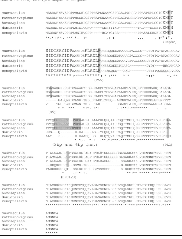
Figure 11 Sequence alignment of Mus musculus, Rattus novergicus, 42 Homo sapiens, Danio rerio and Xenopus levis C/EBPα proteins
ABBREVATIONS
AFB1 aflatoxin B1
AML acute myeloid leukemia
APC apolipoprotein C
APS ammonium per sulphate
bp basepair
BRCA2 breast cancer 2
BR-LZ basic region-leucine zipper Brm brahma
C- terminus carboxy- terminus
C/EBP CCAAT/enhancer binding protein
CBP CREB binding protein
Cdk cyclin dependent kinase
DNA deoxyribonucleic acid
EDTA ethylenediaminetetraecetic acid
eIF2α translation initiation factor 2 alpha eIF4E translation initiation factor 4E
EST expressed sequense tags
EtBr ethidium bromide
ETS1 E twenty six (E26). g gram
GATA1 GATA binding protein 1 (globin transcription factor 1) G-CSF granulocyte colony stimulating factor
GLUT4 glucose transporter 4
GSK-3β glycogen synthase kinase 3 beta HBV hepatitis B virus
HCC hepatocellular carcinoma
HCV Hepatitis C virus
HNF hepatocyte nuclear factor IGF-2 insulin like growth factor-2
KCl potassium chloride LIP liver inhibitory protein
LOH loss of heterozygosity
M6P metelloproteinase 6 phosphate ml milliliter
µl microliter
mRNA messenge RNA
mTOR Target of rapamycin N- terminus amino- terminus
NaCl sodium chloride
NFKB nuclear factor kappa B ORF open reading frame
PAGE polyacrylamde gel electrophoresis PCNA proliferating cell nuclear antigen
PCR polymerase chain reaction
PI3K protein 3 kinase
PKC protein kinase C
PKR protein kinase R
PP2A Protein phosphatase 2A
PPARγ peroxisome proliferating antigen receptor gamma PTEN phosphatase and tensin homologue
Rb retinoblastoma
RNA ribonucleic acid
RUNX1 Runt-related transcription factor 1
SCD1 stearoyl-CoA desaturase
SSCP single strand conformation polymorphism
TBE tris-boric acid-EDTA
TBP TATA binding protein
TE transactivation elements
TFIIB transcription factor II B
Tris Tris (hydroxymethyl)-methylamine UV ultraviolet
1. INTRODUCTION
1.1 Hepatocellular carcinoma (HCC)
Hepatocellular carcinoma is a type of liver cancer derived from hepatocytes and it is among the most common types of cancers in worldwide with more than 564 000 incidence in 2000 (Parkin et al., 2001). HCC incidence shows variations between different geographic areas with the highest incidence in Africa and Far East, where the hepatitis B virus (HBV) is endemic and aflatoxin B1 (AFB1) exposure is high (Feitelson et al., 2002). The average incidence of HCC is ten times higher in Asia than in Western countries but due to hepatitis C virus (HCV) infection and alcohol consumption, it is expected to rise in 20 years in West (Parkin et al., 2001). Males develop HCC approximately two times more than women do (Beasley and Hwang, 1984).
Life expectancy of HCC is about 6 months from the time of diagnosis and survival rate is <3% for untreated cancer over 5 years. It can be cured by surgical resection by partial hepatectomy but the recurrence rate is very high (Feitelson et al., 2002). HCC is the fifth common type of cancer worldwide but its third leading cause of cancer death; the high mortality rate of HCC is due to asymptomatic feature of HCC progression and unresponsiveness to therapy (Block et al., 2003).
1.1.1 Etiology of HCC
HBV and HCV infections, AFB1exposure, alcohol, hemochromatosis, alpha 1-antitrypsin deficiency, tyrosinemia, glycogen storage disease, etc are the main risk factors for development of HCC (Anthony, 2001). HBV and HCV infections are responsible from 70-75% and 10-15% of HCC worldwide respectively (Parkin et al., 2001; Anthony, 2001). HBV is the main risk factor in the Asia and Africa and HCV infection is generally seen in Japan, Europe and America (Bruix et al., 2004). Consumption of AFB1 contaminated food enhances the HCC risk in China and South Africa. Although alcohol is not a carcinogen itself, it may act as cofactor with viruses and chemicals and alcoholic consumption rises the risk in the Western Europe (Bruix et al., 2004). Inherited metabolic diseases are rare but some may have a high risk in development of HCC (Anthony, 2001).
HBV and HCV infections result in either acute infection or an unresolved, long-term persistence. Chronic infection is asymptomatic for years but eventually fatigue, malasia and other symptoms specific for hepatitis appear (Lok et al., 2001). HBV and HCV infections cause chronic hepatitis, cirrhosis and HCC. Several mechanisms for the malignant transformation properties of HBV and HCV have been proposed although the exact mechanism still is not clear. HBV is a DNA virus from hepadnavirus family. HBV infects all age groups but chronic infection usually occurs in perinatal period, during infancy or early childhood. HBV uses RNA intermediate and reverse transcriptase for replication. According to the one of the most accepted mechanism, HBV integrates into the host genome randomly and transcribe X protein which is thought to be important in oncogenesis. Hbx transactivates the cellular oncogenes c-myc, c-jun and c-fos by inducing protein kinase C and nuclear factor kappa B pathways (Feitelson, 1999) and inactivates p53 and Rb (Andrisani and Barnabas, 1999). Another mechanism suggests that the integrated viral genome increases the genomic instability causing insertions, deletions and duplications (Szabo et al., 2004).
HCV is a RNA virus from a flavivirus family. HCV is believed not to be cytotoxic virus but cause hepatitis through reaction of the host immune system against virus infected cells (Szabo et al., 2004). Unlike HBV, HCV does not integrate into the host genome. It changes the transcription activity of NFκB and stat-3 proteins (Waris and Siddiqui, 2003).
Figure 1: Mechanisms involved in HBV- and HCV-related chronic liver disease and HCC. Oxidative stress, up- and down-regulation of several growth factors and cytokines
lead to chronic inflammation, cell death and proliferation. HBV integration into host genome and viral proteins, especially Hbx of HBV and the core protein of HCV may involve in HCC development (Szabo et al., 2004).
AFB1 is a metabolite of Aspergillus flavus that contaminate food stored in humid conditions. There is a correlation between areas of high aflatoxin exposure (southern Africa and Qidong, China) and a high incidence of HCC. The characteristic genetic change associated with AFB1 is G >T transversion at codon 249 of p53, affects the p53 gene in > 50% of the tumors in AFB1 endemic areas (Puisieux and Ozturk, 1997).
1.1.2 Molecular pathogenesis of HCC
Chronic hepatitis and cirrhosis are the setting in which HCC develops frequently and they are considered to be a precancerous lesions. In cirrhotic and hepatic liver, hepatocytes are killed and inflammatory cells invade the liver, these changes alter the phenotype of the liver and cause increase in oxidative stress. HCC develops in 10 to 30 years after the infection with the HBV and HCV (preneoplastic phase). Most of the changes in this phase are quantitative changes like increase in the expression of transforming growth factor β (TGFβ) and insulin-like growth factor-2 (IGF-2). These epigenetic changes lead to accelerated proliferation and formation of dysplastic hepatocytes that have structural aberrations in genes and chromosomes. HCC develops through the accumulation of irreversible structural aberrations during dysplastic phase (Thorgeirsson and Grisham, 2002). 30 % of dysplastic nodules evolve into HCC in 1-5 years (Borzio et al., 2003). Early nodules of HCC are small, well differentiated and fibrotic tissue formed during the cirrhosis surrounds the nodules. Increase in expression of metalloproteinases leads to break down of cellular matrix and accelerate tumor growth (Feitelson et al., 2002).
1.1.3 Genetics of HCC
Development of HCC is a multistep process during which changes in structure of the genes and chromosomes accumulate, and at least four different genetic events are required for malignant transformation (Buendia, 2000). Different HCC nodules show different genetic abnormalities, suggesting heterogeneity on the molecular level (Feitelson et al., 2002). Although the major viral and environmental risk factors leading to HCC development are known, the underlying genetic mechanisms are currently unknown. Point mutations, allelic gains/losses, gene arrangements and methylation specific changes result in activation of oncogenes and/or inactivation of tumor suppressor genes.
1.1.3.1 Loss of heterozygosity (LOH)
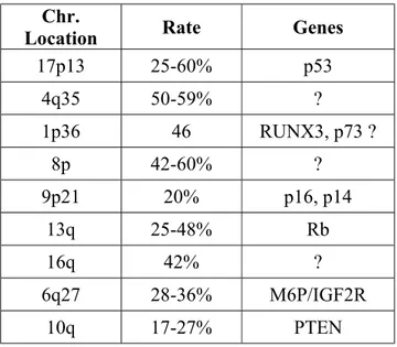
The major genetic abnormalities in HCC are the allelic gains and losses. Allelotype studies by microsatellite analysis and comparative genomic hybridization revealed regions of loss of heterozygosity (LOH) in liver tumors. Table 1 shows the common regions where the allelic losses are detected by several different studies (Nagai et al., 1997; Boige et al., 1997; Marchio et al., 1997) . In addition to these regions there are other LOH regions reported, 14q, 18q, 1q, 22q, 16p and 15q (Jou et al., 2004). The frequency of LOH may show regional variations and it may correlate with the other alterations. It has been shown that LOH at 4q is associated with the mutations in p53 (Rashid et al., 1999) and LOH at 4q and 16p is predominant in Qidong where the aflatoxin contamination is common (Fujimoto et al., 1994). Some of the tumor suppressor genes located at the LOH regions are identified but the genes at 4q, 8p and 16q are needed to be identified.
Table 1: High frequency of loss of heterozygosity (LOH) in HCC
Chr.
Location Rate Genes
17p13 25-60% p53 4q35 50-59% ? 1p36 46 RUNX3, p73 ? 8p 42-60% ? 9p21 20% p16, p14 13q 25-48% Rb 16q 42% ? 6q27 28-36% M6P/IGF2R 10q 17-27% PTEN
1.1.3.2 Other genetic and epigenetic alterations in HCC
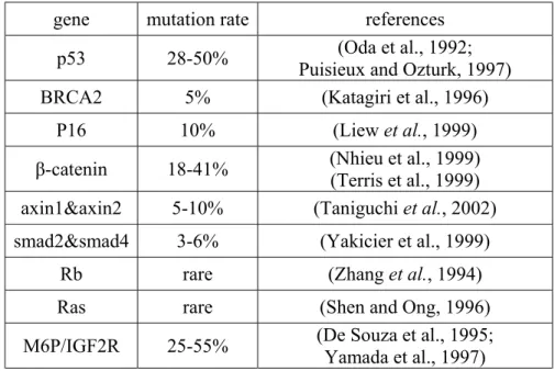
Recent studies identified several genetic and epigenetic alterations in HCC although these represent the fraction of abnormalities present in HCC. These genetic and epigenetic alterations generally result in disruption of p53, Wnt or Rb-p16 pathways. Rb, p16 and cyclin D1 are important in cell cycle regulation and they control G1 to S phase transition. Amplification and overexpression of cyclin D1, an oncogene and key regulator of cell cycle progression, are seen 10-13 % of HCC cases (Tannapfel et al., 2000). Rb and p16 are the tumor suppressor genes and their mutations are not generally found in HCC although hypermetylation and allelic loss is observed frequently (Zhang et al., 1994; Liew et al., 1999). Aberrations in this pathway results in the increase proliferation rate due to loss of control on cell cycle.
p53 is activated in response to DNA damage and halts the cell cycle for DNA repair or apoptosis. Mutations in p53 are observed in most of the cancers and approximately 30% of HCCs have mutated p53. The frequency of p53 mutations is 20% in North America and 67% in Africa due to differences in HBV infection and aflatoxin exposure (Tannapfel and Wittekind, 2002). Mutation at codon 249, which leads to change in arginine to serine, is the hot spot mutation and associated with the aflatoxin exposure (Ozturk, 1995). Hypermethylation of p14, which stabilize p53, is seen in 40% of HCC (Anzola et al., 2004) with no abnormalities in p53.
Aberrations in Wnt pathway were first observed in colon cancers, which has 80% mutations in APC. Mutations in APC lead to accumulation of β-catenin in nucleus. APC mutations are not observed in HCC however, β-catenin mutations are seen in 18-41% of HCC (Nhieu et al., 1999). β -catenin mutations inhibit the phosphorylation by glycogen synthase-3 kinase β (GSK3-β) and stabilize β-catenin. Nuclear accumulation of β-catenin increases the transcription of c-myc, cyclin D1 and fibronectin and matrix
metalloproteinases and cellular proliferation (Buendia, 2000). Mutations in the axin gene, another player of Wnt pathway, are responsible for the nuclear accumulation of β-catenin in about 7% of additional HCC cases(Taniguchi et al., 2002).
Table 2: Mutations in tumor suppressor genes and oncogenes in HCC
gene mutation rate references
p53 28-50% Puisieux and Ozturk, 1997) (Oda et al., 1992; BRCA2 5% (Katagiri et al., 1996)
P16 10% (Liew et al., 1999)
β-catenin 18-41% (Nhieu et al., 1999) (Terris et al., 1999) axin1&axin2 5-10% (Taniguchi et al., 2002)
smad2&smad4 3-6% (Yakicier et al., 1999)
Rb rare (Zhang et al., 1994)
Ras rare (Shen and Ong, 1996)
M6P/IGF2R 25-55% (De Souza et al., 1995; Yamada et al., 1997)
In addition to these three pathways, alterations in several other pathways are also observed, for example, up regulation of mitogen activated protein kinase pathway (MAPK) (Okabe et al., 2001) and alteration in TGF-β pathway (Yakicier et al., 1999). Table 2 summarizes the oncogenes and tumor suppressor genes mutated in HCC. Multiple pathways and molecules are involved in transformation of hepatocytes and an understanding of the molecular mechanisms of the HCC development is important for development of new markers for tumor staging and therapeutic intervention.
1.2 CCAAT- Enhancer Binding Proteins (C/EBPs)
CCAAT/ enhancer–binding proteins are a family of transcription factors that are involved in several responses like control of cellular proliferation, differentiation, metabolism and inflammation (Ramji and Foka, 2002). The first identified member is the C/EBPα found in Steve McKnight lab in 1988. So far, six members of the C/EBP family have been identified. These members are designated by Greek letters as C/EBPα, β, γ, δ, ε and ζ as proposed by Cao et al in 1991.
C/EBPs bind to CCAAT box motif found in the promoter regions of several genes through DNA binding domain and leucine zipper domains at the C terminus. The consensus binding site, RTTGCGYAAY (R = A or G, and Y = C or T), are similar with small variations within the C/EBP family members (Osada et al., 1996). C/EBPs have an activation domain, leucine zipper domain and basic DNA binding domain. The leucine zipper domain is the most conserved region that is important for the dimerization of C/EBP proteins. Dimerization is necessary for DNA binding and because of high homology at this region; different C/EBP members can form heterodimers with each other. These heterodimers are able to bind same consensus sequence on DNA, only exception is the C/EBP ζ which can form heterodimers with other members but interacts with the different sequence on several other genes promoters due to presence of two proline residues in the basic region (Ron and Habener, 1992).
The N terminus of C/EBP family members contains the activation domains that interact with the basal transcription machinery. The N termini of the C/EBPs are not highly conserved (only >20% homology exists). Homology exists between the two conserved motifs, box A and box B, which interacts with transcription binding protein (TBP) and TBP associated factor TFIIB (Nerlov and Ziff, 1995).
1.2.1 C/EBPα
C/EBPα is the first identified member of the C/EBP family. C/EBPα gene has one exon that encodes a 2.7kb mRNA translated into two isoforms; 42kDa and 30kDa due to leaky ribosome scanning. The human C/EBPα is expressed in a tissue restricted manner with high expression in placenta, liver, lung, skeletal muscle, pancreas, small intestine, colon and peripheral blood leukocytes but no or low expression is detected in brain, kidney, thymus, testis and ovary (Antonson and Xanthopoulos, 1995). C/EBPα activates the transcription of several genes in hepatocytes, adipocytes and hematopoietic cells. The C/EBPα gene is conserved among species, especially in its DNA binding region. It has 90% homology to the rat C/EBPα, 100% identity in DNA binding region (Hendricks-Taylor and Darlington, 1995).
1.2.2 C/EBPα isoforms
C/EBPα mRNA generates two isoforms, 42 kDa and 30 kDa, by differential use of translation initiation codons (figure 2). The C/EBPα mRNA has four AUG start codons. When the conditions are favorable, the activity of the translation initiation factors, elF2α and elF4E, increases and translation start from first AUG (suboptimal initiation site). Translation from this site generates small upstream open reading frame and translation is reinitiated from fourth AUG that generates p30 isoform. When the activity of these translation initiation factors are low, translation is initiated from second AUG (optimal initiation site) which generates p42 isoform (Calkhoven et al., 2000).
P42 isoform has three activation domains. Activation domains, TEI and TEII interact with components of the basal transcription machinery TBP/TFIIB and histone
and TEII are not present in P30 form, which lacks the N terminal 117 amino acids. The third activation domain (TEIII) which recruits chromatin remodeling complex SWI/SNF (Pedersen et al., 2001) and DNA binding domain are present in both isoforms (figure 3).
Generation of C/EBPα isoforms are tightly regulated by PKR and mTOR signaling pathways and the ratio of these isoforms determine the cell fate (Calkhoven et
al., 2000). p42 to p30 ratio regulates the proliferation and differentiation control. p42
isoform has the ability to block proliferation and induces adipogenic and granulocytic differentiation unlike p30 isoform, which is not antimitotic and although it induces early adipocyte differentiation, it inhibits terminal differentiation (Lin et al., 1993). P30 isoform binds DNA less efficiently comparing to p42 and acts as dominant negative mutant (D'Alo' et al., 2003).
Figure 2: Generation of C/EBPα isoforms. Two C/EBPα isoforms, p42 and p30, are
formed by alternative use of translation initiation codons, which is regulated by mTOR and PKR pathways.
Figure 3: Functional domains of C/EBPα isoforms. p42 isoform has the all three
transactivation elements, TE-I, TE-II, TE-III while the p30 isoform lacks the TE-I and TE-II. Basic region and leucine zipper domain are present in both isoforms.
1.2.3 C/EBPα knockout mice
Wang et al generated C/EBPα knockout mice that die soon after birth due to hypoglycemia and impaired energy homeostasis in liver and adipose tissue (Wang et al., 1995). C/EBPα is the transcriptional regulator of genes important in energy metabolism and differentiation in adipocytes and hepatocytes and C/EBPα -/- mice have reduced expression of genes involved in glycogenesis such as glycogen synthase, phosphoenolpyruvate carboxykinase and glucose-6-phosphatase (Wang et al., 1995; Lee et al., 1997). C/EBPα -/- mice also have abnormality in control of hepatic growth and lung development. The liver architecture of the mice lacking C/EBPα is impaired, resembling regenerating liver after partial hepatectomy or pseudoglandular hepatocellular carcinoma (Flodby et al., 1996). C/EBPα -/- mice show induced hepatic proliferation and expression of c-myc, c-jun, β-actin and α-fetoprotein are increased several folds. In
the G1/S phase of the cell cycle (Flodby et al., 1996). Heterozygous mice do not have any obvious abnormalities neither in energy metabolism nor in differentiation and proliferation.
Targeted disruption of C/EBPα prevents neutrophil differentiation. C/EBPα -/- mice do not have mature neutrophils and eosinophils in the blood or fetal liver; however, the other blood cells are not affected. The defect in the differentiation of neutrophils is due to reduced expression of granulocyte colony-stimulating factor receptor (Zhang et al., 1997).
1.2.4 The role of C/EBPα in adipocyte differentiation
C/EBPα is expressed at high levels in terminally differentiated adipocytes and it has role in the differentiation of adipocytes together with peroxisome proliferators antigen receptor γ (PPARγ) (Rosen et al., 2002). When the expression of C/EBPα is inhibited by antisense RNA in 3T3-L1 preadipocyte cell line, adipocyte specific genes are not expressed and differentiation is blocked (Lin and Lane, 1994). Adipocyte differentiation is induced by the rapid and transient increase in the expression of C/EBPβ and C/EBPδ upon treatment with the differentiation inducers. C/EBPβ and C/EBPδ induce the expression of C/EBPα, which leads to cell cycle arrest and transcriptional activation of many genes involved in adipocyte differentiation such as GLUT4, SCD1, leptin and 422/aP2 (Darlington et al., 1998). PPARγ and C/EBPα induce each other’s expression and promotes and maintains differentiated state (Rosen et al., 2002).
1.2.5 The role of C/EBPα in lung development and lung cancer
C/EBPα is expressed in bronchial cells and type II pneumocytes in the lung and regulates expression of several genes involved in lung differentiation. C/EBPα knockout mice shows abnormal proliferation of type II pneumocytes (Flodby et al., 1996). C/EBPα is proposed to be a candidate tumor suppressor gene in lung cancer since its expression is downregulated in large proportion of lung cancers (Halmos et al., 2002).
1.2.6 The role of C/EBPα in granulopoiesis and in acute myeloid leukemia
Granulopoiesis is the formation of mature neutrophil granulocytes from immature myeloblasts. Transcription factors, which activate lineage specific genes, are important in the granulopoiesis. One of these transcription factors is the C/EBPα expressed in the stem and myeloid progenitor cells but not in other cells in the hematopoietic system. C/EBPα expression is initiated during the commitment of multipotential precursors to the myeloid lineage and its expression is up regulated during granulocytic differentiation, and is rapidly downregulated during the monocytic pathway (Scott et al., 1992; Radomska et al., 1998). C/EBPα activates the transcription of granulocyte specific genes; receptors for the growth factors, macrophage colony-stimulating factor, granulocyte colony colony-stimulating factor (G-CSF) (Smith et al., 1996; Zhang et al., 1996).
The importance of C/EBPα in granulocytic differentiation comes from the evidences from knockout mice studies and mutations of C/EBPα in acute myeloid leukemia (AML). C/EBPα knockout mice have disruption in granulopoiesis and do not have any mature neutrophils while the other blood cells are not affected (Zhang et al., 1997).
Mutations of C/EBPα in AML is first described by Pabst et al. and later by other groups (Pabst et al., 2001b; Gombart et al., 2002; Barjesteh van Waalwijk van Doorn-Khosrovani et al., 2003; Snaddon et al., 2003). C/EBPα is mutated approximately 9% of the AML patients, especially M2 subtype where the C/EBPα mutation is 20%. There are two types of common mutations seen in AML patients. First one is the in-frame insertion, deletion or substitution mutation in the leucine zipper domain or in the region between the basic region and leucine zipper dimerization domain. These mutations cause the disruption of α helical configuration and prevent C/EBPα to bind DNA, however, they do not act as dominant negative mutants (Gombart et al., 2002). Second type of mutations is insertions and deletions in the N terminal region, which results in frameshift in the p42 isoform but the transcription of p30 isoform is not affected. Therefore, second type of mutation results in increased level of p30 isoform, which has less efficieny to bind DNA and is dominant negative inhibitor of wild type p42 (Pabst et al., 2001b).
In addition to decrease in efficiency in DNA binding, C/EBPα mediated repression of E2F may explain the molecular mechanism of mutations in AML (Nerlov, 2004). E2F is the transcription factor that regulates the cell cycle progression genes and c-myc. E2F activation leads to induction of cell cycle genes and upregulation of c-myc, which inhibits granulopoiesis and favors proliferation. C/EBPα and c-myc expression are reciprocally regulated and expression of C/EBPα downregulates c-myc expression through repressing E2F transactivation domain (Johansen et al., 2001). Two regions of C/EBPα are important in E2F repression, basic region (E2F interacting domains) and N terminal region. Therefore, mutations in the amino acids that interact with E2F and P30 isoform, which lacks the N terminal 117-amino acids, cannot repress the E2F activity and cannot down regulate c-myc (D'Alo' et al., 2003).
Mutations in C/EBPα have not been found in patients with the t(8;21), inv 16 (Timchenko et al., 1998) and t(15;17) translocations in which the function or expression of C/EBPα is repressed by other mechanisms (Nerlov, 2004). AML1-ETO, the fusion product of t(8;21) translocation, repress the C/EBPα expression by inhibiting positive autoregulation of the C/EBPα promoter (Pabst et al., 2001a). PML-RARα fusion protein
of acute promyelocytic leukemia is reported to repress C/EBPα activity by trapping it in the cytoplasm (Truong et al., 2003). Therefore, inactivation of C/EBPα by either mutation or other mechanisms is common in AML and thought to be important in malignant transformation.
1.2.7 The role of C/EBPα in hepatocyte proliferation and differentiation
The liver enriched transcription factors (C/EBP, HNF1, HNF3, HNF4 and HNF6) accomplish the transcription of hepatocyte specific genes through interacting promoter/enhancer sites. Distinct classes of transcription factors regulate the liver development, differentiation and regeneration. C/EBPα has role in liver differentiation regulating transcription of genes involved in hepatic glycogen synthesis, gluconeogenesis and lipid homeostasis and it negatively regulates hepatocyte proliferation (Costa et al., 2003).
C/EBPα is one of the key regulators of cell growth and inhibits the cell proliferation in cultured cells and in liver (Hendricks-Taylor et al., 1995; Diehl et al., 1996). The expression of C/EBPα is reduced during hepatocyte proliferation in regenerating liver and in hepatocellular carcinoma (Mischoulon et al., 1992; Diehl and Yang, 1994; Flodby et al., 1995; Tomizawa et al., 2002). Although the C/EBPα function of growth inhibition is well established, the molecular pathways of this function are unknown. Several mechanisms for C/EBPα growth inhibitory function have been proposed. It is proposed that the growth inhibition function of C/EBPα in liver is not to be due to reduced transcriptional activity but due to protein-protein interactions (Wang et al., 2001; Porse et al., 2001).
One possible pathway for C/EBPα mediated growth inhibition is the stabilization of p21 protein by C/EBPα (Timchenko et al., 1996). p21 is cyclin-dependent kinase inhibitor and stops the cell cycle progression at G1 phase. C/EBPα directly interacts with p21 and protects it from proteolytic degradation (Timchenko et al., 1997) and it is proposed to cooperate with p21 to inhibit CDK2 activity in vitro (Harris et al., 2001). In mouse hepatocytes that express C/EBPα, high levels of p21 protein is present but the level of p21 protein is low in hepatocytes that do not have C/EBPα (Serfas et al., 1997). However, other studies suggest that the p21 is not important for C/EBPα mediated growth inhibition since p21 knockout mice do not show any alteration in hepatocyte proliferation (Deng et al., 1995) and C/EBPα inhibits cell proliferation in the absence of p21 (Muller et al., 1999).
In addition to p21, C/EBPα interacts with several other proteins, which have role in the cell cycle progression and transcription. One of these proteins is cyclin-dependent kinases, which regulate the cell cycle progression through association with cyclins. Cdk2-cyclin E, A and cdk4-Cdk2-cyclinD mediate the S phase transition of cell cycle through Rb-dependent repression of E2F. In liver, C/EBPα directly interacts with cdk2 and cdk4 and inhibits their functions through disrupting their association with cyclins (Wang et al., 2001). C/EBPα also reduces the protein level of cdk4 by mediating proteasome-dependent degradation of cdk4 (Wang et al., 2002). C/EBPα knockout liver has the increased activities of the cdk2 and cdk4 consistent with increased rate of proliferation (Wang et al., 2001). C/EBPα also interacts with the SWI/SNF chromatin-remodeling complex during the regulation of genes involved in differentiation. C/EBPα cannot inhibit the proliferation in cells, which are defective in SWI/SNF (Muller et al., 2004).
C/EBPα- mediated repression of E2F is believed to be important for proliferation arrest. E2F regulates the transcription of genes, which are involved in DNA synthesis and mitosis and E2F-dependent transcription is regulated by Rb-mediated phosphorylation. E2F interacts with Rb and Rb-like proteins, p107 and p130, at different stages of cell
cycle. In quiescent cells, p130-E2F complex is seen and p107-E2F complex is seen in dividing cells. C/EBPα disrupts the p107-E2F complex through interacting with p107 (Timchenko et al., 1999).
Iakoava et al suggested the switch in the mechanism of C/EBPα-dependent growth arrest as the mice gets older from cdk inhibition to E2F repression and combines the two models. In liver of the young mice, C/EBPα interacts with cdk2, this interaction represses the E2F and c-myc expression. Ageing leads to increase in the Brahma protein (Brm), which replaces the cdk2 and binds to C/EBPα, leading to formation of C/EBPα-Rb-E2F complex. This complex represses the E2F dependent expression of c-myc and it is not disrupted after partial hepatectomy. This explains the reduced proliferative capacity of liver by aging (Iakova et al., 2003). But this complex involves the Rb and it has been previously shown that the growth-inhibitory function of the C/EBPα is independent of Rb (Hendricks-Taylor et al., 1995).
Since C/EBPα is a negative regulator of cell proliferation, there must be a mechanism, which blocks the growth inhibitory activity of C/EBPα in liver tumors. In a recent study Wang et al. shows that the growth inhibitory activityof C/EBPα is blocked in liver tumors by activation of the PI3K/Akt pathway. Activation of this pathway leads to PP2A accumulation in nucleus where PP2A dephosphorylate ser 193 on mouse C/EBPα. Phosphorylated ser 193 (ser 190 for human C/EBPα) is necessary for C/EBPα to interact with cdks and E2F-Rb-Brm complexes (Wang et al., 2004).
2. AIM
HCC is a genetically heterogeneous disease and several genetic and epigenetic alterations have been reported to be involved in malignant transformation of hepatocytes. However, there is much left for the understanding critical pathways and genes deregulated.
C/EBPα is a negative regulator of cell proliferation and highly expressed in normal hepatocytes. Mutations, inactivation and/or reduced expression of C/EBPα were reported in AML and lung cancer. It has also been shown that the activation of PI3K/Akt pathway inhibits the growth inhibitory function of C/EBPα in liver tumors. All of these findings together with C/EBPα knockout mice (that show phenotype resembling hepatocellular carcinoma) study have strongly implied that C/EBPα is a possible candidate tumor suppressor gene for the development of HCC. Therefore, we think that the genetic alterations in C/EBPα might contribute the malignant transformation of hepatocytes.
The aim of this study is to check whether the genetic abnormalities of C/EBPα gene are involved in hepatocellular carcinoma.
3. MATERIALS AND METHODS 3.1 Tumor samples
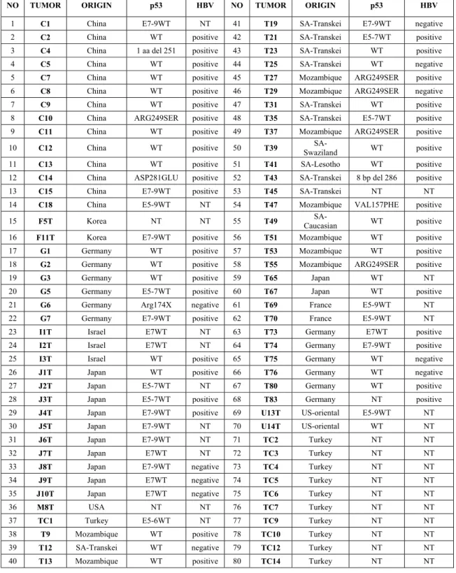
Genomic DNA of 70 tumors were chosen from a previous HCC sample collection (Unsal et al., 1994). In addition, genomic DNA from 10 Turkish HCC patient tumors and 142 controls blood were used. The patients are from South Africa (n=12), China (n=14), Mozambique (n=9), Japan (n=12), Germany (n=12), Korea (n=2), Israel (n=3), France (n=2), USA (n=3) and Turkey (n=11). Ten samples have the p53 mutation and 43 samples are HBV positive (table 3).
3.2 Cell lines and tissue culture reagents
DMEM, trypsin/EDTA and penicillin / streptomyocin were obtained from Biochrom, RPMI 1640 was obtained from Kibbutz Beit Haenek Israel and fetal bovine serum was obtained from Sigma. Tissue culture flasks, petri dishes, 15 ml polycarbonate centrifuge tubes with lids and cryotubes were purchased from Costar Corp (Cambridge, England). 13 HCC cell lines; Snu182, Snu398, Snu387, Snu423, Snu475, Snu449, PLC, Focus, HepG2, Hep40, Mahlavu, Hep3B, SK Hep1; 7 breast cancer cell lines; MDA-MB-453, T-47-D, BT-474, BT-20, MCF-7, MDA-468,HME-1 and 12 colorectal cancer cell lines; metaSW480, CO115, WiDr, CCL231, SW480, LS411, KM12, CCL225, CCL229, HTB38, HRT-8 and CCL222 were used in this study. Breast cancer cell line DNAs were kindly supplied by Dr. Işık Yuluğ.
3.3 Solutions
Ethidium Bromide (EtBr)
10mg/ml in water (stock solution) 30ng/ml (working solution)
Table 3: Characteristics of tumor samples
NO TUMOR ORIGIN p53 HBV NO TUMOR ORIGIN p53 HBV
1 C1 China E7-9WT NT 41 T19 SA-Transkei E7-9WT negative 2 C2 China WT positive 42 T21 SA-Transkei E5-7WT positive 3 C4 China 1 aa del 251 positive 43 T23 SA-Transkei WT positive 4 C5 China WT positive 44 T25 SA-Transkei WT negative 5 C7 China WT positive 45 T27 Mozambique ARG249SER positive 6 C8 China WT positive 46 T29 Mozambique ARG249SER negative 7 C9 China WT positive 47 T31 SA-Transkei WT positive 8 C10 China ARG249SER positive 48 T35 SA-Transkei E5-7WT positive 9 C11 China WT positive 49 T37 Mozambique ARG249SER positive 10 C12 China WT positive 50 T39 Swaziland SA- WT positive 11 C13 China WT positive 51 T41 SA-Lesotho WT positive 12 C14 China ASP281GLU positive 52 T43 SA-Transkei 8 bp del 286 positive 13 C15 China E7-9WT positive 53 T45 SA-Transkei NT NT 14 C18 China E5-9WT NT 54 T47 Mozambique VAL157PHE positive 15 F5T Korea NT NT 55 T49 Caucasian SA- WT positive 16 F11T Korea E7-9WT positive 56 T51 Mozambique WT positive 17 G1 Germany WT positive 57 T53 Mozambique WT positive 18 G2 Germany WT positive 58 T55 Mozambique ARG249SER positive 19 G3 Germany WT positive 59 T65 Japan WT NT 20 G5 Germany E5-7WT positive 60 T67 Japan WT positive 21 G6 Germany Arg174X negative 61 T69 France E5-9WT NT 22 G7 Germany E7-9WT positive 62 T70 France E5-9WT NT 23 I1T Israel E7WT NT 63 T73 Germany E7WT positive 24 I2T Israel E7WT NT 64 T74 Germany E7-9WT positive 25 I3T Israel WT positive 65 T75 Germany WT negative 26 J1T Japan WT positive 66 T76 Germany WT negative 27 J2T Japan E5-7WT NT 67 T80 Germany WT positive 28 J3T Japan E5-7WT positive 68 T83 Germany NT positive 29 J4T Japan E7-9WT positive 69 U13T US-oriental E5-9WT NT 30 J5T Japan E7-9WT NT 70 U14T US-oriental WT NT 31 J6T Japan E7-9WT NT 71 TC2 Turkey NT NT
32 J7T Japan E7WT NT 72 TC3 Turkey NT NT
33 J8T Japan E7-9WT negative 73 TC4 Turkey NT NT 34 J9T Japan E7WT negative 74 TC5 Turkey NT NT 35 J10T Japan E7WT negative 75 TC6 Turkey NT NT
36 M8T USA NT NT 76 TC7 Turkey NT NT
37 TC1 Turkey E5-6WT NT 77 TC9 Turkey NT NT 38 T9 Mozambique WT positive 78 TC10 Turkey NT NT 39 T12 SA-Transkei WT negative 79 TC12 Turkey NT NT 40 T13 Mozambique WT positive 80 TC14 Turkey NT NT
10X TBE Buffer Solution
108g Tris
55g Boric Acid
8.3g EDTA
dissolved in 1lt of deionized water.
6X Loading Buffer Solution 30% Glycerol
0.04% Bromphenolblue
0.04% Xylene Cyanol
∆dH2O
Agarose Gel Solution for PCR
50 ml 1X TBE
0.75g Agarose
Acrylamide/ Bisacrylamide 30% stock 29g Acrylamide 1g Bisacrylamide
dissolved in 100 ml ∆dH2O
12% Non-Denaturing Polyacrylamide Gel Solution
10X TBE 10 ml
30% acry./bisacry. 40 ml
∆dH2O 50 ml
10X Phosphate-buffered saline (PBS)
NaCl 80 g
KCl 2 g
Na2HPO4 14.4 g
KH2PO4 2.4 g
dissolved in 1 lt of water and pH is adjusted to 7.4.
3.4 DNA isolation
DNA is isolated from blood, cell lysates and paraffin embedded liver tissue by using Qiagen DNeasy Tissue Kit (#69506) following manufacturer recommendation.
3.5 Mutation screening
3.5.1 Polymerase chain reaction (PCR)
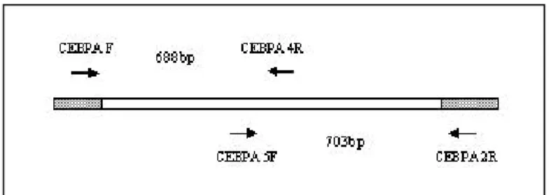
Two sets of primers were used for amplification of the single C/EBPα exon.
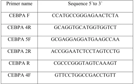
Table 4: Sequences of primers used in PCR and sequencing
Primer name Sequence 5`to 3`
CEBPA F CCATGCCGGGAGAACTCTA CEBPA 4R GCAGGTGCATGGTGGTCT CEBPA 5F GCGAGGAGGATGAAGCCAA CEBPA 2R ACCGGAATCTCCTAGTCCTG CEBPA R CGCCCGGGTAGTCAAAGT CEBPA 4F GTTCCTGGCCGACCTGTT PCR conditions
PCR was performed by Qiagen Hot Star Polymerase (#203203) with primers CEBPA 5F and CEBPA 2R and Qiagen Taq Polymerase (#201203) with primers CEBPA F and CEBPA 4R .
1X Buffer
1X Q- Solution
15mM dNTP mix 20 pmol Forward Primer 20 pmol Reverse Primer 1.25u Taq Polymerase
100ng DNA
PCR conditions for amplification of CEBPA F/ CEBPA 4R product: 5 minutes denaturation at 94 oC, 35 cycles of 1-min at 94 oC, 40 sec at 50 oC 100 sec at 72 oC. 10 min at 72 oC.
PCR conditions for amplification of CEBPA 5F/ CEBPA 2R product: 15 minutes denaturation at 95 oC, 35 cycles of 1-min at 94 oC, 40 sec at 50 oC 100 sec at 72 oC. 10 min at 72 oC.
3.5.2 Agarose gel electrophoresis
5 ml of PCR product was mixed with 1 ml of 6 x loading buffer and loaded into 1.5 % agarose gel. They were run at 100V for 40 minutes and then visualized under UV light.
3.5.3 Automated sequencing
PCR products were purified with Qiagen PCR Purification Kit (#28106) and sequencing was performed by using Amersham Dynamic ET Terminator Cycle Sequencing Kit (#US8 1050) and Perkin Elmer Big Dye Terminator Kit vs 3.1
(# 4337455) with CEBPA F, CEBPA R, CEBPA 4F, CEBPA 5F and CEBPA 2R primers and analyzed in ABI 377 and ABI 310 sequencer.
3.6 Heteroduplex analysis 3.6.1 PCR
PCR was performed by using CEBPA 5F and CEBPA 4R primers with Qiagen Taq Polymerase. PCR conditions ; 5 min denaturation at 94 oC followed by 35 cycles of 1 min at 94 oC, 20 sec at 52 oC and 30 sec at 72 oC. After 35 cycles, a last step at 72 oC for 10 min was added.
3.6.2 PAGE
250 µl of 10 % Ammonium persulfate (APS) and 25 µl of Temed is added to 25 ml of 12 % PAGE solution. The solution is poured into gel apparatus and left for polymerization for 1 hour. 10 µl of PCR product is mixed with 2 µl of 6 x loading buffer and loaded into an electrophoresis apparatus and run at 120 V for 4 hours. After electrophoresis, gel is stained with 0.5 µg / ml EtBr for 30 min and visualized under UV light.
3.7 Restriction analysis
Restriction digestion was performed by SmaI (Fermentas #ER0661), ApaI (Fermentas #ER1411), BssSI (New England Biolabs # R0587S) and Mae III (Roche # 822 248) enzymes.
SmaI Digest ApaI Digest
1 x Buffer Y+ / TangoTm 1 x buffer B
1unit of SmaI enzyme 5 units of ApaI ezyme PCR product PCR product
∆ H2O ∆ H2O
BssSI Digest MaeIII Digest
1 x NEB3 1x MaeIII Buffer
1 unit of BssSI enzyme 2 units of MaeIII enzyme PCR product PCR product
∆ H2O ∆ H2O
incubated at 37 0C overnight. incubated at 55 0C overnight.
Restriction products were run at 1.5 % agarose gel and visualized under UV light.
4. RESULTS
4.1 Sequencing analysis in HCC and breast cancer cell lines
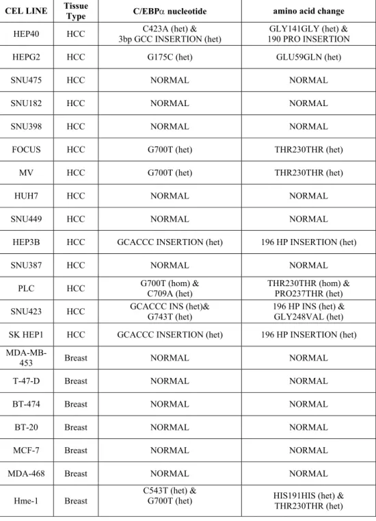
13 HCC and 7 breast cancer cell lines were sequenced and results are summarized in table 5. Three HCC cell lines, Hep 3B, SNU 423 and SK HEP1, have heterozygous 6bp insertion, which leads to insertion of histidine and proline amino acids at codon 196 (Figure 6b). In addition to this insertion, SNU 423 cell line has glycine to valine change at codon 248 (Figure 5b). Hep 40 cell line has heterozygous 3 bp insertion leading extra proline at position 190 (Figure 6a) and C to A transversion at position 423 which does not cause amino acid change (data not shown). PLC cell line has C to A transversion at nucleotide position 709, results in proline to threonine change at amino acid position 237 (Figure 5a). Hep G2 cell line is heterozygous for glutamic acid to glutamine change at 59 due to G to C substitution (Figure 5c). Three cell lines, PLC, Focus and Mahlavu, have G to T substitution at nucleotide position 700, which does not result in amino acid change (data not shown). In breast cancer cell lines, two alterations were detected. First one is the G700T polymorphism and second is the C543T transition in HME-1 cell line. This second substitution does not cause amino acid change either. Except for these two polymorphisms, no alteration was observed in breast cancer cell lines.
Sequencing were performed both by forward and reverse primers and alterations were confirmed by repeating sequencing.
Nucleotide numbering is based on Genbank sequence # NM_004364 (gi: 28872793)
Table 5: HCC and breast cancer cell lines sequencing results
CEL LINE Tissue Type C/EBPα nucleotide amino acid change
HEP40 HCC 3bp GCC INSERTION (het) C423A (het) & 190 PRO INSERTION GLY141GLY (het) &
HEPG2 HCC G175C (het) GLU59GLN (het)
SNU475 HCC NORMAL NORMAL
SNU182 HCC NORMAL NORMAL
SNU398 HCC NORMAL NORMAL
FOCUS HCC G700T (het) THR230THR (het)
MV HCC G700T (het) THR230THR (het)
HUH7 HCC NORMAL NORMAL
SNU449 HCC NORMAL NORMAL
HEP3B HCC GCACCC INSERTION (het) 196 HP INSERTION (het)
SNU387 HCC NORMAL NORMAL
PLC HCC G700T (hom) & C709A (het) THR230THR (hom) & PRO237THR (het) SNU423 HCC GCACCC INS (het)& G743T (het) 196 HP INS (het) & GLY248VAL (het)
SK HEP1 HCC GCACCC INSERTION (het) 196 HP INSERTION (het)
MDA-MB-453 Breast NORMAL NORMAL
T-47-D Breast NORMAL NORMAL
BT-474 Breast NORMAL NORMAL
BT-20 Breast NORMAL NORMAL
MCF-7 Breast NORMAL NORMAL
MDA-468 Breast NORMAL NORMAL
Hme-1 Breast
C543T (het) &
G700T (het) HIS191HIS (het) & THR230THR (het)
a.
b.
c.
Figure 5: Electrophorograms showing substitutions in HCC cell lines. a. PLC cell
line has a heterozygous substitution, C to A transversion at nucleotide position 709. b.SNU 423 cell line has heterozygous G to T transversion at nucleotide position 743. c. G175C substitution in Hep G2 cell line.
a.
b.
c.
Figure 6: Electrophorograms showing insertions in HCC cell lines. a. Heterozygous
3 bp insertion in Hep 40 cell line. b. The sites of 6 and 3 bp insertions in C/EBPα protein. c. Heterozygous 6 bp insertion in Hep 3B cell line.
+ HP +Pro
4.2 Hereroduplex analysis using PAGE
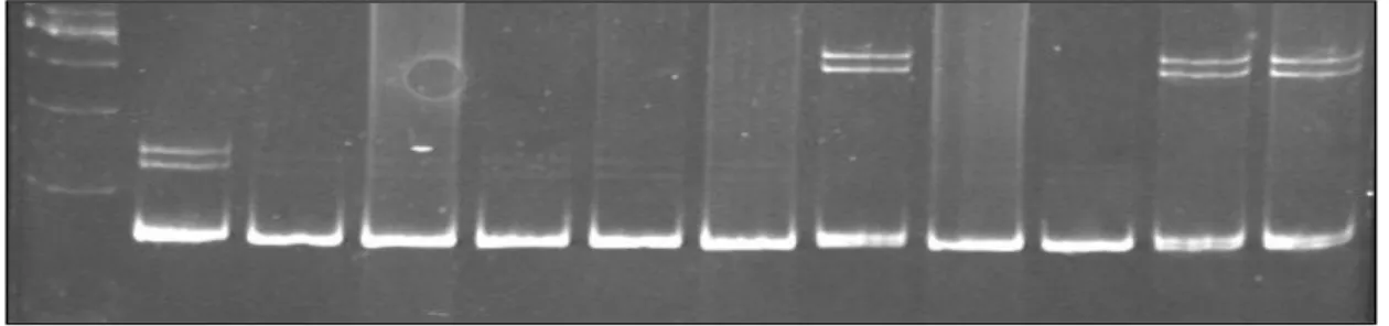
As three of the HCC cell lines have the 6 bp insertion and one cell line has 3 bp insertion, we decided to screen colorectal cancer cell lines, HCC tumors and control samples for these insertions. 167 bp fragment containing these sites were amplified and run on 12% PAGE (figure 7). One of the colorectal cancer cell lines, CCL231, has 6 bp insertion. 80 tumors were screened and twelve of them have 6 bp GCACCC insertion, whereas none of them has 3 bp GCC insertion (table 6). Among twelve tumors, two of them are homozygous for this insertion while the others are heterozygous. Normal DNA samples of six tumors having 6 bp insertion (C8N, C10N, C11N, C12N, C14N, N66) were also screened and they also posses this alteration (figure 7 and data not shown). Among 142 controls, 14 of them have the 6 bp insertion and none of them has 3 bp insertion. These results were confirmed by sequencing.
Figure 7: Polyacrylamide gel showing insertions and wild types. Lane1: Molecular
size marker Lane2: Hep 40 cell line with 3bp insertion. Lane3-7,9,10: C1,C2, T19, T21,J3, TC1 and U13T HCC tumor samples with no insertion. Lane 8: C8 tumor sample with 6 bp insertion. Lane11: C10 tumor sample with 6 bp insertion. Lane12: C10N normal sample with 6bp insertion.
Table 6: Sequencing and PAGE analysis in HCC tumors
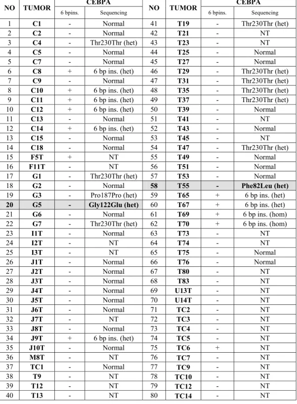
CEBPA CEBPA NO TUMOR
6 bpins. Sequencing NO TUMOR 6 bpins. Sequencing
1 C1 - Normal 41 T19 - Thr230Thr (het)
2 C2 - Normal 42 T21 - NT
3 C4 - Thr230Thr (het) 43 T23 - NT
4 C5 - Normal 44 T25 - Normal
5 C7 - Normal 45 T27 - Normal
6 C8 + 6 bp ins. (het) 46 T29 - Thr230Thr (het)
7 C9 - Normal 47 T31 - Thr230Thr (het)
8 C10 + 6 bp ins. (het) 48 T35 - Thr230Thr (het)
9 C11 + 6 bp ins. (het) 49 T37 - Thr230Thr (het)
10 C12 + 6 bp ins. (het) 50 T39 - Normal
11 C13 - Normal 51 T41 - NT
12 C14 + 6 bp ins. (het) 52 T43 - Normal
13 C15 - Normal 53 T45 - NT
14 C18 - Normal 54 T47 - Thr230Thr (het)
15 F5T + NT 55 T49 - Normal
16 F11T - NT 56 T51 - Normal
17 G1 - Thr230Thr (het) 57 T53 - Normal
18 G2 - Normal 58 T55 - Phe82Leu (het)
19 G3 - Pro187Pro (het) 59 T65 + 6 bp ins. (het)
20 G5 - Gly122Glu (het) 60 T67 + 6 bp ins. (het)
21 G6 - Normal 61 T69 + 6 bp ins. (hom)
22 G7 - Thr230Thr (het) 62 T70 + 6 bp ins. (hom)
23 I1T - Normal 63 T73 - NT 24 I2T - NT 64 T74 - NT 25 I3T - NT 65 T75 - Normal 26 J1T - Normal 66 T76 - Normal 27 J2T - Normal 67 T80 - NT 28 J3T - Normal 68 T83 - NT 29 J4T - Normal 69 U13T - NT 30 J5T - Normal 70 U14T - NT 31 J6T - Normal 71 TC2 - NT 32 J7T - NT 72 TC3 - NT 33 J8T - Normal 73 TC4 - NT 34 J9T + 6 bp ins. (het) 74 TC5 - NT 35 J10T - Normal 75 TC6 + NT 36 M8T - NT 76 TC7 - NT 37 TC1 - Normal 77 TC9 - NT 38 T9 - NT 78 TC10 - NT 39 T12 - NT 79 TC12 - NT 40 T13 - NT 80 TC14 - NT
4.3 Sequencing results in HCC tumors
50 HCC tumors were sequenced and results are shown in table 6. Tumor G5 has heterozygous G to A substitution at nucleotide position 365, which cause glycine to glutamine change at codon 122 (figure 9c). T55 tumor has heterozygous C to A substitution, which leads to phenylalanine to leucine change at codon 82 (figure 9a).This substitution is not present in normal liver tissue of this patient (figure 9b). 6 bp insertion is present in ten of the tumors and nine of the tumors have Thr230Thr polymorphism. Homozygous 6 bp insertion in T69 and T70 was confirmed by sequencing (Figure 8a).
a.
b.
Figure 8: Electrophorograms showing homozygous 6bp insertion in HCC tumor. a.
a.
c.
b.
Figure x T55 and N54
c.
Figure 9: Electrophorograms showing substitutions in HCC tumors. a. T55 HCC
tumor with heterozygous C246A substitution. b. N54 normal sample without substitution. c. G5 HCC tumor with heterozygous G365A substitution.
4.4 Restriction analysis
In order to check the presence of the substitutions, found in HCC cell lines and tumors, in normal control population, we screened 50 healthy individual bloods from Turkey by restriction analysis. G743T and G175C substitutions in Snu 423 and Hep G2 cell lines cause loss of ApaI and BssSI restriction sites respectively. For C709A alteration in PLC cell line, no restriction site has been found. G365A substitution in G5 leads to loss of SmaI restriction site and C246A in T55 creates of MaeIII restriction site. These substitutions were not present in 50 normal individuals or in additional 7 breast cancer cell lines.
Figure 10: Restriction digestion results a. MaeIII digestion of T55 HCC tumor, N54
normal tissue and control samples. b. BssSI digestion of Hep G2 cell line and control samples. c. SmaI digestion of G5 HCC tumor, G5N normal sample and control sample. d. ApaI digestion of Snu 423 cell line and control samples.
688 bp 420 bp 268 bp 688 bp 490 bp 198 bp 338 bp 301 bp 223 bp 127 bp 705 bp 452 bp 253 bp T55 N54 Ctrl 1 Ctrl 2 Ctrl 3 a. b. c. d. Hep G2 Ctrl 1 Ctrl 2 Ctrl 3 Ctrl 4 G5 G5N Ctrl 1 Snu 423 Ctrl 1 Ctrl 2 Ctrl 3
5. DISCUSSION
In liver, C/EBPα interacts with several proteins, which are important in cell cycle progression such as Cdk2, Cdk4, E2F, p21 and Brm and it is believed that the C/EBPα regulates the cell proliferation through forming complexes with these proteins (Iakova et
al., 2003). C/EBPα has a dominant antiproliferative role in hepatocytes and
overexpression of this gene results in the block of proliferation in HCC cell lines (Diehl
et al., 1996;Watkins et al., 1996). Therefore, tumor cells must find out a way for the
inactivation of C/EBPα to escape from its growth inhibitory function. C/EBPα is located on 19q13, where no LOH has been reported in HCC. Although, reduced expression of C/EBPα has been shown in HCC tumors by one group (Tomizawa et al., 2002; Tomizawa et al., 2003), other studies did not confirm these results (Wang et al., 2004). The finding showing inactivation of C/EBPα by PI3K/Akt pathway in liver tumors confirms that the antiproliferative role of C/EBPα is important in hepatocyte growth control (Wang et al., 2004). Since the mutation of C/EBPα causes AML and knockout mice have increased proliferation and liver morphology resembling hepatocellular carcinoma, we thought that the mutations in C/EBPα might be involved in hepatocellular carcinoma.
Direct automated sequencing and heteroduplex analysis methods were used for the screening of the HCC, breast and colorectal cancer cell lines and HCC tumors. Although, there are other methods for mutation detection like single strand conformation polymorphism (SSCP) since the C/EBPα is a small gene with single exon, we decided to perform sequencing to detect mutations. In addition, the size of the amplified product should not be bigger than 250bp for SSCP but C/EBPα gene is high in GC content and it is not possible to design primers in each 250bp. Moreover, SSCP can detect only 80% of all mutations. To detect the 6bp and 3bp insertion, we used heteroduplex analysis that is quicker and cheaper for the screening of large number of samples.
In HCC cell lines, the most common alteration is 6 bp insertion, which leads to insertion of histidine and proline amino acids, in the transcription activation domain 3 (TEIII). Three out of 14 HCC cell lines and one colorectal cancer cell line have this insertion but none of the breast cancer cell lines has it. Hep 40 cell line has 3 bp insertion located a few base pairs upstream of 6 bp insertion region, this 3 bp insertion is not present in any of breast and colorectal cancer cell lines. Therefore, we thought that these insertions might be specific for hepatomas and decided to screen HCC tumors for these insertions by heteroduplex analysis. The 6 bp insertion is present in 12 tumors out of 80 whereas, none of them has the 3 bp insertion. 6 bp insertion seems to be common in China (35%) and Japan (25%) but not in Africa.
To examine whether this 6 bp insertion is somatic, we analyzed the matched histologically-normal tissue from 6 patients. All 6 patients normal DNA showed 6 bp insertion indicating that this insertion is not somatic or arise early in preneoplastic phase. In 2001, Pabst et al. identified ten mutations associated with AML in C/EBPα gene. Since then, the presence of the C/EBPα mutations has been assessed in various patients but GCACCC insertion was not previously reported. We performed a database search against the EST and polymorphism databases and no such polymorphism has been identified by multiple sequencealignment. We further examined 142 control samples from Turkey and 14 of them were found to have 6 bp insertion but none of them had 3 bp insertion. Therefore, this 6 bp insertion seems to be a polymorphism that is common in China, Japan and also in Turkey. Two samples from France is homozygous for this insertion, it is possible that these samples seem homozygous due to LOH in other allele. Therefore, it would be interesting to see whether LOH is present in these two samples.
These insertions are located in a functionally important region of C/EBPα, where it interacts with the Cdk2/Cdk4 and Brm. This region was also reported as growth inhibitory region in mouse liver (Wang et al., 2001).Whether these insertions cause any defect in C/EBPα interaction with other proteins and/or in growth-inhibitory function of C/EBPα needs to be addressed by functional analysis.
Apart from the insertions, HCC cell lines and tumors have heterozygous substitutions. Snu 423 cell line has glycine to valine change at codon 248 in addition to 6bp insertion.
In this change, the size of new amino acid, valine, is much larger than glycine, which is the smallest amino acid. The "classic" oncogenic mutation in ras is also a glycine to valine change at position 12. This mutation causes ras to be held in the activate state that sends growth signals to the cell and also causes the gene to be transformed from a proto-oncogene to an proto-oncogene (Nagata et al., 1992).
In PLC, proline to threonine change is present at codon 237. This change is in the site, where the glycogen synthase kinase 3 (GSK3) and protein kinase C (PKC) phosphorylate serine and threonine residues. Therefore, this change may result in abnormal phosphorylation of C/EBPα. Phosphorylation is important for the C/EBPα activity and may affect protein-protein interactions (Behre et al., 2002; Wang et al., 2004; Ross et al., 2004).
As mentioned before there are two conserved motifs in N terminal part of the C/EBPα, box A and box B, which interact with the TBP/ TFIIB and p300/CBP (Nerlov
et al., 1995). The glutamic acid to glutamine alteration in Hep G2 cell line is in the box
A motif and phenylalanine to leucine alteration found in tumor T55 is in the box B motif. These residues are conserved between the human, rat, mouse, zebrafish and xeonopus C/EBPα and between other C/EBP members (figure 11). Moreover, the alteration in T55 is present only in the tumor but not in the normal DNA of this patient showing that, this change is somatic. It can be speculated that these changes may lead to reduced transcriptional activation due to disruption of box A and box B motifs. The
alteration in T55 tumor also disrupts the binding region of Rb like protein, p107. This may lead to increase in E2F-p107 complex and expression of DNA synthesis and mitosis related genes. The glycine (a nonpolar side chain aa.) to glutamic acid (acidic side chain aa.) change in G5 is located in the TEIII, Cdk2/Cdk4 and Brm interacting region and may affect the conformation of protein. Although the non-tumor DNA sample from the liver of same patient also display this change, lack of this alteration in control population and predicted functional importance strongly suggest its somatic nature. One possible reason for the presence of this change in normal liver tissue of the same patient is that we did not microdissect the tumor and normal cells before the DNA extraction. The inclusion of normal stromal cells in the tumor sample or tumor cells in normal stroma can easily mask the genetic changes.
In tumor sample G3, G561C substitution is present; this substitution does not change amino acid and in Hep 40 cell line, in addition to 3bp insertion there is another substitution, C423A, which does not cause amino acid change either. These polymorphisms were not reported before. Except the change in PLC, all other substitutions that leads to amino acid change are checked whether present in normal population or not and none of the controls has any of these alterations and alterations described in this study were not reported before. Thus the most of the alterations, found in our study, are likely to be somatic.
It should be noted that except the two samples with homozygous 6 bp insertion, all other alterations are heterozygous. Being the fact that, C/EBPα +/- mice does not show any abnormality, these alterations are expected to act as dominant negative mutants in order to cause hepatocellular carcinoma. Dominant-negative mutations in C/EBPα were reported in AML patients (Pabst et al., 2001b). These mutations generally result in the frameshift in the p42 form and increase the translation of p30 form. However, none of the alterations described here cause frameshift. Thus, functional studies with mutant forms of C/EBPα should be done to learn if these alterations have dominant negative effects.
The alterations that we have found are different from the mutations reported in AML patients. Most of the mutations in AML are clustered in DNA binding domain, which cause the disruption of C/EBPα dimerization and DNA binding, and N terminal region leading to expression of p30 isoform and these mutations block the differentiation in hematopoietic lineage (Nerlov, 2004). However, alterations that we described are located generally in the protein-protein interacting domain of C/EBPα. This is consistent with findings showing that, C/EBPα inhibits proliferation through protein-protein interactions in liver, unlike myeloid cells where the transcriptional activity of C/EBPα block proliferation (Wang et al., 2001;Porse et al., 2001). However, the cdk2/cdk4, Brm and p21 interacting domains are also present in p30 isoform of C/EBPα and this isoform does not cause growth arrest (Lin et al., 1993). Therefore, additional transcription activation may be required. Mutations in Hep G2 cell line and T55 HCC tumor, which are in box A and boxB motif respectively, might be important in this sense. These two alterations are present only in the p42 isoform but not in p30 isoform.
Ours is the first study showing alterations in the C/EBPα in hepatocellular carcinoma. There is one study which carried out mutation screening in only six hepatomas and they detected only the G700T polymorphism that we have also found (Gombart et al., 2002). 6 bp insertion present in HCC cell lines and HCC tumors is considered to be a polymorphism since the normal population also has it. Because the alteration in PLC was not checked in control population, we do not know now whether it is a polymorphism or not although it is not reported in polymorphism databases and not present in 50 tumors sequenced. The other alterations are not likely to be a polymorphism since we could not detect them in control population.Therefore, mutations of C/EBPα are seen in HCC (8%) with a frequency similar to that in AML (9%). The functional analysis of the alterations described here should be performed in future.
As mentioned before, in addition to mutations in C/EBPα, there are other ways in AML for the inactivation of C/EBPα like repression of C/EBPα expression by t(8:21) fusion product (Pabst et al., 2001a). Therefore, transcriptional, translational or
posttranslational mechanisms may modulate the C/EBPα protein level and function during HCC. Activation of other oncogenic pathways, such as MAPK pathway can modulate mRNA and protein levels of C/EBPα (Hemati et al., 1997). Phosphorylation and dephosphorylation may also play role in the modulation of C/EBPα function. Phosphorylation of C/EBPβ by PKC has been shown to attenuate its binding to DNA (Mahoney et al., 1992). Since there are PKC phosphorylation sites on C/EBPα, this phosphorylation may lead to similar consequences for C/EBPα. Another possible mechanism for the inhibition of the C/EBPα function could be the increase in the expression of the dominant-negative proteins such as p30 or LIP ( Liver Inhibitory Protein) which is the C/EBPβ isoform formed by alternative use of initiation codons in C/EBPβ mRNA. Therefore, the other pathways such as MAPK, mTOR or PKC can be evaluated to see their roles in regulation of C/EBPα in hepatocellular carcinoma in future.
In this study we discovered previously unidentified genetic polymorphisms in the C/EBPα geneand further confirmed their presence by population studies. Since C/EBPα plays a crucial role in cellular homeostasis, the discovery and understanding of any genetic variationin the C/EBPα gene present in the human population would be valuable. Further studies are required to determine if these genetic polymorphisms are involved in disease susceptibility. 6 bp insertion present in the HCC cell lines and tumors is frequent. Although the same insertion is found in the control population, it is possible that this insertion will cause predisposition to hepatocellular carcinoma. Therefore, large number of tumor and control samples can be screened for this insertion to check this hypothesis.