See discussions, stats, and author profiles for this publication at: https://www.researchgate.net/publication/41395658
IVIG- responsive multiple cranial neuropathy: A pharyngo-facial variant of
Guillain-Barré syndrome
Article in Acta neurologica Belgica · December 2009
Source: PubMed CITATIONS 8 READS 215 4 authors, including: Mehmet zülküf Önal LIV HOPITAL, Ankara, Turkey 27PUBLICATIONS 293CITATIONS SEE PROFILE Ersin Tan Hacettepe University 120PUBLICATIONS 1,490CITATIONS SEE PROFILE
All content following this page was uploaded by Ersin Tan on 16 May 2014.
Abstract
We report a case with symptoms of facial swelling, bilateral facial paralysis, dysphagia and aspiration. On electrophysiological studies, the right facial nerve was not excitable. The left facial nerve compound muscle action potential (CMAP) amplitude was severely dispersed and latency was mildly prolonged, consistent with demyelina-tion. Cerebrospinal fluid examinations were normal. Anti-ganglioside antibodies and tumor markers were negative. Bickerstaff brainstem encephalitis, stroke, diabetes melli-tus, vasculitis, sarcoidosis, Sjögren’s syndrome, Melkers-son-Rosenthal Syndrome, trauma, infectious diseases, toxicity, neoplasm, facial onset sensory and motor neuronopathy (FOSMN) and other degenerative diseases were excluded. Intravenous immunoglobulin therapy resolved symptoms of lower cranial nerve dysfunction. Clinically incomplete improvement of bilateral facial paralysis was observed. We conclude that IVIg therapy may improve the symptoms of multiple cranial nerve palsies due to pharyngo-facial variant of Guillain-Barré syndrome.
Key words: Bilateral facial diplegia; Guillain-Barré syndrome; dysphagia; neuropathy; intravenous immuno -globulin therapy.
Introduction
Facial diplegia is characterized by loss of volun-tary movements of bilateral facial muscles. Although unilateral facial or lower cranial nerve palsies may frequently be seen, multiple cranial neuropathies, in-volving bilateral facial and lower cranial nerves are rarely reported (Shuaib and Becker, 1987; Spillane et al., 1991). Clinical localization may be due to dis-eases affecting the supranuclear structures, subarach-noid space, skull base, nuclear lesions, cranial nerves, the neuromuscular junction or the muscle. Usually post-infectious autoimmune diseases such
as Guillain-Barré Syndrome, diabetes mellitus, stroke, vasculitis, neoplasm, trauma, infectious dis-eases, toxicity, congenital or degenerative diseases may be the cause (Weintraub, 1976; Gorman et al., 1977; Beal, 1990; Keane, 1994).
Case report
An 82 years old housewife noticed swelling and weakness on her right eyelid and the right side of her face. Within 3 days she noticed the same symptoms on the left hemiface and started to have difficulty swallowing with occasional aspiration. Her medical history disclosed hypothyroidism. She denied any infection or taking any new medication within the last month. On examination she had facial swelling, facial drooping, bilateral ptosis and tongue was fis-sured. The patient was alert and oriented. Bilateral facial paresis prominent on the right and weakness of bilateral cranial nerves IX and X, with decreased gag reflex and dysarthria was noted. There was no sensory or motor deficit in her extremities. Deep ten-don reflexes were normal. There was no pathological reflex. Brain MRI revealed multiple, non-specific, ischemic-gliotic lesions in the periventricular white matter. On electrophysiological studies, sensory potentials were absent in both sural nerves. Sensory nerve conduction velocities were slow in median and ulnar nerves (Table 1a). Distal motor latencies were prolonged and motor nerve conduction velocities were slow. The compound muscle action potential (CMAP) amplitude was low in the right peroneal nerve. The right facial nerve was inexcitable. CMAP amplitude was low in the left facial nerve (Table 1b). The left facial nerve CMAP amplitude was severely dispersed and latency was mildly prolonged (Fig. 1). There was total denervation of the right facial mus-cles and partial denervation of the left facial musmus-cles.
IVIG- Responsive Multiple Cranial Neuropathy: a pharyngo-facial variant of
Guillain-Barré Syndrome
Isin UNAL-CEVIK1, Mehmet Zulkuf ONAL1, Zeki ODABASI2and Ersin TAN3
1Ufuk University, Faculty of Medicine, Department of Neurology, Ankara; 2Gulhane Military Medical Academy, Department of Neurology, Ankara and 3Hacettepe University, Faculty of Medicine, Department of Neurology, Ankara, Turkey
318 I. UNAL-CEVIK ET AL.
There was chronic partial denervation of the right upper extremity distal muscles (Table 1c). Cerebro -spinal fluid studies, performed 3 weeks after symp-tom onset, were normal. Routine blood tests, ESR, CRP, thyroid function tests, Vit B12, folic acid levels,
blood ACE and urine calcium level were normal. Thorax CT was negative for sarcoidosis. Vasculitis markers, tumor markers, VDRL, HIV, hepatitis, Lyme, Brucella, ganglioside antibodies of anti-GM1, anti-GQ1b and anti-GD1b were negative. Skin
Table 1a
Sensory Nerve Conduction Studies
Nerve/Sites SNAP (µV) Normal values CV Normal values
(antidromic SNAP-µV) m/s (CV-m/s)
R MEDIAN Digit II-Wrist 15.6 20 34.3 41.3
L MEDIAN-Digit II-Wrist 15.1 20 32.1 41.3
R ULNAR-Digit V-Wrist 4.1 15 30.1 39.3
L ULNAR-Digit V-Wrist 10.4 15 30.6 39.3
R SURAL - Lat Malleolus-calf NP 8 34.7
L SURAL - Lat Malleolus-calf NP 8 34.7
CV (m/s): conduction velocity (meter/second), SNAP (µV): sensory nerve action potential (microvolt), NP: no potential, R: right, L: left.
Table 1b
Motor Nerve Conduction Studies
CMAP (mV): compound muscle action potential (millivolt), CV (m/s): conduction velocity (meter/second), NP: no potential, R: right, L: left. Normal values (Odabasi et al., 1996).
Nerve/Sites Latency (ms) CMAP (mV) CV m/s Normal values (Latency-ms) Normal values (CMAP-mV) Normal values (CV-m/s) R MEDIAN- ABP Wrist-Elbow 4.8 3.7 37 3.6 5 50 L MEDIAN- ABP Wrist-Elbow 5.35 4.9 43.1 3.6 5 50 R ULNAR- ADM Wrist- Elbow 4.15 5.9 46.0 2.5 5 50.6 L ULNAR- ADM Wrist- Elbow 3.55 7.6 42.6 2.5 5 50.6 R FACIAL-frontalis 1. Ant Ear NP 3.1 2.2 L FACIAL-frontalis 1. Ant Ear 5.15 1.3 3.1 2.2
R FACIAL - Orb Oculi
1. Ant Ear NP 3.1 2.2
L FACIAL - Orb Oculi
1. Ant Ear 3.45 0.8 3.1 2.2
R FACIAL-orb oris
1. Ant Ear NP
L FACIAL-orb oris
1. Ant Ear 4.5 0.7 2.7 3.5
R COMMON PERONEAL - EDB
biopsy from right inferolateral angle of the orbit, revealed actinic (solar) keratosis, without any sign of specific inflammation, neoplasm nor granulo-matosis.
The patient was fed enterally through a naso -gastric tube. Due to acute onset of symptoms accompanied by demyelination, without any
identi-fiable cause, an immune-mediated process was con-sidered. A five day course of intravenous immun-globulin (IVIg) 0.4 g/kg/day was given. Two weeks later, she was able to eat foods and drink liquids without aspiration. Her facial diplegia partially responded to IVIg treatment. She was stable for the next 12 months.
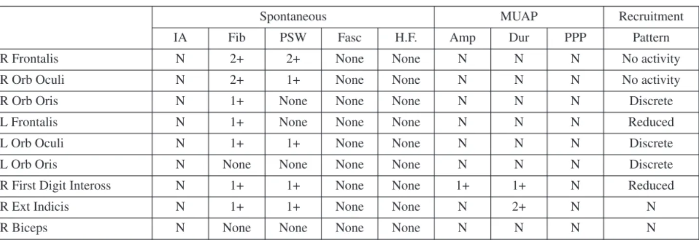
Table 1c Needle EMG
IA: Insertional Activity, Fib: Fibrillation, PSW: positive sharp wave, Fasc: Fasciculation, H.F.: High frequency, MUAP: Motor unit action potential, Amp: Amplitude, Dur: duration, PPP: polyphasic potential, R: right, L: left.
Spontaneous MUAP Recruitment
IA Fib PSW Fasc H.F. Amp Dur PPP Pattern
R Frontalis N 2+ 2+ None None N N N No activity
R Orb Oculi N 2+ 1+ None None N N N No activity
R Orb Oris N 1+ None None None N N N Discrete
L Frontalis N 1+ None None None N N N Reduced
L Orb Oculi N 1+ 1+ None None N N N Discrete
L Orb Oris N None None None None N N N Discrete
R First Digit Inteross N 1+ 1+ None None 1+ 1+ N Reduced
R Ext Indicis N 1+ 1+ None None N 2+ N N
R Biceps N None None None None N N N N
FIG. 1. — After stimulation of the left facial nerve, compound muscle action potential (CMAP) recorded from left orbicularis oculi
amplitude is severely dispersed and latency was mildly prolonged, consistent with demyelinization. (Scale of time: 2 milliseconds and amplitude: 1 milliVolt).
320 I. UNAL-CEVIK ET AL. Discussion
Stroke, vasculitis, glial tumors, metastasis, infec-tions, trauma, demyelinization, and degenerative dis-ease may produce clinical signs of supranuclear, nuclear or infranuclear involvement. Foix-Chavany-Marie (bi-opercular) syndrome is characterized by faciopharyngoglossomasticatory diplegia (Mariani et al., 1980). Brain MRI of our patient did not dis-play a lesion responsible for the clinical picture. GBS is an acutely evolving areflexic, demyelinating neuropathy with albuminocytologic dissociation. Variants of GBS may include asymmetric, pure motor, prominent sensory, preserved muscle strech reflexes, pharyngeal-cervical-brachial, paraparetic, facial diplegia with paresthesia, pure sensory neu-ropathy, pure autonomic neuneu-ropathy, Miller-Fisher syndrome, axon loss variants, acute motor axonal neuropathy (AMAN) and acute motor and sensory axonal neuropathy (AMSAN) (Shuaib and Becker, 1987; Sinsawaiwong and Thampanitchawong, 2000; Levin, 2004; Curtis et al., 2008). Isolated cranial nerve involvement without prominent signs of GBS is considered as rare variant of this disease (Dididze, 2009). The cranial, bulbar and sensory variants are usually associated with antibodies to the disialylated gangliosides (Willison and Yuki, 2002; Mas-Lazaro et al., 2008). Bickerstaff’s brainstem encephalitis causes motor axonal neuropathy of facial and bulbar muscles, consciousness disturbances, ophtalmople-gia and ataxia (Odaka et al., 2003). Serum anti-GQ1b IgG is usually positive. Our patient had negative antiganglioside antibodies. CSF studies were normal. Contrast-enhanced brain MRI dis-closed no sign of inflammation or neoplasm. We excluded the diagnosis of Bickerstaff’s brainstem encephalitis , paraneoplastic diseases, diabetes mel-litus, vasculitis and infectious diseases.
Melkersson-Rosenthal Syndrome causes facial swelling and facial palsy(Mukherjee and Dongre, 1973). Tongue is often fissured. An unusual case pre-senting with facial, glossopharyngeal and vagal nerve palsies is reported (Khandpur et al., 2006). Our patient’s skin biopsy did not support the diag-nosis.
Facial onset sensory and motor neuronopathy (FOSMN) is characterized by paraesthesia and numbness in trigeminal distribution and slowly pro-gressive weakness of head, neck and upper extrem-ities as if a syringomyelia-like syndrome(Vucic et al., 2006; Hokonohara et al., 2008). The presenting feature of all cases was hypoesthesia and paraesthe-sia in the lower face and mouth. Within 2-6 years, the sensory symptoms progressed to involve entire face, scalp, upper extremities, upper back and chest.
Dysphagia was reported in half of the patients after 5 years of symptom onset. Facial nerve palsy of the lower motor neuron type was seen in one patient. Neurophysiologically generalized sensorymotor neuronopathy of caudally decreasing severity was demonstrated. All patients were treated with im-munosuppressive or immunomodulatory therapy without any benefit. This syndrome is considered to be a slowly progressive neurodegenerative disorder (Vucic et al., 2006). Interestingly, Hokonohara et al. reported a similar case who partially responded to immunotherapy(Hokonohara et al., 2008). Responses to immunotherapies may be best ex-plained by an immunological mechanism resembling chronic inflammatory demyelinating polyneuropathy (Kokubun and Hirata, 2007; Hokonohara et al., 2008). Our patient only had acute onset of multiple lower cranial nerve palsies without any symptom in the trigeminal nerve, scalp, trunk or upper extremi-ties resembling syringomyelia.
Therapeutic approach in cranial neuropathies is based on the underlying disease. Facial diplegia may affect patient’s appearance and has a risk of corneal injury. Bilateral lower cranial nerve dysfunction has the increased risk of aspiration pneumonia, dehydra-tion, electrolite imbalance and malnutrition. IVIg treatment is a good choice for immune-mediated neuropathies(Nobile-Orazio and Terenghi, 2005). Based on electrophysiological studies and appropri-ate differential diagnosis, our patient has been treappropri-ated early with IVIg. We believe that this patient had a pharyngo-facial variant of Guillain-Barré syndrome who responded to IVIg treatment.
REFERENCES
Beal MF. Multiple cranial-nerve palsies:a diagnostic challenge. N Engl J Med. 1990;322(7):461-463. Curtis CE, Barnes EV, Dupiche CA. Guillain-Barre
syn-drome variant:presenting with myalgias and acute facial diplegia. Mil Med. 2008;173(5):507-508. Dididze MN. Clinical variants of Guillain-Barre
syndrome:some aspects of differential diagnosis. Georgian Med News. 2009;(166):48-51.
Gorman JB, Nagler B, Botton JE. Facial diplegia and its causes. Va Med. 1977;104(8):544-546.
Hokonohara T, Shigeto H, Kawano Y, Ohyagi Y, Uehara M, Kira J. Facial onset sensory and motor neuronopathy (FOSMN) syndrome responding to immunotherapies. J Neurol Sci. 2008;275(1-2): 157-158.
Keane JR. Bilateral seventh nerve palsy:analysis of 43 cases and review of the literature. Neurology. 1994;44(7):1198-1202.
Khandpur S, Malhotra AK, Khanna N. Melkersson-Rosenthal syndrome with diffuse facial swelling
and multiple cranial nerve palsies. J Dermatol. 2006;33(6):411-414.
Kokubun N, Hirata K. Neurophysiological evaluation of trigeminal and facial nerves in patients with chronic inflammatory demyelinating polyneuropathy. Muscle Nerve. 2007;35(2):203-207.
Levin KH. Variants and mimics of Guillain Barre Syndrome. Neurologist. 2004;10(2):61-74. Mariani C, Spinnler H, Sterzi R, Vallar G. Bilateral
peri-sylvian softenings: bilateral anterior opercular syn-drome (Foix-Chavany-Marie synsyn-drome). J Neurol. 1980;223(4):269-284.
Mas-Lazaro C, Garcia-Pastor A, az-Insa S, Molto-Jorda JM, Lacruz-Ballester L.
cervical-brachial variant of Guillain-Barre
syndrome: a well-defined clinical condition with a heterogeneous immunological profile]. Rev Neurol. 2008;47(11):579-581.
Mukherjee PK, Dongre RC. A case of acquired facial diplegia, macular oedema and lingua plicata (“Melkersson-Rosenthal syndrome”). Indian J Ophthalmol. 1973;21(1):36-39.
Nobile-Orazio E, Terenghi F. IVIg in idiopathic autoim-mune neuropathies:analysis in the light of the latest results. J Neurol. 2005;252 Suppl 1:I7-13. Odabasi Z, Joy JL, Claussen GC, Herrera GA, Oh SJ.
Isaacs’ syndrome associated with chronic inflam-matory demyelinating polyneuropathy. Muscle Nerve. 1996;19(2):210-215.
Odaka M, Yuki N, Yamada M, Koga M, Takemi T, Hirata K, Kuwabara S. Bickerstaff’s brainstem
en-cephalitis:clinical features of 62 cases and a sub-group associated with Guillain-Barre syndrome. Brain. 2003;126(Pt 10):2279-2290.
Shuaib A, Becker WJ. Variants of Guillain-Barre syn-drome: Miller Fisher syndrome, facial diplegia and multiple cranial nerve palsies. Can J Neurol Sci. 1987;14(4):611-616.
Sinsawaiwong S, Thampanitchawong P. Guillain - Barre’ syndrome following recombinant hepatitis B vac-cine and literature review. J Med Assoc Thai. 2000;83(9):1124-1126.
Spillane L, Roberts JR, Meyer AE. Multiple cranial nerve deficits after ethylene glycol poisoning. Ann Emerg Med. 1991;20(2):208-210.
Vucic S, Tian D, Chong PS, Cudkowicz ME, Hedley-Whyte ET, Cros D. Facial onset sensory and motor neuronopathy (FOSMN syndrome):a novel syn-drome in neurology. Brain. 2006;129(Pt 12):3384-3390.
Weintraub MI. Facial diplegia. Arch Otolaryngol. 1976; 102(5):311-312.
Willison HJ, Yuki N. Peripheral neuropathies and glycolipid antibodies. Brain. 2002;125(Pt 12): 2591-2625.
Assoc. Prof. Dr. Isin Unal-Cevik, M.D., Ph.D., Ufuk University, Faculty of Medicine, Department of Neurology, Balgat, 06520-Ankara (Turkey) E-mail: [email protected]
View publication stats View publication stats