Turk J Gastroenterol2014; 25: 118-9
Diagnostic usefulness of technetium-99m-pertechnetate SPECT in a
patient with Meckel’s diverticulum
To the Editor,
Meckel’s diverticulum (MD) is the most common con-genital anomaly of the gastrointestinal tract. It is en-countered in 2-3% of the population, and the lifetime complication rate is 2-4% (1). Bleeding from MD is rare in adulthood (2).
We present an adult case with MD with lower gas-trointestinal bleeding and diagnosed by techne-tium-99m-pertechnetate single-photon emission com-puted tomography (SPECT).
A 20-year-old male presented with sudden onset pain-less hematochezia. Complete blood count revealed
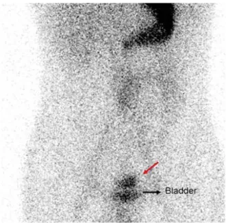
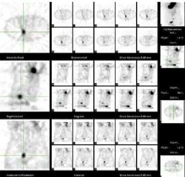
anemia (hemoglobin: 7 gr/dL). Upper gastrointestinal endoscopy was normal. Colonoscopy revealed that the cecum and ascending colon were smeared with bright red blood, without any active bleeding. MD was suspected and technetium-99 m-pertechnetate scin-tigraphy was performed. Dynamic images revealed posteriorly localized focal increased activity superior to the urinary bladder (Figure 1). Postmicture posteri-or static images better visualized this focus (Figure 2). SPECT imaging acquired for accurate anatomic local-ization demonstrated focal uptake posterior and su-perior to the urinary bladder (Figure 3). Surgical explo-ration revealed MD located 60 centimeters proximal to the terminal ileum. Histopathologic examination confirmed MD.
Adress for Correspondence: Gülbanu Erkan, Department of Gastroenterology, Ufuk University Faculty of Medicine, Ankara, Turkey E-mail: [email protected]
Received: 31.5.2012 Accepted: 22.7.2012
© Copyright 2014 by The Turkish Society of Gastroenterology • Available online at www.turkjgastroenterol.org • DOI: 10.5152/tjg.2014.3705
Figure 1. Tc-99 m pertechnetate Meckel scintigraphy. Dynamic images show posteriorly localized focal increased activity supe-rior of the bladder (red arrow). It is thought to be in relation with the bladder (ureter?).
Figure 2. Tc-99 m pertechnetate Meckel scintigraphy. Postmic-ture, posterior static images show posteriorly localized focal in-creased activity, superior of the bladder (black arrow). It is still suspicious but primarily thought to be ectopic gastric mucosa (Meckel’s diverticulum, red arrow).
118
Let
In the diagnosis of MD, ultrasound, x-ray, and computed to-mography display non-specific findings unless there is obstruc-tion or intussuscepobstruc-tion (2). While technetium-99 m pertech-netate scans detect ectopic gastric mucosa in MD in 90% of pediatric cases, this ratio is 46% in adults due to less gastric mu-cosa in the diverticulum (3). Traditional planar images in tech-netium-99 m-pertechnetate scintigraphy may be diagnostic in the appropriate clinical setting. When the index of suspicion for MD is high and planar imaging is negative or ambiguous, SPECT images may be helpful (4,5). Mesenteric angiography, capsule endoscopy, and double-balloon enteroscopy may also be beneficial, but they are not suitable in the hemodynamically unstable patient with active bleeding (2).
Bleeding from MD is unusual in adult patients (2); so, clinical diagnosis depends on a high index of suspicion. Therefore, all patients who present with massive, painless lower gastrointes-tinal bleeding without risk factors for bleeding and in whom endoscopy and colonoscopy fail to localize a bleeding site, MD should be suspected and scintigraphic evaluation should be performed. Traditional planar images in technetium-99
m-pertechnetate scintigraphy are diagnostic for MD in most of the cases. If planar imaging is equivocal or ambiguous, SPECT imaging may be beneficial for diagnosis and precise localiza-tion of MD. A positive SPECT study can aid in planning of the surgical procedure. Diagnostic procedures must be performed promptly, followed by surgical resection, in order to avoid com-plications and mortality.
Ethics Committee Approval: N/A. Informed Consent: N/A.
Peer-review: Externally peer-reviewed.
Author contributions: Supervision - B.D.; Materials - G.E., E.O., S.O., O.A.; Data Collection&/or Processing - A.C.; Analysis&/or Interpretation - G.E, E.O, O.A.; Literature Search - G.E.; Writing - G.E.; Critical Reviews - O.A., H.D., E.O., D.T., M.C., S.O.
Conflict of Interest: No conflict of interest was declared by the au-thors.
Financial Disclosure: The authors declared that this study has re-ceived no financial support.
Gülbanu Erkan1, Mehmet Çoban1, Aysun Çalışkan1, Duygu Tokbay2, Bülent Değertekin1, Süleyman Özdemir3, Emel Öztürk2,
Handan Doğan5, Ömür Ataoğlu4
1Department of Gastroenterology, Ufuk University Faculty of Medicine, Ankara, Turkey 2Department of Nuclear Medicine, Ufuk University Faculty of Medicine, Ankara, Turkey 3Department of General Surgery, Ufuk University Faculty of Medicine, Ankara, Turkey 4Micro-Pat, Pathology Center, Ankara, Turkey
5 Department of Pathology, Ufuk University Faculty of Medicine, Ankara, Turkey
REFERENCES
1. Levy AD, Hobbs CM. From the archives of the AFIP. Meckel diver-ticulum: radiologic features with pathologic correlation.
Radio-graphics 2004; 24: 565-587. [CrossRef]
2. Yang JF, Sun LM, Wang XF, Dai N. Massive gastrointestinal bleed-ing from Meckel diverticulum with ectopic pancreatic tissue. Chin Med J (Eng) 2011; 124: 631-33.
3. Kiratli PO, Aksoy T, Bozkurt MF, Orhan D. Detection of ectopic gas-tric mucosa using 99mTc pertechnetate: review of the literature.
Ann Nucl Med 2009; 23: 97-105. [CrossRef]
4. Dillman JR, Wong KK, Brown RK, Frey KA, Strouse PJ. Utility of SPECT/CT with Meckel’s scintigraphy. Ann Nucl Med 2009; 23:
813-5. [CrossRef]
5. Connolly LP, Treves ST, Bozorgi F, O’Connor SC. Meckel’s diverticu-lum: demonstration of heterotopic gastric mucosa with techne-tium-99m-pertechnetate SPECT. J Nucl Med 1998; 39: 1458-60. Figure 3. SPECT imaging demonstrated focal uptake in the right
lower quadrant, localized posterior and superior to the urinary bladder.
119
Erkan et al. Diagnostic usefulness of spect in meckel diverticulumTurk J Gastroenterol 2014; 25: 118-9
Let