R E S E A R C H A R T I C L E
Open Access
Arthroscopic treatment of focal
osteochondral lesions of the first
metatarsophalangeal joint
Ersin Kuyucu
1,4*, Harun Mutlu
2, Serhat Mutlu
3, Baris Gülenç
1and Mehmet Erdil
1Abstract
Background: Although arthroscopic surgical treatment of the first metatarsophalangeal (MTP) joint involves painful sesamoid excision, synovectomy, debridement, and partial cheilectomy, no gold standard treatment technique has been defined in the literature for hallux rigidus and focal osteochondral lesions. This study aimed to assess the arthroscopic treatment for early-grade focal osteochondral lesions of the first MTP joint and to determine the impact of arthroscopic microhole drill surgery on foot function and activities of daily living in a group of patients who failed conservative treatment.
Methods: This prospective study included 14 patients with hallux rigidus and focal osteochondral lesions of the first MTP joint who underwent surgery in 2014 and were followed on a regular basis thereafter.
Results: The patients had mean preoperative VPS (visual pain scale) and AOFAS (American Orthopedic Foot and Ankle Society)-Hallux scores of 8.14 ± 0.86 SD and 48.64 ± 4.27, respectively; the corresponding postoperative values of both scores were 1.86 ± 0.66 SD and 87.00 ± 3.70, respectively. Both VPS and AOFAS-Hallux scores changed significantly.
Discussion: In this prospective study, we explored the impact of arthroscopic microhole drill surgery on foot function and activities of daily living in patients with focal osteochondral lesions of the first MTP joint. Our results showed significant improvements in VPS and AOFAS scores with this treatment.
Conclusions: An arthroscopic microhole drill technique can be used with impressive functional scores and without any complications in patients who failed conservative therapy for hallux rigidus with focal chondral injury.
Keywords: Metatarsophalangeal joint, Hallux rigidus, Arthroscopy Background
First defined by Watanabe in 1972 [1, 2], arthroscopic treatment of the first metatarsophalangeal (MTP) joint was later detailed by Bartlett in 1988 [3]. Thanks to ad-vancements in arthroscopic techniques and technology, arthroscopy of the first MTP joint is now used for both diagnosis and treatment of a variety of clinical condi-tions, such as hallux valgus, gout, and hallux rigidus [4]. Although arthroscopic surgical treatment of the first MTP joint involves painful sesamoid excision, synovectomy, de-bridement, and partial cheilectomy, no gold standard
treatment technique has been defined in the literature for hallux rigidus and focal osteochondral lesions.
This study aimed to assess arthroscopic treatment, 1 of the surgical treatment options for early-grade focal osteochondral lesions of the first MTP joint, and deter-mine the impact of arthroscopic microhole drill surgery on foot function and activities of daily living in a group of patients who failed conservative treatment.
Methods
This prospective study included 14 patients with hallux rigidus and focal osteochondral lesions of the first MTP joint who underwent surgery in 2014 and were followed on a regular basis thereafter. An initial recommendation for conservative treatment composed of footwear modification,
* Correspondence:[email protected]
1Orthopedics and Traumatology, Istanbul Medipol University, Istanbul, Turkey 4TEM Avrupa Otoyolu Göztepe Çıkışı No:1, Bağcilar, Istanbul, Turkey
Full list of author information is available at the end of the article
© The Author(s). 2017 Open Access This article is distributed under the terms of the Creative Commons Attribution 4.0 International License (http://creativecommons.org/licenses/by/4.0/), which permits unrestricted use, distribution, and reproduction in any medium, provided you give appropriate credit to the original author(s) and the source, provide a link to the Creative Commons license, and indicate if changes were made. The Creative Commons Public Domain Dedication waiver (http://creativecommons.org/publicdomain/zero/1.0/) applies to the data made available in this article, unless otherwise stated.
analgesic use, and physical therapy for at least 6 months was offered to all patients. Patients who failed this conser-vative therapy, had pain, showed full-thickness cartilage in-jury on magnetic resonance imaging (MRI), and were followed on a regular basis participated in the study after providing written informed consent. Patients with Coughlin-Shurnas grade 4 hallux rigidus, osteochondral kissing lesions, or an indication for osteotomy and/or chei-lectomy apart from arthroscopy were excluded, as were those who did not attend regular follow-up visits. Patients were also excluded if they underwent any foot operation or had another foot deformity such as flat foot, excessive foot pronation, or moderate or severe hallux valgus. All patients were operated on by surgeons (ME, EK) experienced in their field. The mean postoperative follow-up period was 16.43 ± 1.86 SD months. Eight (57.1%) patients were female and 6 (42.9%) were male. The median age of the patients was 44.07 years (range, 38–49).
The Coughlin and Shurnas classification [5] was used to determine the hallux rigidus grade; the Outerbridge classification [6] was used to grade cartilage lesions. Foot function before and after surgery was assessed by the American Orthopedic Foot and Ankle Society (AOFAS) score [7]. The visual pain scale (VPS) was used for rating pain [8]. All patients were informed about the study and provided written informed consent.
Statistical analysis
Statistical analyses were done with the NCSS 2007 (Number Cruncher Statistical System, Kaysville, Utah, USA) software package. Descriptive statistics included mean, standard deviation, median, frequency, ratio, minimum, and maximum. Intra-group comparison of non-normal distribution parameters was done with the Friedman test, and post hoc paired comparisons were done with the Wilcoxon signed-rank test. A p value of less than 0.01 was considered statistically significant.
Surgical technique
Patients were administered spinal anesthesia and placed in a supine position. An arthroscopic intra-articular water pressure system with pump was considered suffi-cient for hemostasis, and thus, no pneumatic tourniquet was used. A noninvasive joint distraction technique was applied. Before marking arthroscope entry points, 2– 3 cc isotonic saline was administered intra-articularly to ensure capsule retention. Standard dorsolateral and dor-somedial portals were established 2–4 mm medial and lateral to the extensor hallucis longus tendon, and no additional portals were required. First, the dorsomedial portal site at the level of the joint was determined with the help of a needle. Following skin incision using a no. 15 scalpel, the joint was accessed by blunt soft tissue dis-section using a hemostat and it was visualized with a
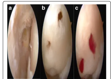
2.0-mm 30° oblique arthroscope (Fig. 1). The dorsolat-eral portal was prepared with the same technique, and the arthroscope and manual devices were alternatively used through both portals. A 2.0 shaver, probe, and a straight punch were used as the manual devices. A syno-vectomy was done first to have a better view of the sur-gical field (Figs. 1 and 2). Cartilage was examined with the probe, and the joint was irrigated with abundant iso-tonic saline. After measuring the lesion size, the cartilage was intervened using the microhole drill method (Figs. 3 and 4). After ensuring adequate bleeding, the procedure was terminated, and the entry portals were sutured with 3/0 rapid suture.
Postoperative joint loading was permitted to the max-imum tolerated point. Active and passive joint move-ments were not restricted on the first postoperative day. All patients were discharged 1 day after surgery. Control examinations were performed at 3 and 6 weeks and at 3, 6, 12, and 18 months.
Results
This study included 27 patients with hallux rigidus and osteochondral injury of the first MTP joint who under-went arthroscopic surgical treatment of the first MTP joint (Fig. 5). Five patients had Coughlin-Shurnas grade 4 hallux rigidus and were excluded from the study, 5 pa-tients were excluded due to having an osteochondral kis-sing lesion, and 3 patients were excluded for not having attended regular follow-up after the third month. After excluding the above patients, the study was completed with 14 patients. Six patients were male and 8 were fe-male. The mean age was 44.07 ± 3.40 years (Table 1).
Fig. 1 a First view of the MTP joint by the 30° scope. The blue arrow is the metatarsal head. The red arrow is the proximal phalangeal joint surface. b Synovectomy and debridement of the joint by the shaver
The mean follow-up duration was 16.43 ± 1.86 months. All patients were operated on by two experienced sur-geons (ME, EK).
The mean hallux valgus angle was 13.29° ± 1.93 SD, and the mean intermetatarsal angle was 9.14° ± 0.86 SD. Apart from joint arthroscopy, no soft tissue procedure or any procedure requiring osteotomy was done on any patient. The median operative duration was 27.8 min (range, 19– 56). While 9 patients had Coughlin-Shurnas grade 2 hallux rigidus with moderate pain and joint flattening affecting less than 50% of the joint, 5 patients had grade 3 hallux rigidus with severe pain and joint stiffness (Table 2). Arthroscopically, all patients had Outerbridge grade 4 full-thickness cartilage injury. Only 1 patient had diabetes mel-litus controlled with oral antidiabetic medication.
The patients had mean preoperative VPS and AOFAS-Hallux scores of 8.14 ± 0.86 SD and 48.64 ± 4.27, respect-ively; the corresponding postoperative values of both
scores were 1.86 ± 0.66 SD and 87.00 ± 3.70, respectively. Both VPS and AOFAS-Hallux scores changed statisti-cally significantly (p < 0.01) (Tables 3 and 4).
None of the patients developed postoperative complications.
Discussion
In this prospective study, we explored the impact of arthroscopic microhole drill surgery on foot functions and activities of daily living in patients with focal osteo-chondral lesions of the first MTP joint. Our results indi-cated significant improvements in VPS and AOFAS scores with this treatment.
Hallux rigidus is the most common first-line pathology of the foot after hallux valgus [9]. This condition affects people at a younger age than hallux valgus, and the primary complaints are pain and movement limitations [9, 10]. Pain is usually the first symptom in the initial stages of hallux
Fig. 5 MRI of the 39-year-old man right foot. Sagittal and axial view of the MTP joint
Fig. 4 a Chondral defect. b Chondral surface after the microhole drill. c Migration of the blood from the tunnels
Fig. 3 a Chondral surface after the microhole drill. b Migration of the blood from the tunnels
Fig. 2 a Curettage of the chondral lesion. b Microhole drill of the chondral lesion
rigidus. At this stage, conservative therapy consisting of nonsteroid anti-inflammatory drug (NSAID) use and footwear modification aims to suppress synovitis and joint inflammation and reduce pain [10–12]. However, surgical treatment may be preferred when conservative therapy fails in these young and active patients [11–14]. We first applied conservative treatment in the whole patient group, and we proceeded with surgery when that treatment failed. Hallux valgus can be managed via open or arthroscopic surgery, depending on patient characteristics and disease stage [14, 15]. Derner and Aldo showed that arthroscopic surgery was more bene-ficial than open surgery by being minimally invasive, having a shorter recovery period, requiring no special rehabilitation program, and allowing patients to return to daily activities quickly [16–18]. We achieved fast re-turn to work with the arthroscopic approach in suitable patients since this approach caused no complications and allowed joint loading with as much weight as toler-ated on the same day of surgery as mentioned in the lit-erature [16, 17]. Pain reduction is another important marker for treatment. Our study demonstrated signifi-cantly lowered VPS scores at both first month follow-up and the last control visit compared to the preoperative period in a homogenous patient population involving only patients with hallux rigidus (Table 4).
Arthroscopic treatment of the MTP joint is used alone or in conjunction with metatarsal osteotomies for the treatment of a wide array of conditions such as synovitis, hallux rigidus, gout, and degenerative hallux valgus [16]. However, there is still insufficient information about the principles of the evaluation and treatment of these pa-tients. Whereas 3 patients in Ahn’s study [17] and 5 pa-tients in the series published by van Dijk [18] had this condition, no study to date has specifically addressed it. Moreover, there is no study that addresses patients hav-ing hallux rigidus with only focal osteochondral lesions.
Satisfactory improvement of hallux rigidus has been reported with arthroscopic cheilectomy and debridement without a need for revision [19]. However, the goal of this technique is to remove and clear only the injured part that restricts motion, but not to treat other intra-articular pathologies. As the potential for spontaneous healing is slim in the case of chondral injury, treatment with either the microfracture or microhole drill niques should be considered. The microhole drill tech-nique we used in this study is based on the principle of opening 4–6-mm-long tunnels to enable stem cells to migrate to the injured area and achieve healing with dif-ferentiation in full-thickness chondral injuries with ex-posed subchondral bone [20, 21]. As it was previously reported that the thickness and quality of the newly formed cartilage are better with the microhole drill tech-nique than with the microfracture techtech-nique [21], we chose the former technique. The most notable indication of the success of this surgical technique was the ability of the patient to use his/her foot with comfort in daily activities; likewise, we demonstrated significant increases in AOFAS scores following surgery.
Postoperative complications are another problem. While a secondary surgery may be required after open surgeries, especially when cheilectomy is selected [22], complications necessitating additional surgery may occur at a rate of 6% after arthroplasty or arthrodesis [23]. Lin and Murphy’s trial reporting clinical-radiological pro-gression and numbness in the first web space at a rate as high as 40% after cheilectomy in a patient group with hallux rigidus indicates the fact that surgical procedures that appear simple may not be as innocuous as perceived [24]. No minor or major complications occurred at early
Table 2 Hallux lesional evaluations
Grade Number Coughlin and Shurnas Classification Grade 2 9 Intermetatarsal angle Grade 3 5
Table 3 AOFAS scores oof the patients
Preoperative Postoperative Difference pb AOFAS Mean±SD 48.64±4.27 87.00±3.70 38.35±6.48 0.001**
Min/Max (Median)
43-59 (49) 80-95 (85)
b
Wilcoxon Signed Ranks *p<0.01
Table 1 The demographic properties of the study subjects
Min-Max (Median) Mean±SD Age (years) 38-49 (45) 44.07±3.40 Follow-up period (months) 13-18 (17.5) 16.43±1.86
n %
Sex Male 6 42.9 Female 8 57.1
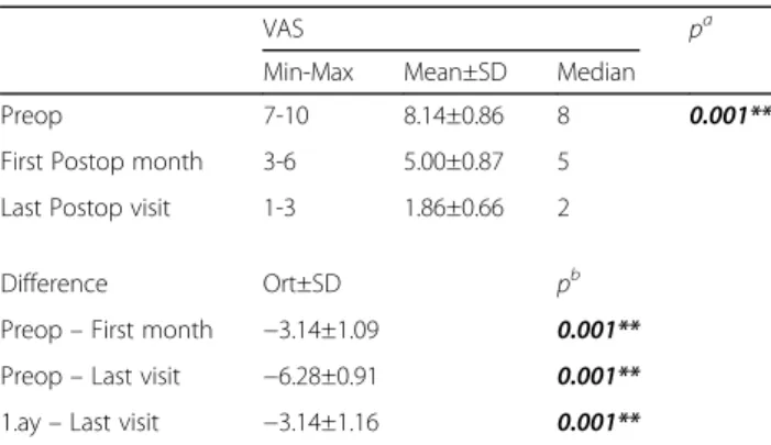
Table 4 Comparison of VAS Scores
VAS pa
Min-Max Mean±SD Median
Preop 7-10 8.14±0.86 8 0.001** First Postop month 3-6 5.00±0.87 5
Last Postop visit 1-3 1.86±0.66 2 Difference Ort±SD pb Preop– First month −3.14±1.09 0.001** Preop– Last visit −6.28±0.91 0.001** 1.ay– Last visit −3.14±1.16 0.001**
a
or late periods with our arthroscopic microhole drill technique; rather, there occurred a rapid relief of pain and a quick return to daily activities.
The strongest aspects of our study are its prospective cohort study design and a homogenous study sample. Our most notable limitations, on the other hand, are the lack of a comparison group with hallux rigidus managed by either open surgery or conservative therapy, as well as a short follow-up period. Another limitation of our study is its small sample size.
Conclusions
In conclusion, an arthroscopic microhole drill technique can be applied with impressive functional scores and with-out any complications in persons who failed conservative therapy for hallux rigidus with focal chondral injury. There is a need for comparative studies with long-term follow-up in this patient population.
Abbreviations
AOFAS:American Orthopedic Foot and Ankle Society;
MTP: Metatarsophalangeal; NSAID: Nonsteroidal anti-inflammatory drug; VPS: Visual pain scale
Acknowledgements
No funding resources or acknowledgements. Availability of data and materials
Data sharing is not applicable to this article, as no data sets were generated or analyzed during the current study.
Authors’ contributions
EK organized the study and writing. HM and SM carried out the writing. ME organized the surgeries. BG analyzed the data. All authors read and approved the final manuscript.
Competing interests
The authors declare that they have no competing interests. Consent for publication
Written informed consents were obtained from the patients for publication of this cases and accompanying images. A copy of the written consent is available for review by the Editor-in-Chief of this journal.
Ethics approval and consent to participate
This study was unanimously approved by the Medipol University Local Ethics Committee with the decision number 12002.2017.
Publisher’s Note
Springer Nature remains neutral with regard to jurisdictional claims in published maps and institutional affiliations.
Author details
1Orthopedics and Traumatology, Istanbul Medipol University, Istanbul, Turkey. 2
Orthopedics and Traumatology, Taksim Ilkyardım Training and Education Hospital, Istanbul, Turkey.3Orthopedics and Traumatology, Kanuni Sultan
Süleyman Training and Education Hospital, Istanbul, Turkey.4TEM Avrupa Otoyolu Göztepe Çıkışı No:1, Bağcilar, Istanbul, Turkey.
Received: 10 January 2017 Accepted: 5 April 2017
References
1. Lucas DE, Hunt KJ. Hallux rigidus: relevant anatomy and pathophysiology. Foot Ankle Clin. 2015;20(3):381–9.
2. Watanabe M. Selfoc-arthroscope (Watanabe no. 24 arthroscope). Monograph. Tokyo: Teishin Hospital; 1972. p. 46–53.
3. Bartlett DH. Arthroscopic management of osteochondritis dissecans of the first metatarsal head. Arthroscopy. 1988;4(1):51–4.
4. Siclari A, Piras M. Hallux metatarsophalangeal arthroscopy: indications and techniques. Foot Ankle Clin. 2015;20(1):109–22. doi:10.1016/j.fcl.2014.10.012. Epub 2014 Dec 29.
5. Coughlin MJ, Shurnas PS. Hallux rigidus. Grading and long-term results of operative treatment. J Bone Joint Surg Am. 2003;85-A(11):2072–88. 6. Cameron ML, Briggs KK, Steadman JR. Reproducibility and reliability of the
Outerbridge classification for grading chondral lesions of the knee arthroscopically. Am J Sports Med. 2003;31(1):83–6.
7. Malviya A, Makwana N, Laing P. Correlation of the AOFAS scores with a generic health QUALY score in foot and ankle surgery. Foot Ankle Int. 2007; 28(4):494–8.
8. Jensen MP, Chen C, Brugger AM. Interpretation of visual analog scale ratings and change scores: a reanalysis of two clinical trials of postoperative pain. J Pain. 2003;4(7):407–14.
9. Beeson P, Phillips C, Corr S. Hallux rigidus: a cross-sectional study to evaluate clinical parameters. Foot (Edinb). 2009;19(2):80–92. doi:10.1016/j. foot.2008.12.001. Epub 2009 Apr 17.
10. Haddad SL. Hallux rigidus. In: Kelikian AS, editor. Operative treatment of the foot and ankle. Connecticut: Appleton & Lange; 1999. p. s:127–46. 11. Camasta CA. Hallux limitus and hallux rigidus. Clinical examination, radiographic
findings, and natural history. Clin Podiatr Med Surg. 1996;13(3):423–48. 12. Kunnasegaran R, Thevendran G. Hallux rigidus: nonoperative treatment and
orthotics. Foot Ankle Clin. 2015;20(3):401–12. doi:10.1016/j.fcl.2015.04.003. Epub 2015 Jun.
13. Delman C, Kreulen C, Sullivan M, Giza E. Proximal phalanx hemiarthroplasty for the treatment of advanced hallux rigidus. Foot Ankle Clin. 2015;20(3): 503–12. doi:10.1016/j.fcl.2015.05.002.
14. Ferguson CM, Ellington JK. Operative technique: interposition arthroplasty and biological augmentation of hallux rigidus surgery. Foot Ankle Clin. 2015;20(3):513–24. doi:10.1016/j.fcl.2015.05.003.
15. Rajczy RM, McDonald PR, Shapiro HS, Boc SF. First metatarsophalangeal joint arthrodesis. Clin Podiatr Med Surg. 2012;29(1):41–9. doi:10.1016/j.cpm. 2011.11.001.
16. Derner R, Naldo J. Small joint arthroscopy of the foot. Clin Podiatr Med Surg. 2011;28:551–60. doi:10.1016/j.cpm.2011.05.004.
17. Ahn JH, Choy W-S, Lee K-W. Arthroscopy of the first metatarsophalangeal joint in 59 consecutive cases. J Foot Ankle Surg. 2012;51:161–7. 18. van Dijk CN, Veenstra KM, Nuesch BC. Arthroscopic surgery of the
metatarsophalangeal first joint arthroscopy. J Arthrosc Relat Surg. 1998; 14(8):851–5.
19. Iqbal MJ, Chana GS. Arthroscopic cheilectomy for hallux rigidus. Arthroscopy. 1998;14(3):307–10.
20. Hongmei C, Anik C, Hoemann CD, et al. Characterization of subchondral bone repair for marrow-stimulated chondral defects and its relationship to articular cartilage resurfacing. Am J Sports Med. 2011;39:1731.
21. Chen H, Hoemann CD, Sun J, et al. Depth of subchondral perforation influences the outcome of bone marrow stimulation cartilage repair. J Orthop Res. 2011;29(8):1178–84.
22. O’Malley MJ, Basran HS, Gu Y, et al. Treatment of advanced grades of hallux rigidus with cheilectomy and phalangeal osteotomy. J Bone Joint Surg Am. 2013;95(7):606–10. doi:10.2106/JBJS.K.00904.
23. Kennedy JG, Chow FY, Dines J, et al. Outcomes after interposition arthroplasty for treatment of hallux rigidus. Clin Orthop Relat Res. 2006;445:210–5. 24. Lin J, Murphy GA. Treatment of hallux rigidus with cheilectomy using a
dorsolateral approach. Foot Ankle Int. 2009;30(2):115–9. doi:10.3113/FAI. 2009.0115.