Ankara Üniversitesi Tıp Fakültesi Mecmuası 2008, 61(3)
CERRAHİ BİLİMLER / SURGICAL SCIENCES
Olgu Sunumu / Case Report
Association of Rectal and Bladder “Ears”: Is It A Transient Variant
of Normal, or A Pathological Entity?
Rektum Ve Mesane Kulağı Birlikteliği: Geçici Bir Varyant mı Yoksa Patolojik Bir Durum mu?
Rahşan Vargün
1, Bilal Alper
2, Selçuk Yüksel
3, Suat Fitöz
4, Meltem Bingöl-Koloğlu
51Özel Medicana International Ankara Hastanesi 2İskenderun Doğum ve Çocuk Bakımevis Hastanesi 3Bursa Devlet Hastanesi
4Ankara Üniversitesi Tıp Fakültesi, Radyodiagnostik Anabilim Dalı 5Ankara Üniversitesi Tıp Fakültesi, Çocuk Cerrahisi Anabilim Dalı
168
Received: 09.10.2007 • Accepted: 08.12.2008
Corresponding author
Uzm. Dr. Rahşan Vargün Özel Medicana Ankara Hastanesi Phone : +90 (312) 292 92 92/54310 Fax : +90 (312) 220 35 78 E-mail address : [email protected]
In a three-month-old boy with right renal multicystic dysplasia and inguinal hernia, rectal and bladder “ears” were encountered during evaluation for chronic constipation and vesicoureteral refl ux. The etiology and clinical importance of these radiological observations are discussed. Key Words: Rectal “ears”, bladder “ears”, child
Sağ multikistik displastik böbrek ve kasık fıtığı nedeniyle takip edilen üç aylık erkek bebekte, kro-nik kabızlık ve vezikoüreteral refl ü açısından yapılan değerlendirmede, rektum ve mesane kulağı tespit edildi. Bu radyolojik bulguların etiyolojisi ve klinik önemi tartışılmak istendi.
Anahtar Kelimeler: Rektal kulak, mesane kulağı, çocuk
Rectal and bladder “ears” which has been regarded as transient protrusions of rectum and blad-der wall are very rare entities de-scribed only in infants. Etiology and clinical importance of these radiological observations are un-clear in the scarce number of re-ports in literature (1-4). Although it is reported that bladder “ears” usually observed during voiding cystourethrograms and if not rec-ognized may cause bladder injury during inguinal hernia repair ( 1), there is no knowledge about the rectal “ears” other than they may be observed intermittently during barium enema (3, 4). The case of a 3-month-old boy with right renal multicystic dysplasia, and both rec-tal and bladder “ears” is herein re-ported in which we discussed the etiology and clinical importance of these radiological findings.
Case Report
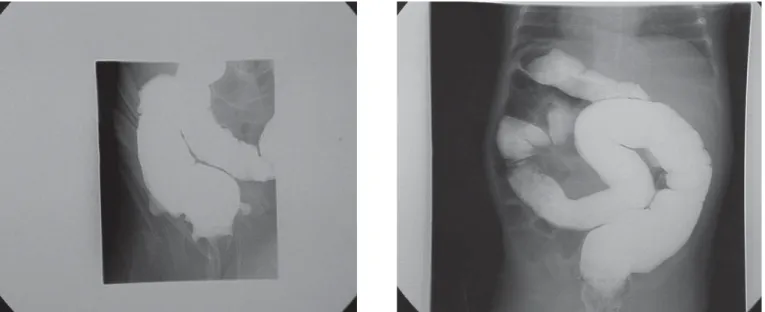
A three-month-old boy with right renal multicystic dysplasia was referred to our department for right inguinal hernia repair and intractable constipation. There was infrequent stooling once a week while breast-feeding. Laxa-tives were commenced at the age of 2- months but were found to be decreasingly effective. Follow-ing Follow-inguinal hernia repair barium enema and anorectal manometry was performed to rule out Hir-schsprung’s disease. At fluoros-copy, lateral protrusion of the rectal wall which was intermittent and appeared with contractions of levator ani was observed (Fig-ure1a, 1b). Rectoanal inhibitory reflex was present on anal mano-metry. Voiding cystourethrography which was performed to evaluate vesicoureteral reflux revealed lat-eral protrusions of the bladder
Journal of Ankara University Faculty of Medicine 2008, 61(3)
169
Rahşan Vargün, Bilal Alper, Selçuk Yüksel, Suat Fitöz, Meltem Bingöl-Koloğlu
through the internal inguinal ring (Figure 2). Patient’s constipation was resolved 2 months after the commencement of a regular en-ema plan in addition to laxatives. He was given antibiotic prophylax-is for urinary tract infections. The patient was still doing well on his 6-month follow-up.
Discussion
There is no knowl-edge in literature answering the question if rectal and bladder “ears” are variant of nor-mal anatomy or real pathological entities requiring treatment. Moreo-ver the role of al-tered embryogene-sis leading to rectal and bladder “ears” is a matter of spec-ulation, and there is no clue indicat-ing an embryologic cause of these le-sions.Bladder “ears” are rarely seen in the first six months of life on voiding cystourethrograms and represents a transient extraperitoneal protru-sion of bladder wall into internal ring (1). They are frequently bila-teral and disappear with voiding and complete filling of the blad-der. In infants, bladder assumes a more abdominal position, which places it in close proximity to
in-ternal inguinal ring. With growth, the pelvis becomes more devel-oped, and bladder assumes a more pelvic position. Therefore, bladder “ears” are rarely observed in adults. Knowledge of this entity is important to surgeons during inguinal herniotomy because of bladder injury may occur in pres-ence of bladder “ears” (1).
As far as we know there are three re-ports of rectal “ears” observed in a 3- month-old, 6 –month-old and 16 month-old female all with histo-ry of constipation. Depending on these reports, rectal “ears” are in-termittent protrusion of rectal wall observed synchronous with con-traction of levator ani (2- 4). The appearance and location of these rectal wall protrusions are similar to the more commonly observed bladder “ears” and represent rec-tal “ears”. The similarities between our case and these reported cases are the presentation with constipa-tion and same radiological obser-vation. An explanation for rectal “ears” could be that different lo-calization of peritoneal reflections around rectum allowing more mo-bility than normal. Contraction of levator ani therefore pulls rectum
Figure 2: Voiding cystourethrography, oblique radiography reveals
anterolateral protrusions of bladder through internal inguinal ring.
Ankara Üniversitesi Tıp Fakültesi Mecmuası 2008, 61(3)
170 Association of Rectal and Bladder “Ears”: Is It A Transient Variant of Normal, or A Pathological Entity?
anteriorly and leads to transient herniation of rectal wall into deep inguinal ring (4). The weakness of perirectal supportive tissues resulting in extensive mobility of rectal wall and leading protrusion during first year of life might be an-other explanation for rectal “ears”. Depending on literature and our case it is evident that rectal “ears” may cause constipation. The rea-son why they cause constipation is unclear. It might be speculated that they may interfere propulsion of rectal contractions resulting in constipation. The association of both rectal and bladder “ears” in our patient raises the query if there is a real ethological connec-tion between bladder and rectal
“ears”. There are many similarities between rectal and bladder “ears” such as intermittent protrusion of rectal or bladder wall into deep in-guinal ring and transient presen-tation in the first year of life. The only difference between them is the lack of anatomic confirmation of rectal “ears” whereas protru-sion of bladder “ears” to internal inguinal ring and bladder injury during inguinal hernia repair has been reported (1). Both weakness of supportive perirectal tissue or different orientation of peritoneal reflexions might result in rectal and bladder “ears” in the same pa-tients. Voiding cystourethrography which revealed bladder “ears” was indicated in our case because of
the presence of the renal pathol-ogy and urinary infection.
From clinical point of view rec-tal “ears” might cause refractory chronic constipation in the first year of life. Therefore rectal “ears” should be considered in differtial diagnosis of constipation en-countered in infancy. Conservative treatment of constipation might be sufficient because they usually disappear by growth and develop-ment. Bladder “ears” should be suspected during inguinal hernia repair in infants to avoid bladder injury. During radiological evalua-tions care should be taken not to mistake bladder and rectal “ears” for diverticula.
REFERENCES
1- Chung Hui-Ming, Yu Tsan-Jung. Bladder rupture after inguinal her-niotomy. Peditar Surg Int 1999; 15:
584-585.
2- Kassner EG, Schussheim A, Gor-don DH. Rectal ears. J Can Assoc Radiol. 1975; 26:125-127.
3- Saxena AK, Choudhary S. Rectal ears. J Postgrad Med 2006; 52:139. 4- Morrison SC. Rectal “ears”. Pediatr