Infection and Drug Resistance
Dove
press
751 R E V I E W
open access to scientific and medical research Open Access Full Text Article
Majocchi’s granuloma: current perspectives
Hazal Boral1
Murat Durdu2
Macit Ilkit1
1Division of Mycology, Department
of Microbiology, Faculty of Medicine, University of Çukurova, Adana, Turkey;
2Department of Dermatology, Faculty
of Medicine, Başkent University Adana Hospital, Adana, Turkey
Abstract: Majocchi’s granuloma (MG) is a rare fungal infection of the dermis that is mainly
caused by dermatophytes (in ≥95% of cases); the most frequently identified cause is anthropo-philic Trichophyton rubrum. In the rest of the cases, the causes are non-dermatophytic fungi such as Aspergillus species. This review aimed to provide information about the current perspec-tives on MG regarding its clinical characteristics, predisposing factors, laboratory diagnosis, and treatment strategies. Although the lower extremities were reported to be the most common site of infection, facial involvement has been predominant in the past 5 years. Our literature research showed that the most common predisposing factor (55%) is the use of topical steroid creams without potassium hydroxide examination during treatment of erythematous squamous dermatoses. A reliable diagnosis of MG is based on histopathological examination, including fungal culture and molecular analyses. MG should be treated not only with topical agents but also with systemic antifungal agents that are continued until the lesions are completely resolved. In systemic treatment, the most preferred drug is terbinafine, because of its efficacy, side effects, and safety.
Keywords: dermatomycosis, histopathology, immunosuppression, predisposing factor, Tricho-phyton rubrum
Introduction
Dermatophytes are highly specialized keratinophilic and keratinolytic fungi that con-sist of seven genera, including Trichophyton, Microsporum, Epidermophyton, and the recently introduced Arthroderma, Paraphyton, Nannizzia, and Lophophyton.1 Although
dermatophytes are the most common human fungal pathogens worldwide, these fungi are neglected because 1) they uncommonly cause a life-threatening disease;2 2) in vitro
resistance to the first choice of antifungal drugs has been reported, but it is not very common;3 and 3) most of the effective antifungal drugs are accessible in most countries.4
However, besides the ability of these fungi to cause infections in both immunosup-pressed and immunocompetent individuals, a growing population of individuals with diabetes and immunosuppression, improvements in medical device technology, and the prolonged life spans of these patients make these fungi more noticeable.5
Majocchi’s granuloma (MG) is an inflammatory and granulomatous, dermatophytic infection that is classified into two forms, depending on the affected individual’s health situation and clinical picture. The first form is mainly observed in healthy individuals and is defined as a perifollicular, papular form induced by penetrating trauma that is mostly observed in the lower extremities. The second form is granulomatous, related Correspondence: Macit Ilkit
Division of Mycology, Department of Microbiology, Faculty of Medicine, University of Çukurova, Adana 01330, Turkey Tel +90 532 286 0099 Fax +90 322 457 3072 Email [email protected] Year: 2018 Volume: 11
Running head verso: Boral et al
Running head recto: Trichophytic granuloma DOI: http://dx.doi.org/10.2147/IDR.S145027
This article was published in the following Dove Press journal: Infection and Drug Resistance
to immunosuppression, seen in a nodular form, and usually appears on the upper extremities.6 The main cause of MG
is Trichophyton rubrum, followed by T. mentagrophytes,
T. violaceum, and T. tonsurans. However, several fungi, such
as T. interdigitale, Microsporum canis, Nannizzia gypsea (former M. gypseum), Epidermophyton floccosum, and
Aspergillus species, can also cause MG.7−10
Throughout our review of the epidemiological charac-teristics and treatment strategies of MG,11 we noticed that
the number of cases of MG has been rising over the past 5 years. In addition, we are aware that there is some confusion regarding the classification of this invasive infection. Hence, in this review, we aimed to provide up-to-date information about the current knowledge on MG, including demographic characteristics, clinical features, predisposing factors, and diagnostic and treatment strategies for the disease.
Search strategy
We searched PubMed (MEDLINE) and Google Scholar for MG cases that were published in the English-language litera-ture between August 2011 and November 2017, using the key words “Majocchi’s granuloma,” “trichophytic granuloma,” and “dermatophytic granuloma.” Other types of invasive or disseminated dermatophyte infections were excluded from the present review. The clinical and demographic characteris-tics of 33 patients with MG from 32 articles were evaluated.
Invasive dermatophytosis and MG
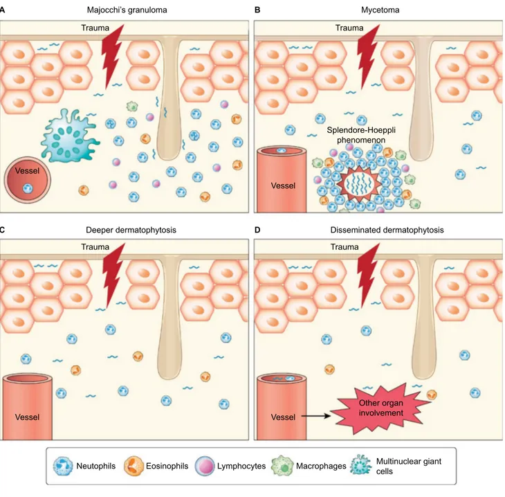
Although dermatophytes require keratin for nutrition, in some circumstances, they can be isolated from the deeper layers of the skin.12−16 In our previous review, we classifiedthese infections as follows: 1) MG (nodular, granulomatous perifolliculitis); 2) deeper dermal dermatophytosis; 3) dis-seminated dermatophytosis; and 4) mycetoma and pseudo-mycetoma (Figure 1).11
Importantly, in the case of dermis invasion by dermato-phytes, the immune response determines the clinical picture as follows: 1) a granulomatous inflammation around the hair follicle is called MG. Histopathologically, MG demonstrates a nodular perifollicular granulomatous infiltrate of lymphoid cells, macrophages, epithelioid cells, multinucleated giant cells, and neutrophils. Unlike superficial dermatophytoses, fungal hyphae and spores can be detected not only on the surface of the epidermis but also within or around the hair follicles (Figure 1);16 2) in mycetoma, dermal fungal elements
are surrounded by an eosinophilic material, including antigen- antibody complexes and debris from host inflammatory cells (Splendore-Hoeppli reaction); 3) dermal invasion and a mild
immune response without perifollicular granulomatous inflammation or a Splendore-Hoeppli reaction are called deep dermal dermatophytosis; 4) the clinical picture that consists of vascular involvement and dissemination to other organs is called disseminated dermatophytosis (Figure 1). In this latest form, fungi can be isolated from sputum, blood, or other tissue samples in addition to skin biopsy samples.14,16−18
Recently, Rouzaud et al19 reported the clinical and
histo-logical differences between deep dermatophytosis and MG. However, a patient’s immune status, the type and location of the lesion, and direct microscopic examination with potassium hydroxide (KOH) may not be helpful to reliably diagnose MG.
Pathogenesis
Dermatophytes degrade the keratin in nonliving keratinized tissues to survive. However, in the case of MG, the fungi must survive in the dermal and subcutaneous tissues. Although the underlying mechanisms of the pathogenesis of MG are not well understood, there are some proposals for this mecha-nism, and they all rely on several factors that are associated with the host and microorganism.
The first and most important host factor is a physical skin barrier that prevents fungal skin infections.20 Physical trauma
of the skin due to shaving or scratching and immunosup-pression are believed to cause fungal invasion. This invasion occurs because of damage to the epidermal barrier’s integrity and follicular disruption; thus, microorganisms, along with keratin and necrotic materials, can enter the dermis. Fungi must hide from the host’s immune system, and they cause an inflammatory response during infection. Fungal LysM domain-associated proteins mask chitin on the fungal cell wall and regulate fungal growth and development.21 Fungi also
have several enzymes, such as lipases, esterases, and collage-nases.22 Moreover, the microorganisms express several genes
that encode the key components of the glyoxylate pathway (i.e. isocitrate lyase and malate synthase) and excrete a large amount of sulfite to degrade the components of the skin.23,24
Dermatophytes can cause deep and invasive infections under some acquired or congenital immunosuppressive conditions. For instance, disseminated dermatophytosis might be associated with lymphopenia, reduced complement C3 and C4, and hypogammaglobulinemia.25 Additionally,
the deficiency of autosomal-recessive caspase recruitment domain-containing protein 9, which has effects on the signal transducer and activator of the transcription 3 pathway and interleukin (IL)-17 and IL-22 secretion, was also reported in 17 patients with deep dermatophytosis.26−28
Host factors also affect the characteristics of the infec-tion. In a patient with pancytopenia, dermal dermatophytosis without granuloma- or dermatophyte-related sepsis might develop. However, in a patient with partial immunosuppres-sion, granuloma, abscesses, and mycetoma may occur.12,14 The
host also uses several mechanisms to control the infection. Antimicrobial peptides such as cathelicidins and defensins protect the patient against fungi, and they also promote epi-dermopoiesis to clear the infection.29,30 In addition, natural
killer cells, neutrophils, and macrophages also respond to dermatophytosis. Therefore, therapeutic immunosuppression causes lower cellular immunity and ingestion/killing rate of fungal spores.31
Source of infection and possible
predisposing factors
The available data in the literature provide some predictions about the source of infection and predisposing factors of
Figure 1 Pathogenesis of invasive dermatophytosis.
Notes: Physical trauma impairs the epidermal barrier. Penetration of the dermatophytes into the skin causes a granulomatous, inflammatory response, including neutrophils
(N), eosinophils (E), lymphocytes (T), macrophages (M), and multinuclear giant cells (MGC). Majocchi’s granuloma (A), mycetoma (B), deeper dermatophytosis (C), and
disseminated dermatophytosis (D). Disseminated dermatophytosis Splendore-Hoeppli phenomenon Deeper dermatophytosis Trauma Trauma Trauma
Majocchi’s granuloma Mycetoma
Trauma Vessel Vessel Vessel Other organ involvement Multinuclear giant cells Macrophages Lymphocytes Eosinophils Neutophils Vessel C D A B
MG. It commonly occurs in the presence of chronic derma-tophytoses, such as tinea unguium and tinea corporis.13,28,32−36
These infections may be a source of MG in cases wherein the skin barrier is destroyed. Moreover, shaving the legs or pubic area, sexual contact, and occupation should also be investigated.7,8,37,38 In this review, four of the patients were
thought to have been in contact with an animal, suggesting that animal exposure was a predisposing factor of MG. Three of these patients had been in contact with guinea pigs.34,39,40
Guinea pigs are often cryptic carriers,41 and the clinician
should consider the zoophilic characteristics of dermato-phytes and whether the patient has a pet or is in frequent contact with animals.
Preexisting dermatophytosis is a major risk factor of MG. Consistently, Kershenovich et al42 reported that the anatomic
regions that are involved in preexisting dermatophytosis are the possible origins of MG. This was also evident in our review, which reflected that 10/24 immunocompetent and 7/9 of immunosuppressed patients had prior or concomitant der-matophytic infections. The lesions that were reported in these patients were related to MG, except those in two patients. One was an immunocompetent male patient who had lesions on his suprapubic and inguinal regions and a tinea barbae as a prior dermatophytosis.7 However, the patient had a history of
unpro-tected sexual exposures in Thailand. The other was a healthy 58-year-old man who had tinea pedis prior to developing MG lesions on his left forearm, but the source of infection could not be determined because he did not have a history of any local or general immunosuppressive conditions or animal exposure.43
Long-term use of steroids, chemotherapy, and antineo-plastic therapy or other immunosuppressive conditions may also lead to MG.32,44−47 In particular, steroids have been used
successfully to treat many lethal diseases, such as pemphigus. However, many cases with steroid use-related sepsis have also been recorded. Steroids affect the functions of macro-phages and neutrophils and reduce the Th1-mediated immune response. After steroid use, lesions may become atypical, and complete resolution of fungal infections may be delayed.36,48
Our analyses revealed that the use of steroids (n=21) was the most common underlying condition.
The increase in organ transplantation led to the extensive use of some immunosuppressive drugs such as tumor necrosis factor alpha inhibitors (e.g. adalimumab). Although these drugs can reduce the side effects of steroids, an MG patient who had used adalimumab was reported.49 Similarly, BRAF
inhibitors (e.g. vemurafenib) are considered promising treat-ments for aggressive skin tumors; however, they may also facilitate MG development.32
Dermatophytic infections occur frequently in human immunodeficiency virus- or acquired immune deficiency syndrome (AIDS)-positive patients; however, there is no consensus on whether there is any relationship between the lymphocyte count and cutaneous dermatophytosis.50,51
When it occurs, dermatophytosis can be either atypical or widespread in these patients. In addition to invasive and chronic, resistant infections, MG can also develop.52
Among the cases that were searched for in this review, only one case with T. tonsurans had AIDS.53 Several other
possible predisposing factors, such as chemotherapy (n=1) and transplantation [solid organ (n=3) and facial tissue allotransplantation (n=1)], were also addressed in some studies.
Clinical characteristics
The clinical pictures of MG in healthy individuals and immunosuppressed patients differ. A perifollicular, papular form that is induced by penetrating trauma is mostly seen in healthy individuals. On the other hand, the granulomatous form is related to immunosuppression and is seen in nodular forms.6 In addition to the papular and nodular forms, plaques,
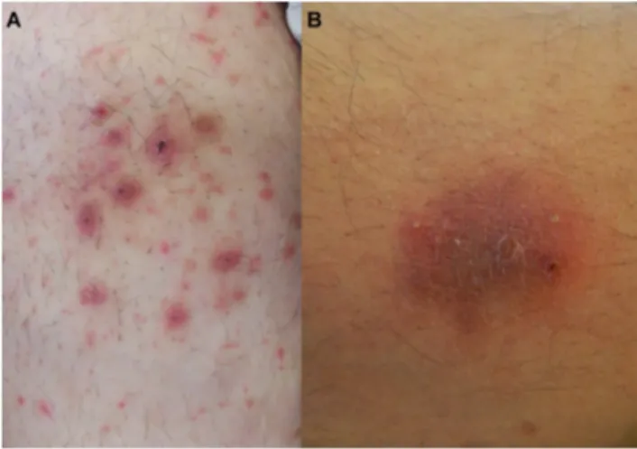
patches, and multiple forms, with or without a crust, can also be seen on the lesions (Figure 2A and B).
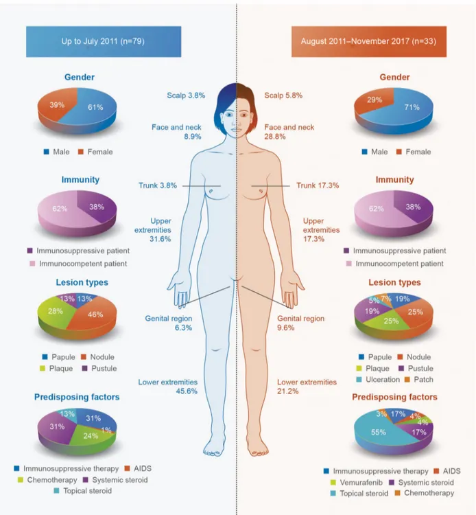
We reviewed 32 studies including 33 cases (21 men and 12 women) that were published in the English language lit-erature between August 2011 and November 2017. The mean age of the patients was 38 years (range: 3–65 years), and the mean duration of the infections was 9 months (range: 3 days–60 months). The clinical characteristics of patients with MG are shown in Figure 3. The majority of the patients in
Figure 2 Multiple erythematous papules and nodules with scales and/or crusts
are located on the anterior surface of the abdomen in a patient with Majocchi’s granuloma (A). Erythematous plaque with pustules, scales, and crusts on the lateral
both the immunocompetent and immunosuppressed groups had multiple lesions: 16/24 and 7/9, respectively. The most affected area was the face (37.5%) among all immunocom-petent patients and the lower extremities among the immu-nosuppressed patients (66.7%). Although multiple types of lesions (n=22) appeared, the most predominant forms were nodules (n=19) and plaques (n=19). Immunocompetent
patients mostly had plaques (62.5%) and nodules (54.2%), whereas immunosuppressed patients had nodules (66.7%) and papules (55.6%). In addition, erythroderma and pal-moplantar hyperkeratosis have been reported in a patient with AIDS.53
The number of reported MG cases has increased remark-ably in the past 6 years (n=33), compared to that between
Figure 3 Clinical characteristics of the patients with MG reported in the literature: location of the lesions, sex, immunity, predisposing factors, and type of lesion. Abbreviation: MG, Majocchi’s granuloma.
1883 and 2011 (n=79). Moreover, the frequency of facial involvement was also prominently higher (36.4%) than that previously reported (6.3%).11
Laboratory diagnosis
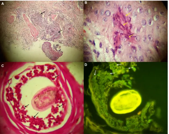
Diagnosing MG requires detection of not only dermatophytes but also perifollicular granulomatous inflammation. The most commonly used method for displaying fungal hyphae and spores is KOH examination (n=18; 7/18 had negative results). However, KOH examination is insufficient for distinguishing superficial and invasive dermatophytoses. As mentioned pre-viously, perifollicular granulomatous inflammation should be demonstrated for the diagnosis of MG (Figures 4A and B).37
However, in this review, we noted that a histopathological examination was not performed in nine patients with MG. Further, in four patients, the staining techniques of histologi-cal examination were not mentioned.7,28,43,46,54
The stains used in histopathological examinations are very important. Although histopathological examination is the “gold standard” method for demonstrating granulomatous infiltration, hematoxylin-eosin (HE) staining, which is used
in routine histopathology, may be insufficient in detecting fungal elements. The most frequently used staining methods for eliminating this deficiency are the periodic acid-Schiff (PAS) (n=18) and Grocott-Gomori’s methenamine silver (GMS) (n=5) methods.49,54,55
PAS staining is more preferable in the histopathological examination of samples containing suspected fungal infec-tion than GMS because it is easy to perform and has higher sensitivity and negative predictive values (Figure 4C).56
When the fungal elements on the suspected samples are numerous, HE staining can also be helpful. However, when there are few fungal elements, they may be overlooked.57
HE-stained preparations can be examined under an immunofluo-rescence microscope, and fungi are shown as autofluorescent particles (Figure 4D).58 On the other hand, GMS staining can
be more helpful because it has greater contrast than PAS staining. However, there are no adequate data to conclude that GMS staining is superior to PAS staining. Although GMS staining has an advantage over PAS because it has better powers of detection on low- and intermediate-power microscopy and better contrast to detect fungal elements
Figure 4 Histopathological findings of a patient with MG.
Notes: (A) The histopathology showed perifollicular, granulomatous inflammation (arrows). (B) Hyphae (arrows) are seen with great magnification. (C) Perifollicular spores
(arrows) were positively stained with PAS staining. (D) In the HE-stained slides, spores (arrows) showed autofluorescence under an immunofluorescence microscope. (A,
HE ×100; B, D, HE ×1000; C, PAS ×1000).
easily, it is time and temperature dependent, requires an expert technician, and contains hazardous compounds such as chromic acid.56,59,60
As histological examination is not sufficient for the identification of a fungus, especially in immunosuppressed patients, it is important to use molecular-based techniques, such as internal transcribed spacer (ITS) sequencing, for identifying fungal species.1,55,61,62 A fungal culture is required
both to detect fungal pathogens and to recognize the spe-cies eventually by combining histological analysis and ITS sequencing. Among the studies that were reviewed, fungal culturing was performed for 28 cases, only 1 of which was negative.63 In only seven patients, the isolates were identified
molecularly using ITS primers. Performing more than one technique, that is, culturing and microscopy, almost always leads to the detection of fungal elements.10,35,53,64−66
Etiological agents
The causes of dermatophytosis depend on the geographic region. Consistently, the etiology of MG may also differ. However, T. rubrum is the most isolated fungal agent of MG in both immunocompetent and immunosuppressed individu-als worldwide. Additionally, T. interdigitale, T. tonsurans,
T. violaceum, M. canis, M. ferrugineum, N. gypsea, and E. floccosum were also reported.7,8,10,35,49,66 In this review, the
most common fungal isolate was T. rubrum (n=15), followed by T. mentagrophytes (n=5), T. interdigitale (n=2), N. gypsea (n=2), T. tonsurans (n=2), and T. violaceum (n=1) (Table 1). Non-dermatophytic fungi such as those belonging to the genera Phoma and Aspergillus were also reported as etiological agents of MG.9,67 Among the 33 cases that were
reviewed, only one study reported the presence of a non-dermatophytic but keratinophilic fungus, a Malbranchea species, in an immunocompetent patient who had eczema as the underlying disease.68
Differential diagnosis
MG can be confused with diseases that also cause chronic erythematous papules and nodules. Due to the presence of pain in these lesions, they are usually perceived as symptoms of bacterial infections, and this confusion results in patients receiving antibiotic treatment. Other chronic infections (e.g. mycobacterial infections, deep fungal infections, dis-seminated toxoplasmosis, and cutaneous leishmaniasis) may also be misleading.11 In addition to histopathology, bacterial,
fungal, and parasitic examinations, as well as polymerase chain reaction and other molecular diagnostic tools, are cru-cial for reliable organism detection. Notably, when the lesion involves the face, it can imitate granulomatous rosacea and granuloma faciale. Painful nodules also imitate erythema nodosum, thrombophlebitis, and erythema induratum bazin. In immunosuppressed patients, it is important to distinguish MG from some tumoral diseases such as Kaposi sarcoma and lymphoma.36
Treatment
Oral potassium iodide, local X-radiation, and topical 2-dimethylamino-6-(β-diethylaminoethoxy)-benzothiazole (Asterol®) were used to treat MG before antifungal treatments
were discovered. Antifungal drugs are used topically and/or systemically. Although topical antifungal therapy is usually sufficient for the treatment of superficial dermatophytoses, systemic treatment is also required to treat tinea capitis, onychomycosis, invasive dermatophytoses, and widespread superficial dermatophytoses. The selection of antifungal drug changes with the discovery of novel antifungal drugs. Although ketoconazole was frequently used previously, about half of the patients reported today are treated with terbinafine (250 mg/day).8,13,36,40,43,47,54,68−71 Other systemic
antifungal drugs are itraconazole (100–200 mg/day),32,44,53,72
griseofulvin (250–500 mg/day),10,35,63 voriconazole,73 and
posaconazole.28 Antifungal therapy should be continued
until the lesions are completely resolved. Depending on the severity of the disease, the duration of MG treatment varies from 1 to 6 months.55,69
Rallis et al37 reported a patient who did not respond
to systemic itraconazole treatment, but responded well to systemic terbinafine treatment. Liu et al73 reported the case
of a patient with a mixed infection of T. rubrum and
Kleb-siella pneumoniae and treated this case first with systemic
antibiotics (combined cefoselis and levofloxacin for 7 days) and then voriconazole (200 mg, twice daily). Although some newer antifungal drugs were developed after terbinafine, the interaction of novel antifungal drugs is higher than that Table 1 The causative fungi that were isolated from patients with
MG
Causative fungi Frequency (%)
Dermatophytic fungi 96.4 Trichophyton rubrum 55.6 T. mentagrophytes 18.5 T. interdigitale 7.4 T. tonsurans 7.4 T. violaceum 3.7 Nannizzia gypsea 7.4 Non-dermatophytic fungus 3.6 Malbranchae sp. 3.6
of terbinafine. Post-inflammatory pigmentation, atrophic scarring, and alopecia may develop following the use of antifungal treatment.7,34,36,37,47,70
Conclusion
MG is an uncommon fungal infection that is mostly related to local physical trauma of the skin, followed by disruption of the hair follicles. It may occur in both immunosuppressed and immunocompetent individuals. The source of MG can be a prior dermatophyte infection, exposure to infected or asymptomatic animals or humans, and local or general immunosuppressive conditions.
The diagnosis of MG should be verified by histological examinations, and PAS or GMS staining reveals evidence of the infection. Recognizing the fungal species using conven-tional and/or molecular methods is also crucial, particularly in immunosuppressed patients. Additionally, understanding the clinical, epidemiological, and histological characteristics of the infection depends on an accurate and reliable clinical and mycological diagnosis. MG can mimic several other infections; therefore, it is important to differentiate MG and begin treatment as soon as possible. Topical antifungal agents do not respond to treatment, and systemic antifungal agents should be applied at a proper dose and for an appro-priate duration. Further studies in this field should focus on proposing a guideline that includes the current diagnostic and management procedures of MG.
Author contributions
All authors contributed towards data analysis, drafting and critically revising the paper and agree to be accountable for all aspects of the work.
Disclosure
The authors report no conflicts of interest in this work.
References
1. de Hoog GS, Dukik K, Monod M, et al. Toward a novel multilocus phylogenetic taxonomy for the dermatophytes. Mycopathologia. 2017;182(1–2):5–31.
2. Bongomin F, Gago S, Oladele RO, Denning DW. Global and multi-national prevalence of fungal diseases – estimate precision. J Fungi. 2017;3(4):pii,E57.
3. Yamada T, Maeda M, Alshahni MM, et al. Terbinafine resistance of Trichophyton clinical isolates caused by specific point mutations in the squalene epoxidase gene. Antimicrob Agents Chemother. 2017;61(7):e00115–17.
4. Kneale M, Bartholomew JS, Davies E, Denning DW. Global access to antifungal therapy and its variable cost. J Antimicrob Chemother. 2016;71(12):3599–3606.
5. Scheen A, van Gaal L. Combating the dual burden: therapeutic targeting of common pathways in obesity and type 2 diabetes. Lancet Diabetes Endocrinol. 2014;2(11):911–922.
6. Radentz WH, Yanase DJ. Papular lesions in an immunocompromised patient. Trichophyton rubrum granulomas (Majocchi’s granuloma). Arch Dermatol. 1993;129(9):1189–1190, 1192–1193.
7. Gallo JG, Woods M, Graham RM, Jennison AV. A severe transmissible Majocchi’s granuloma in an immunocompetent returned traveler. Med Mycol Case Rep. 2017;18:5–7.
8. Parmar NV, Asir GJ, Rudramurthy SM. Atypical presentation of Majoc-chi’s granuloma in an immunocompetent host. Am J Trop Med Hyg. 2017;96(1):1–2.
9. Saadat P, Kappel S, Young S, Abrishami M, Vadmal MS. Aspergillus fumigatus Majocchi’s granuloma in a patient with acquired immunode-ficiency syndrome. Clin Exp Dermatol. 2008;33(4):450–453. 10. Kanaan IC, Santos TB, Kac BK, Souza AM, Cerqueira AM. Majocchi’s
granuloma – case report. An Bras Dermatol. 2015;90(2):251–253. 11. Ilkit M, Durdu M, Karakaş M. Majocchi’s granuloma: a symptom
complex caused by fungal pathogens. Med Mycol. 2012;50(5):449–457. 12. Inaoki M, Nishijima C, Miyake M, et al. Case of dermatophyte abscess caused by Trichophyton rubrum: a case report and review of the litera-ture. Mycoses. 2015;58(5):318–323.
13. Su HA, Sun PL, Sung WW, et al. Deep dermatophytosis caused by zoophilic strain of Trichophyton interdigitale with successful treatment of itraconazole. Mycopathologia. 2017;182(7–8):715–720.
14. Tirado-González M, Ball E, Ruiz A, et al. Disseminated dermatophytic pseudomycetoma caused by Microsporum species. Int J Dermatol. 2012;51(12):1478–1482.
15. Fukushiro R. Dermatophyte abscess [in Japanese]. In: Fukushiro R (ed). Color Atlas of Dermatophytoses. Tokyo: Kanehara Co.;1999:89–95. 16. Marconi VC, Kradin R, Marty FM, Hospenthal DR, Kotton CN.
Dis-seminated dermatophytosis in a patient with hereditary hemochroma-tosis and hepatic cirrhosis: case report and review of the literature. Med Mycol. 2010;48(3):518–527.
17. Smith KJ, Welsh M, Skelton H. Trichophyton rubrum showing deep dermal invasion directly from the epidermis in immunosuppressed patients. Br J Dermatol. 2001;145(2):344–348.
18. Isa-Isa R, Arenas R, Isa M. Inflammatory tinea capitis: kerion, der-matophytic granuloma, and mycetoma. Clin Dermatol. 2010;28(2): 133–136.
19. Rouzaud C, Chosidow O, Brocard A, et al. Severe dermatophytosis in solid organ transplant recipients: a French retrospective series and literature review. Transpl Infect Dis. 2018;20(1). Epub 2018 Jan 25. 20. Lee WJ, Kim JY, Song CH, et al. Disruption of barrier function in
dermatophytosis and pityriasis versicolor. J Dermatol. 2011;38(11): 1049–1053.
21. Martinez DA, Oliver BG, Gräser Y, et al. Comparative genome analysis of Trichophyton rubrum and related dermatophytes reveals candidate genes involved in infection. mBio 2012;3(5):e00259–12.
22. Boral H, Metin B, Döğen A, Seyedmousavi S, Ilkit M. Overview of selected virulence attributes in Aspergillus fumigatus, Candida albi-cans, Cryptococcus neoformans, Trichophyton rubrum, and Exophiala dermatitidis. Fungal Genet Biol. 2018;111:92–107.
23. Monod M. Secreted proteases from dermatophytes. Mycopathologia. 2008;166(5–6):285–294.
24. Zaugg C, Monod M, Weber J, et al. Gene expression profiling in the human pathogenic dermatophyte Trichophyton rubrum during growth on proteins. Eukaryot Cell. 2009;8(2):241–250.
25. Abdel-Rahman SM. Genetic predictors of susceptibility to dermato-phytoses. Mycopathologia. 2017;182(1–2):67–76.
26. Puel A, Picard C, Cypowyj S, Lilic D, Abel L, Casanova JL. Inborn errors of mucocutaneous immunity to Candida albicans in humans: a role for IL-17 cytokines? Curr Opin Immunol. 2010;22(4):467–474. 27. Alves de Medeiros AK, Lodewick E, Bogaert DJ, et al. Chronic and
invasive fungal infections in a family with CARD9 deficiency. J Clin Immunol. 2016;36(3):204–209.
28. Lanternier F, Pathan S, Vincent QB, et al. Deep dermatophytosis and inherited CARD9 deficiency. N Engl J Med. 2013;369(18):1704–1714. 29. Matejuk A, Leng Q, Begum MD, et al. Peptide-based antifungal
therapies against emerging infections. Drugs Future. 2010;35(3):197. 30. van Kilsdonk JWJ, Jansen PAM, van den Bogaard EH, et al. The
effects of human beta-defensins on skin cells in vitro. Dermatology. 2017;233(2–3):155–163.
31. Azad A, Kaufman M, Jayarajan J. Dexamethasone-induced flares of Trichophyton rubrum masquerading as docetaxel cutaneous toxicity: a case report. Cases J. 2009;2:8552.
32. Anforth R, Carlos G, Eiris N, Clements A, Fernandez-Peñas P. Tinea hidden by a vemurafenib-induced phototoxic reaction in a patient with metastatic melanoma taking dexamethasone. Med J Aust. 2015; 203(1):41–42.
33. Rãducan A, Constantin MM. To be or not to be tinea – Misdiagnosis and inappropriate treatment. Dermatovenerol (Buc.). 2017;62:29–32. 34. Trocoli Drakensjö I, Vassilaki I, Bradley M. Majocchi’s granuloma
caused by Trichophyton mentagrophytes in 2 immunocompetent patients. Actas Dermosifiliogr (English Edition). 2017;108(1):e6–e8. 35. Kranseler J, Gupta D. Erythematous annular plaques on a 9-year-old’s
arms and chest. Clin Pediatr. 2018;57(4):488–490.
36. Kim JE, Won CH, Chang S, Lee MW, Choi JH, Moon KC. Majocchi’s granuloma mimicking Kaposi sarcoma in a heart transplant patient. J Dermatol. 2011;38(9):927–929.
37. Rallis E, Katoulis A, Rigopoulos D. Pubic Majocchi’s granuloma unre-sponsive to itraconazole successfully treated with oral terbinafine. Skin Appendage Disord. 2015;1(3):111–113.
38. Molina-Leyva A, Perez-Parra S, Garcia-Garcia F. Case for diagnosis. An Bras Dermatol. 2014;89(5):839–840.
39. Liu C, Landeck L, Cai SQ, Zheng M. Majocchi’s granuloma over the face. Indian J Dermatol Venereol Leprol. 2012;78(1):113–114. 40. Wargo JJ, Berg A, Gudjonsson JE. Ongoing pleuritic chest pain and a
guinea pig: missed pulmonary embolism and Majocchi’s granuloma. Am J Med. 2016;129(9):e165–167.
41. Kraemer A, Hein J, Heusinger A, Mueller RS. Clinical signs, therapy, and zoonotic risk of pet guinea pigs. Mycoses. 2013;56(2):168–172. 42. Kershenovich R, Sherman S, Reiter O, et al. A unique
clinicopatho-logical manifestation of fungal infection: A case series of deep der-matophytosis in immunosuppressed patients. Am J Clin Dermatol. 2017;18(5):697–704.
43. Roy SF, Davar S. Majocchi’s granuloma in a 58-year-old man with treatment-resistant annular scaling plaque. CMAJ. 2017;189(48):e1493. 44. Li FQ, Lv S, Xia JX. Majocchi’s granuloma after topical corticosteroids
therapy. Case Rep Dermatol Med. 2014;2014:507176.
45. Lourdes LS, Mitchell CL, Glavin FL, Schain DC, Kaye FJ. Recurrent dermatophytosis (Majocchi’s granuloma) associated with chemotherapy- induced neutropenia. J Clin Oncol. 2014;32(27):e92–e94.
46. Steiner UC, Trüeb RM, Schad K, et al. Trichophyton rubrum-induced Majocchi’s granuloma in a heart transplant recipient. A therapeutic challenge. JAAD Case Rep. 2012;6(3):70–72.
47. Zhou S, Yang Y, Liu K, Huang S, Hu Y, Zhou X. A case report of widespread Majocchi’s granuloma in a patient with systemic lupus erythematosus. Mycopathologia. 2017;182(5–6):577–581.
48. Gupta V, Bhatia R, Sondhi P, Mahajan R. ‘Ring-within-a-ring’ appear-ance: morphological clue to topical steroid abuse in dermatophytosis. J Eur Acad Dermatol Venereol. 2017;31(1):e2–e3.
49. Chou WY, Hsu CJ. A case report of Majocchi’s granuloma associated with combined therapy of topical steroids and adalimumab. Medicine. 2016;95(2):e2245.
50. Johnson RA. Dermatophyte infections in human immune deficiency virus (HIV) disease. J Am Acad Dermatol. 2000;43(Suppl 5):S135–142. 51. Rodwell GE, Bayles CL, Towersey L, Aly R. The prevalence of derma-tophyte infection in patients infected with human immunodeficiency virus. Int J Dermatol. 2008;47(4):339–343.
52. Ramos-e-Silva M, Lima CM, Schechtman RC, Trope BM, Carneiro S. Superficial mycoses in immunodepressed patients (AIDS). Clin Dermatol. 2010;28(2):217–225.
53. Lyra MR, Muniz Álvarez B, Lanziano AL, et al. Exfoliative erythro-derma and palmoplantar hyperkeratosis associated with Majocchi’s granuloma by Trichophyton tonsurans in a patient with AIDS. Rev Iberoam Micol. 2017;34(3):185–188.
54. Bakardzhiev I, Chokoeva A, Tchernev G, Wollina U, Lotti T. Tinea profunda of the genital area. Successful treatment of a rare skin disease. Dermatol Ther. 2016;29(3):181–183.
55. Su H, Li L, Cheng B, et al. Trichophyton rubrum infection char-acterized by Majocchi’s granuloma and deeper dermatophytosis: case report and review of published literature. Mycopathologia. 2017;182(5–6):549–554.
56. Reza Kermanshahi T, Rhatigan R. Comparison between PAS and GMS stains for the diagnosis of onychomycosis. J Cutan Pathol. 2010;37(10):1041–1044.
57. Al-Amiri A, Chatrath V, Bhawan J, Stefanato CM. The periodic acid-Schiff stain in diagnosing tinea: should it be used routinely in inflam-matory skin diseases? J Cutan Pathol. 2003;30(10):611–615. 58. Elston DM. Fluorescence of fungi in superficial and deep fungal
infec-tions. BMC Microbiol. 2001;1:21.
59. Shiogama K, Kitazawa K, Mizutani Y, Onouchi T, Inada KI, Tsutsumi Y. New Grocott stain without using chromic acid. Acta Histochem Cytochem. 2015;48(1):9–14.
60. D’Hue Z, Perkins SM, Billings SD. GMS is superior to PAS for diagnosis of onychomycosis. J Cutan Pathol. 2008;35(8):745–747.
61. Mirhendi H, Makimura K, de Hoog GS, et al. Translation elongation factor 1-α gene as a potential taxonomic and identification marker in dermatophytes. Med Mycol. 2015;53(3):215–224.
62. Rezaei-Matehkolaei A, Mirhendi H, Makimura K, et al. Nucleotide sequence analysis of beta tubulin gene in a wide range of dermatophytes. Med Mycol. 2014;52(7):674–688.
63. Lewkowicz C, Lakhiani C. Girl’s papular rash will not respond to treat-ment. Int J Contemp Pediatr. 2017;34(9):42–44.
64. Pietrzak A, Tomasiewicz K, Kanitakis J, et al. Trichophyton mentagro-phytes-associated Majocchi’s granuloma treated with cryotherapy. Folia Histochem Cytobiol. 2012;50(3):486–489.
65. Bjekić M, Basara JG. Majocchi’s granuloma in a healthy adult man – a case report. Serb J Dermatol Venerol. 2015;7(1):34–40.
66. Khanna U, Dhali TK, D’Souza P, Chowdhary S. Majocchi’s granuloma of the breast: a rare clinical entity. Actas Dermosifiliogr (English Edi-tion). 2016;107(7):610–612.
67. Sequeira M, Burdick AE, Elgart GW, Berman B. New-onset Majoc-chi’s granuloma in two kidney transplant recipients under tacrolimus treatment. J Am Acad Dermatol. 1998;38(3):486–488.
68. Govind A, L’Etoile N, Vasquez G. The first reported case of Majocchi’s granuloma with Malbranchea sp. in an immunocompetent patient. Case Rep Infect Dis. 2017;2017:9196528.
69. Bressan AL, Silva RSD, Fonseca JCM, Alves MDFGS. Majocchi’s granuloma. An Bras Dermatol. 2011;86(4):797–798.
70. Knoll BM, Hammond SP, Koo S, et al. Infections following facial com-posite tissue allotransplantation – single center experience and review of the literature. Am J Transplant. 2013;13(3):770–779.
71. Wang T, Xu C, Zhou X, et al. Homozygous ALOXE3 nonsense vari-ant identified in a patient with non-bullous congenital ichthyosiform erythroderma complicated by superimposed bullous Majocchi’s granuloma: the consequences of skin barrier dysfunction. Int J Mol Sci. 2015;16(9):21791–21801.
72. Scott-Lang V, Tidman M. Majocchi’s granuloma in an immunocompe-tent child. Arch Dis Child. 2012;97(Suppl 1):A42.
73. Liu HB, Liu F, Kong QT, et al. Successful treatment of refractory Majoc-chi’s granuloma with voriconazole and review of published literature. Mycopathologia. 2015;180(3–4):237–243.
Infection and Drug Resistance
Publish your work in this journal
Submit your manuscript here: https://www.dovepress.com/infection-and-drug-resistance-journal
Infection and Drug Resistance is an international, peer-reviewed open-access journal that focuses on the optimal treatment of infection (bacte-rial, fungal and viral) and the development and institution of preventive strategies to minimize the development and spread of resistance. The journal is specifically concerned with the epidemiology of antibiotic
resistance and the mechanisms of resistance development and diffusion in both hospitals and the community. The manuscript management system is completely online and includes a very quick and fair peer-review system, which is all easy to use. Visit http://www.dovepress.com/ testimonials.php to read real quotes from published authors.