BASIC RESEARCH
Does cigarette smoke exposure lead to histopathological alterations in the
olfactory epithelium? An electron microscopic study on a rat model
Elvan Sahina, Gursel Ortugb, and Alpen Ortug c
aDepartment of Histology and Embryology, School of Medicine, Sakarya University, Korucuk, Sakarya, Turkey;bDepartment of Anatomy,
School of Medicine, Bahcesehir University, Istanbul, Turkey;cDepartment of Anatomy, School of Medicine, Istanbul Medipol University,
Istanbul, Turkey
ABSTRACT
Objective: This study was conducted to examine the influence of smoke exposure of variable duration on the ultrastructure of and histopathologic and morphologic alterations in the olfactory epithelium. Methods: A total of 24 Wistar albino rats were randomly assigned to three groups and fed a standard rat chow and tap water. Experimental rats in groups I and II were exposed to cigarette smoke in a glass cabin over a period of 2 months for 5 or 15 min, respectively, four times daily; control rats (group III) were not exposed to cigarette smoke. After dissection, all tissue specimens were processed using routine procedures for TEM.
Results: Groups I and II exhibited the presence of intraepithelial inflammatory cells and especially deep invaginations in the nuclear membrane of supporting cells. Extended intercellular spaces, cytoplasmic protrusions on the apical surface of supporting cells, atrophy of microvilli and olfactory neuron cilia as well as numerous electron-dense granular structures and lysosome-like structures were observed to an increasing degree from group I to group II. Particularly in group II, both supporting cells and olfactory neurons exhibited a cytoplasmic edema, mitochondrial degeneration, and numerous vacuolar structures, as well as apoptotic and minimal necrotic changes. In this group, hyperplasia of basal cells was also observed.
Conclusion: Our electron microscopic findings show that cigarette smoke leads to toxic degen-erative changes in the rat olfactory mucosa.
ARTICLE HISTORY Received 1 January 2018 Revised 18 May 2018 Accepted 9 July 2018 KEYWORDS Electron microscopy; olfactory mucosa; passive smoking
Introduction
The olfactory epithelium, a pseudostratified, ciliated epithelium, is superiorly located within nasal passages. The epithelium is composed of the following cell types: olfactory cells (bipolar neurons); smell recep-tors; supporting (sustentacular) cells; tall columnar cells, which provide mechanical and metabolic sup-port to olfactory cells and have numerous long micro-villi extending from their apical surface; basal cells; stem cells, from which new olfactory cells and sup-porting cells differentiate; and brush or microvillar (M) cells, functioning in general sensation rather than olfaction.1,2Normally, there are no goblet cells.
The apical pole of each olfactory neuron is thick-ened as a knob-like modified dendrite enlarged into the olfactory vesicle. A number of cilia extend radially from the olfactory vesicle in a plane parallel to the epithelial surface. They are thought to contain receptors for smelling. More proximally, the cell
body widens to accommodate the nucleus and then narrows, in a fusiform fashion, to form the axon. The axonal terminal leaves the epithelial compartment by penetrating the basal lamina to form bundles of nonmyelinated nerve fascicles, the fila olfactoria.1–3 It is known that certain inhalants, particularly cigar-ette smoke, may cause irritation and consequent toxic and neoplastic changes in the respiratory system.4–10As the exposure time increases, the tissue damage becomes more severe.11,12
Smoking is considered a widespread public health problem globally. In addition to other proven results, the deficit in the olfactory sense is six times more common in smokers than in nonsmokers.13,14 The aim of this study was to examine the ultrastructure of olfactory epithelia in rats exposed to cigarette smoke for varying lengths of time and observe histopatho-logical tissue alterations more accurately via trans-mission electron microscopy.
CONTACTAlpen Ortug [email protected] Guney Yerleskesi, Goztepe Mah., Ataturk Cad.No:40, Kavacik, Beykoz, Istanbul 34815 Color versions of one or more of the figures in the article can be found online atwww.tandfonline.com/iusp.
https://doi.org/10.1080/01913123.2018.1499685
Materials and methods
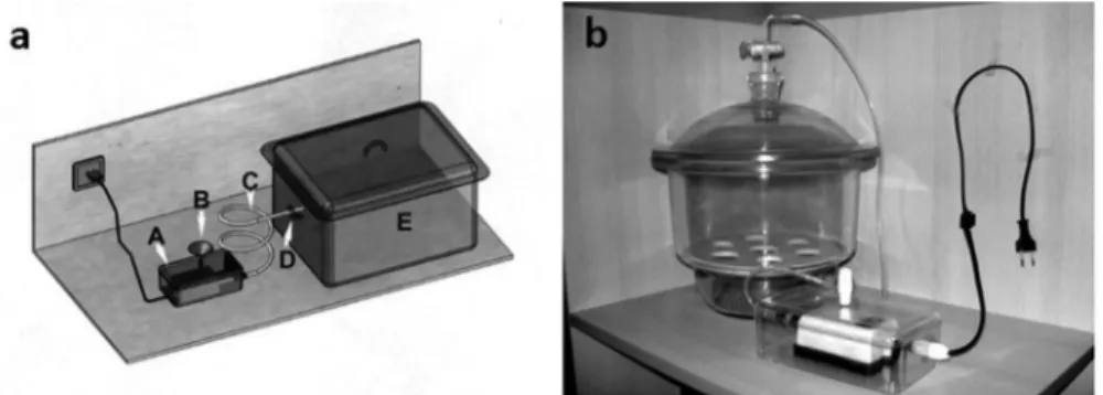
In the present study, 24 adult Wistar albino rats, weighing 250–300 g, were obtained from the Experimental Research and Application Center of Istanbul Medipol University. All animals were handled according to the animal handling protocol approved by the institutional board (IMU-HADYEK). These animals were randomly divided into three groups and maintained under standar-dized conditions of light (12-h light/dark cycle) and temperature (22 ± 2°C), being fed a commer-cial rat diet and fresh tap water for 2 months. Experimental rats were exposed to cigarette smoke four times daily for 2 months in an auto-mated glass cabin [Figure 1] for smoke exposure.15 Control animals (group III) were treated in the same cabin but only exposed to fresh air. The experimental animals of groups I and II were exposed to cigarette smoke for 5 or 15 min, respec-tively. After 2 months, the animals were sacrificed by cardiac perfusion with 2.5% glutaraldehyde in 0.1 M phosphate buffer after being deeply anesthe-tized with ketamine. After the nasal tissues were removed from the animals, the olfactory mucosa was carefully dissected from the bone and placed in the same fixative. Following initial fixation, tissues were postfixed in 2% phosphate-buffered osmium tetroxide, dehydrated in acetone, and embedded in Araldite CY 212. Blocks of tissue were sectioned with glass knives on a Nova LKB Bromma ultramicrotome. Semithin sections were stained with toluidine blue for orientation. Ultrathin sections were stained with uranyl acetate and lead citrate and examined under a Jeol 100 SX electron microscope.16
Results
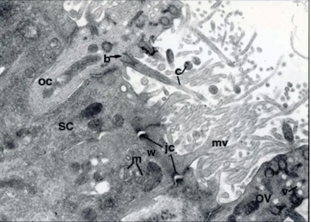
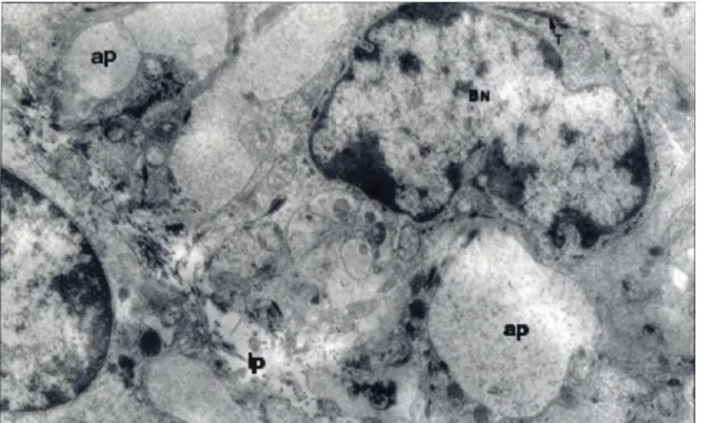
In the control group, olfactory cells (bipolar neu-rons) were characterized by a spindle-shaped form, apical olfactory vesicles, and an electron-lucent cytoplasm. The olfactory vesicles had many sensory cilia. Basal bodies of the cilia were observed within the lateral margins of the vesicle. On the epithelial surface, there were numerous transverse, longitudinal, and tangential profiles of cilia. The thick proximal portion of cilia had a typical “9 + 2” microtubule arrangement. Ciliary narrow regions, containing a few microtubules at the tip, were also observed. Some vesicles, which may have represented a smooth endoplasmic reti-culum (SER) or exocytic or endocytic vesicles, were present in the apical portion of olfactory cells. These cells were linked by junctional com-plexes with adjacent supporting cells. Supporting cells had numerous long, branched microvilli extending from their apical surface. The microvilli were entangled with olfactory cilia and microvilli of neighboring cells on the epithelial surface. The cytoplasm of supporting cells was denser than that of adjacent olfactory neurons. Well-developed terminal webs were observed in the apical portion of supporting cells, separating the organelle-poor apical cytoplasm from a deeper region, where a number of mitochondria were present (Figure 2). Basal cells, small and polygonal or irregular in profile, were located on the basal lamina. Their cytoplasm, surrounding a usually indented hetero-chromatic nucleus, had prominent tonofilaments. Numerous cytoplasmic processes, extending from these cells, appeared to interdigitate with those of neighboring cells and to envelop the axonal
Figure 1.a: Shows technical drawing of the automated exposure apparatus of the experiment. A. Cabin forming negative pressure
and maintains the lite of smoke.B. Cigarette placement handle C. Smoke stream D. On-off controller for stream E. Cabinet for
experiment rats.
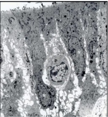
termini of olfactory neurons (Figure 3). In the basal portion of the olfactory epithelium, there were ducts entering the epithelium to deliver the secretion of Bowman’s glands, situated in the lamina propria, to the olfactory surface. Duct cells, squamous in shape, had microvilli and cilia on their luminal surface, as well as cytoplasmic granules (Figure 4).
Prominent ultrastructural alterations were noted in olfactory epithelia of the rats exposed to cigarette smoke. In both experimental groups, degenerating cells and intraepithelial inflammatory cells such as lymphocytes and plasmocytes were observed. In olfactory epithelia from group I, electron-denser supporting cells with an invaginated nuclear mem-brane were encountered among normal ones (Figure 5). In group II, the supranuclear region in supporting cells contained large numbers of round or irregular inclusions filled with an electron-dense osmophilic material (Figure 6). In group III, sup-porting cells exhibited luminal ballooning or
protrusions (Figures 8 and 9). In both supporting cells and olfactory neurons, large amounts of trans-verse or longitudinal SER profiles, mitochondrial degeneration and vacuolization, intracytoplasmic large vacuoles, and cytoplasmic edemas were present to an increasing degree from group I to group II (Figures 7and8). Group II tissues were also parti-cularly notable for numerous apoptotic cells, char-acterized by shrinkage of the cytoplasm, condensation of the chromatin to the periphery of the nucleus, and expanded intercellular spaces (Figure 8). This group showed, in addition, a decrease in the thickness of the microvillous border, lying on the luminal surface (Figure 7), degeneration in olfactory vesicles, from which cilia emerge, and a reduction in the number of microvilli (Figure 6) compared with those in the control group. In both experimental groups, junctional complexes between olfactory neurons and supporting cells on the epithe-lial surface remained intact (Figure 7), and apart from this, many light basal cells with a euchromatic
Figure 2.Electron micrographs from the olfactory epithelium of control rats.OC, olfactory cell; SC, supporting cell; OV, olfactory vesicle; c, cilia; mv, microvilli; b, basal body; v, vesicles; jc, junctional complexes; w, terminal web; m, mitochondria. Uranyl acetate and lead citrate Original Magnification (O.M.). x 800.
Figure 3.An electron micrograph from normal rat olfactory mucosa. BN, the nucleus of the basal cell; ap, axonal process of the olfactory neuron; lp, lamina propria; T, the bundles of tonofilaments. Uranyl acetate and lead citrate (O.M. x 6000).
Figure 4.An electron micrograph from normal rat olfactory mucosa.D, the lumen of a duct entering the olfactory epithelium; N, the nucleus of a duct cell; BN, the nucleus of a basal cell; lp, lamina propria; ap, axonal process; B, a portion of Bowman’s gland. Uranyl acetate and lead citrate-(O.M.).
nucleus and an electron-lucent cytoplasm were encountered among more common dark basal cells (Figure 9).
Discussion
The electron microscopic structure of the normal rat olfactory epithelium, observed in this study, was similar to that reported in the literature.2,17,18 In olfactory epithelia of the experimental groups, swol-len mitochondria, intracytoplasmic vacuolar struc-tures, cytoplasmic edemas, and expanded intercellular spaces were noted. It is known that there are many toxic materials in cigarette smoke.4,19 Upon toxic exposure, cell swelling has been reported to occur, owing to an impairment of the sodium pump mechanism in the cell membrane. In addition, it has been reported that mitochondria are important direct or indirect targets for virtually all types of injurious stimuli, including toxins, and mitochon-drial swelling occurs under toxic conditions.20In our opinion, the vacuolar structures may have occurred owing to mitochondrial swelling and degeneration. Moreover, some researchers have reported that mitochondrial dysfunction, occurring under toxic conditions, causes an increase in cytosolic calcium,
Figure 5.An electron micrograph of the olfactory epithelium from Group I. N, the nucleus of the supporting cell; P, an intraepithelial-plasmocyte. Uranyl acetate and lead citrate, O. M.x 3000.
Figure 6.An electron micrograph of a portion of the olfactory epithelium from Group II.OC, olfactory cell; SC, supporting cell; OV, olfactory vesicle; i, electron-dense inclusions; *, mitochon-drial swelling. Uranyl acetate and lead citrate, O.M. x 5000.
Figure 7.An electron micrograph of a portion of the olfactory epithelium from Group III.SC, supporting cell; OV, olfactory vesicle; i, electron-dense inclusions; *, mitochondrial swelling and degeneration; p, a protrusion of the supporting cell; mv, microvilli. There is a prominent cytoplasmic edema in the supporting cell on the left. Uranyl acetate and lead citrate, O. M. x 8000.
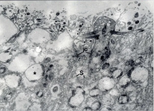
Figure 8.Electron micrographs of apical portions of the olfactory epithelia from Group III. There are a prominent cytoplasmic edema and the luminal ballooning in the supporting cells, and a decrease in the thickness of the microvillous border in comparison with that of the control group. *, vacuolar structures; S, SER-profiles; jc, junctional complexes. Uranyl acetate and lead citrate, O.M. x 8000.
Figure 9.Electron micrographs of the olfactory epithelium from Group III.Many degenerated cell are seen. S, SER-profiles; an, apoptotic nucleus; is, expanded intercellular spaces; m, degenerated mitochondria. Uranyl acetate and lead citrate, O.M. x 4000.
which in turn results in damage to elements of the cytoskeleton.20It has been thought that widening of intercellular spaces and protrusion of the apical cyto-plasm of supporting cells may be related to cytoske-letal damage. In supporting cells of the smoke-treated groups, a large amount of transverse or long-itudinal SER profiles and abundant irregular inclu-sions, containing an osmophilic material, were present. It is known that SER has significant func-tions in detoxification processes.2,17 Under toxic conditions, therefore, excessive development of SER is normal. In our opinion, the irregular, elec-tron-dense inclusions may be secondary lysosomes, lipofuscin granules or aggregates of the olfactory pigment. These structures may reflect an increase in cellular digestion of organelles damaged after smoke exposure or of toxic materials in inhaled cigarette smoke. Apart from these, many mononuc-lear cells such as lymphocytes and plasmocytes were observed in olfactory epithelia of the experimental groups. Under toxic situations, the increase in inflammatory cells may be due to an immunological response to tissue damage.20Moreover, many apop-totic cells were observed in olfactory epithelia of the experimental groups. It has been reported that apop-tosis, or programed cell death, involves the activation of a pathway that leads to a suicide of the cell via a characteristic process wherein the cell becomes more compact, blebbing occurs in the membranes, the chromatin becomes condensed, and DNA is fragmented.21 Apoptosis can be triggered by a vari-ety of stimuli, including toxins.22–24 Particularly in group II, a decrease in the thickness of the micro-villous surface, degeneration of olfactory vesicles, and a reduction in the number of microvilli were noted. Other authors have reported findings similar to ours in the olfactory epithelium treated with pyr-idine, nickel or sulfur dioxide.25–27 Talhaut et al19 have reported that tobacco smoke contains more than 5000 different kinds of toxic chemicals, includ-ing pyridine and nickel. In addition, in the experi-mental groups, many light basal cells with a euchromatic nucleus and a pale cytoplasm were observed among dark ones. It is known that basal cells are stem cells that differentiate to replace olfac-tory neurons and supporting cells lost during a nor-mal turnover or injury, and basal cell hyperplasia follows the olfactory cell loss.11,17,18,28 As basal cells begin to differentiate, their shapes become globose,
and nuclei become euchromatic.18,29In our opinion, the light basal cells may be basal cells differentiating to replace damaged cells after smoke exposure.
In conclusion, we report that cigarette smoke causes serious tissue damage in the rat olfac-tory epithelium and that tissue injuries become more severe with an increasing exposure time.
Acknowledgments
Preliminary data from this article were presented as a poster at the 15th International Congress of Histochemistry and Cytochemistry, May 18-21, 2017, Antalya, Turkey.
Conflict of interest
On behalf of all authors, the corresponding author states that there is no conflict of interest.
Funding
This research did not receive any specific grant from funding agencies in the public, commercial, or not-for-profit sectors.
ORCID
Alpen Ortug http://orcid.org/0000-0002-6813-8351
References
1. Morrison EE, Costanzo RM. Morphology of the human olfactory epithelium. J Comp Neurol.1990;297(1):1–13. doi:10.1002/cne.902970102.
2. Ross MH, Romrell LJ, Kaye GI. Histology, A Text and Atlas. Baltimore: Williams&Wilkins;1995:530–533. 3. Schwob JE, Youngentob SL, Mezza RC. Reconstitution
of the rat olfactory epithelium after methyl
bromide-induced lesion. J Comp Neurol. 1995;359(1):15–37.
doi:10.1002/cne.903590103.
4. Cemeic S, Urbane O. Scanning electron microscopic studies of the nasal mucosa in normal rabbit and rabbit exposed to alcalase. Gegenbaurs Morph Jahrb. 1982;128:405–411.
5. Cho J-H, Kwun Y-S, Jang H-S, Kang J-M, Won Y-S, Yoon H-R. Long-term use of preservatives on rat nasal respiratory mucosa: effects of benzalkonium chloride
and potassium sorbate. Layrngoscope. 2000;110:312–
317. doi:10.1097/00005537-200002010-00025.
6. Dontelwill W, Harke H-P, Baars A, Goertz E. Experimentelle Untersuchungen fiber Aufnahme und Ablagerung von Rauchbestandteilen bei der passiven
Berauchung von Goldhamstem mit Zigarettenrauch.
Arzneim.-Forsch.1971;21:142–143.
7. Frye RE, Schwartz BS, Doty RL. Dose-related effects of cigarette smoking on olfactory function. JAMA.
1990;263:1233–1236.
8. Nadol JB, DeLorenzo AJD. Fine structural localization of acetylcholinesterase in the olfactory mucosa in guina
pig and rabbit. Ann Otol. 1974;83:55–64.
9. Sutherland G, Stapleton JA, Russell MAH, et al. Randomised cntrolled trial of nasal nicotine spray in
smoking cessation. Lancet.1992;340:324–329.
10. Vinke JG, KleinJan A, Severijnen LW, Fokkens WJ. Passive smoking an’allergic’ cell infiltrate in the nasal mucosa of non-atopic children. Int Pediatr Otorhinolaryngol.1999;51 (2):73–81. doi:10.1016/S0165-5876(99)00244-X.
11. Lee KP, Valentine R, Bogdanfify MS. Nasal lesion development and reversibility in rats exposed to aero-sols of dibasic esters. Toxicol Pathol. 1992;20(3):376–
393. doi:10.1177/019262339202000308.
12. Sodeman WA Jr, Sodeman TM. Sodeman’s Pathologic Physiology. 7th ed. Philadelphia: WB Saunders;
1985:461–466.
13. Katotomichelakis M, Balatsouras D, Tripsianis G, et al. The effect of smoking on the olfactory function. Rhinology.2007;45:273.
14. Iskander NM, El-Hennawi DM, Yousef TF, El-Tabbakh MT, Elnahriry TA. Evaluation of the effect of cigarette smoking on the olfactory neuroepithelium of New Zealand white rabbit, using scanning electron
micro-scope. Eur Arch Oto-Rhino-Laryngology. 2017;274
(6):2461–2468. doi:10.1007/s00405-017-4475-1.
15. Koo HJ, Lee HS, Cho KS, Park HY, Roh HJ. Histopathological changes of olfactory epithelium in the rats exposed to cigarette smoke. Korean J
Otorhinolaryngology-Head Neck Surg.2007;50:766–772.
16. Robinson DG, Ehlers U, Herken R, et al. Methods of Preparation for Electron Microscopy-An Introduction for the Biomedical Sciences. 1st ed. Berlin: Springer-Verlag;1987.
17. Fawcett DW. Bloom and Fawcett A Textbook of
Histology. New York: Chapman&Hall;1994:731–734.
18. Jafek BW. Ultrastructure of human nasal mucosa.
Laryngoscope.1983;93:1576–1599.
19. Talhout R, Schulz T, Florek E, Van Benthem J, Wester P, Opperhuizen A. Hazardous compounds in tobacco
smoke. Int J Environ Res Public Health.2011;8(2):613–
628. doi:10.3390/ijerph8020613.
20. Cotran RS, Kumar V, Collins T. Robbins Pathologic Basis of Disease. 6th. Philadelphia: WB Saunders;1999. 21. Lewin B. Genes VII. New York: Oxford University
Press;2000:866–871.
22. Sharma PR, Dugyala RR, Voss KA. Demonstration ofin-situ apoptosis in mouse liver and kidney short-term repeated exposure to fumonisin Bl. J Comp Pathol.1997;117:371–381.
23. Tolleson WH, Dooley KL, Sheldon WG, Thurman JD, Bucci TJ, Howard PC. The mycotoxin fumonisin induces apoptosis in cultured human cell and in livers and
kid-neys of rats. Adv Exp Med Biol.1996;392:237–250.
24. Zhou HR, Harkema JR, Hotchkiss JA, Yan D, Roth RA, Pestka JJ. Lipopolysaccharide and the trichothecene
vomitoxin (deoxynivalenol) synergistically induce
apoptosis in murine lymphoid organs. Toxicol Sci. 2000;53:253–263.
25. Evans JE, Miller ML, Andringa A, Hastings L. Behavioral, histological and neurochemical effects of nickel (II) on the rat olfactory system. Toxicol App
Pharmacol.1995;130:209–220.
26. Min YG, Rhee CS, Choo MJ, Song HK, Hong SC. Histopathologic changes in the olfactory epithe-lium in mice after exposure to sulfur dioxide.
Acta Otolaryngol. 1994;14(4):447–452. doi:10.3109/
00016489409126085.
27. Nikula KJ, Lewis JL. Olfactory mucosal lesions in F344 rats following inhalation exposure to pyridine at threshold limit
value concentrations. Fun-dam Appi Toxicol. 1994;23
(4):510–517. doi:10.1006/faat.1994.1135.
28. Brenneman KA, James RA, Gross EA, Dorman DC. Olfactory neuron loss in adult male CD rats follow-ing subchronic inhalation exposure to hydrogen sul-fide. Toxicol Pathol.2000;28(2):326–333. doi:10.1177/ 019262330002800213.
29. Suzuki Y, Takeda M. Basal cells in the mouse olfac-tory epithelium after axotomy: immunohistochemical and electron-microscopic studies. Cell Tissue Res.
1991;266:239–245.
may not be copied or emailed to multiple sites or posted to a listserv without the copyright holder's express written permission. However, users may print, download, or email articles for individual use.