Case Report
A diagnostic dilemma in a patient with delayed onset
endophthalmitis
Zeynep Eylül Ercan⇑; _Imren Akkoyun; Sirel Gür Güngör; Gürsel Yılmaz
Abstract
We report a case of nonpainful uveitis nine months after an uncomplicated phacoemulsification cataract surgery. Chronic postop-erative endophthalmitis was suspected. Diagnostic vitrectomy and partial capsular bag removal was performed, but the specimens cultured in microbiology laboratory showed no pathogens. Systemic workup came positive for skin Tuberculosis test, and pre-sumed intraocular tuberculosis treatment was started accordingly. Inflammation persisted, so a repeat vitrectomy was performed with removal of the lens implant with the capsule, and this time bedside culture inoculation was performed in operating room, revealing Pseudomonas infection. Delayed-onset postoperative endophthalmitis typically progresses slowly and therefore can be confused with uveitis and treated with steroid and immunosuppressant treatment regimes. Our case confirms both the value of immediate bacterial inoculation and the necessity of aggressive surgical treatment in chronic postoperative endophthalmitis cases.
Keywords: Uveitis, Pseudomonas, Endophthalmitis
Ó 2018 The Authors. Production and hosting by Elsevier B.V. on behalf of Saudi Ophthalmological Society, King Saud University. This is an open access article under the CC BY-NC-ND license (http://creativecommons.org/licenses/by-nc-nd/4.0/).
https://doi.org/10.1016/j.sjopt.2018.06.004
Introduction
Chronic postoperative endophthalmitis may present as chronic uveitis, with recurrent low-grade inflammation long after intraocular surgery. It is usually secondary to Staphylococcus epidermidis and Propionibacterium acnes.1 Main challenge in diagnosing chronic postoperative endoph-thalmitis is to find the offending microorganism, generally low grade and therefore hard to isolate. We report a case of delayed onset endophthalmitis first treated as uveitis, then reported as culture negative in the first diagnostic vitrectomy when samples were sent for inoculation, only to be
diagnosed as chronic endophthalmitis caused by Pseudomonas species when same cultures were prepared in the operating theatre during the repeat vitrectomy. Case report
A 65-year-old female was referred to our clinic for non-painful uveitis in the right eye nine months after an uncompli-cated phacoemulsification cataract surgery with in-the-bag implantation of a hydrophobic acrylic intraocular lens.
Examination of the affected eye revealed a visual acuity of light perception, a pressure of 18 mm Hg, corneal edema
Peer review under responsibility of Saudi Ophthalmological Society,
King Saud University Production and hosting by Elsevier
Access this article online:
www.saudiophthaljournal.com www.sciencedirect.com
Received 16 January 2017; accepted 29 June 2018; available online 9 July 2018.
Department of Ophthalmology, Baskent University, Faculty of Medicine, Ankara, Turkey
⇑ Corresponding author at: Baskent University, Faculty of Medicine Hospital, Department of Ophthalmology, 06490 Ankara, Turkey. Fax: +90 3122237333.
e-mail address:[email protected](Z.E. Ercan).
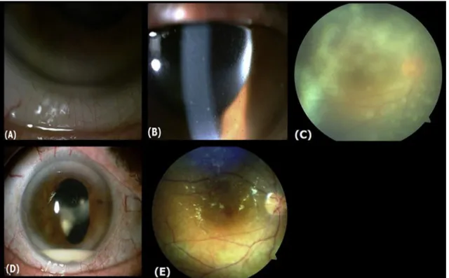
with endothelial folds (Fig. 1A), diffuse fine keratic precipi-tates (Fig. 1B), anterior chamber cells (3+), vitreous haze (3+) (Fig. 1C), and a well-centered, in-the-bag IOL. Chronic postoperative endophthalmitis was suspected. Diagnostic vitrectomy and partial capsular bag removal was performed to exclude chronic endophthalmitis and ocular lymphoma, followed by intraocular injections of Vancomycin (1 mg/0.1 cc), Ceftazidime (2.25 mg/0.1 cc) and Voriconazole (0.05/0.1 cc). However undiluted specimens from anterior chamber and vitreous sent for cultures were negative and pathological examination only showed polymorphonuclear leukocyte infiltration and necrosis, with no signs of lym-phoma. Three weeks postoperatively recurrence of inflam-mation was noted, now with hypopyon formation (Fig. 1D). Her systemic workup showed positive Tuberculosis skin test, so Tuberculosis treatment regime with topical cor-ticosteroids for presumed intraocular Tuberculosis was pre-scribed. Inflammation persisted, so a repeat vitrectomy was performed, this time with complete removal of the lens capsule and implant, followed by combined intraocular antibiotic-antifungal injections and silicone oil tamponade instillation. Anterior chamber and vitreous specimen was also taken prior to antibiotic and antifungal injection, but this time cultures were inoculated immediately in the oper-ating theatre. Anaerobic cultures were found negative, but aerobic cultures of vitreous and explanted intraocular lens showed Pseudomonas species growth sensitive to Amikacin, Aztreonam, Cefepime, Ciprofloxacin, Meropenem and Cef-tazidime. With Systemic and topical Moxifloxacin treatment patient improved symptomatically. Postoperative first month, patient showed no signs of inflammation with the visual acuity of 20/400.
Discussion
Delayed-onset postoperative endophthalmitis can be con-fused with uveitis, prompting steroid treatment to be started. The main challenge is to find the offending microorganism, generally low grade and therefore hard to isolate.2Majority
of these cases is caused by Propionibacterium species, fol-lowed by coagulase-negative Staphylococcus and fungi.3
There have been reports of Pseudomonas species, including Pseudomonas aeruginosa, causing late-onset endophthalmi-tis.4–6However, due to the high virulence and multiple drug resistance of Pseudomonas aeruginosa, chronic postopera-tive endophthalmitis secondary to this organism is rare. In our case, the Pseudomonas isolate was found susceptible to Amikacin, Aztreonam, Cefepime, Ciprofloxacin, Merope-nem and Ceftazidime, with no signs of multidrug-resistance. This made us believe this strain was not Pseudomonas aerug-inosa but a subtype with low virulence.
It is also important to note that the first vitreous specimen, which was sent to microbiology for culture preparation, was found negative. Only with bedside culturing pathogen iden-tification was possible. Also, without the removal of the intraocular lens and total capsulectomy, intraocular wide spectrum antibiotics and antifungals proved to be ineffective. Our case confirms both the value of immediate bacterial inoc-ulation and the necessity of aggressive surgical treatment in chronic postoperative endophthalmitis cases.
Acknowledgement
We thank Prof. Özlem Kurt Azap and Dr. Pelin Börcek for their support and expertise in this patient’s management.
Fig. 1. (A) Anterior segment photograph of cornea, showing endothelial folds due to edema. (B) Keratic precipitates scattered in diffuse pattern. (C) Fundus photograph of grade 3 vitreous haze. (D) Hypopyon formation in the third week of initial vitrectomy. (E) Fundal appearance following the second vitrectomy and silicone oil tamponade.
Financial support
No financial support was received for this article. Conflict of interest
The authors declared that there is no conflict of interest. References
1. Vaziri K, Schwartz SG, Kishor K, Flynn HW. Endophthalmitis: state of the art. Clin Ophthalmol (Auckland, NZ) 2015;9:95–108.
2. Gonzalez-Martin JA, Hiscott P, Palimar P. Chronic postoperative uveitis–a clinicopathological case report. Eye (London, England) 2003;17(5):675–7.
3. Maalouf F, Abdulaal M, Hamam RN. Chronic postoperative endophthalmitis: a review of clinical characteristics, microbiology, treatment strategies, and outcomes. Int J Inflam 2012.https://doi.org/ 10.1155/2012/313248. Article ID 313248.
4. Yu EN, Foster CS. Chronic postoperative endophthalmitis due to pseudomonas oryzihabitans. Am J Ophthalmol 2002;134(4):613–4. 5. Jiraskova N, Rozsival P. Delayed-onset Pseudomonas stutzeri
endophthalmitis after uncomplicated cataract surgery. J Cataract Refract Surg 1998;24(6):866–7.
6. Nagaraj KB, Jayadev C. Pseudomonas aeruginosa endophthalmitis masquerading as chronic uveitis. Indian J Ophthalmol. 2013;61 ():309–10.