210
Kronik Total Oklüzyonu Olan Hastalarda Koroner Kollateral
Dolaşım ile Koroner Arter Ektazisi Arasındaki İlişki
The Relationship Between Coronary Artery Ectasia and Coronary Collateral
Circulation in Patients with Chronic Total Occlusion
Bernas Altıntaş1, Halil Akın2
1Sağlık Bilimleri Üniversitesi, Gazi Yaşargil Eğitim ve Araştırma Hastanesi, Kardiyoloji Kliniği, Diyarbakır, Türkiye 2Lokman Hekim Üniversitesi, Tıp Fakültesi, Kardiyoloji Bölümü, Ankara, Türkiye
ÖZ
GİRİŞ ve AMAÇ: Bu çalışmada, kronik total tıkanıklığı (KTO) olan hastalarda koroner arter ektazisi (KAE) ile koroner koleteral dolaşım (KKD)gelişme arasındaki ilişkiyi araştırdık. YÖNTEM ve GEREÇLER: Şubat 2015 ve Haziran 2018 arasında, stabil angina pektoris ve / veya pozitif efor testi için koroner anjiyografi yapılan ardışık 403 KTO hastası çalışmaya dahil edildi. Koroner kollateral akım, Rentrop nitel sınıflaması kullanılarak bazal anjiyogramlarda derecelendirildi. Çalışma hastaları iyi KKK (Rentrop 2-3) ve kötü KKK (Rentrop 0-1) açısından 2 gruba ayrıldı. Anjiyografik olarak, epikardiyal koroner arterler; segmental veya diffüz 1.5-2 kat genişleme KAE olarak tanımlandı
BULGULAR: Çalışmaya ortanca yaş 68 (61-75) yıl olan ardışık 403 KTO hasta dahil edildi. Güçlü KKD grubunda 168 hasta, zayıf KKD grubunda 235 hasta vardı. KAE, zayıf KKD grubunda 60 (% 25.5), güçlü KKD grubunda 24 hasta (% 14.3) anjiyografik incelemede saptandı (p = 0.006). Ek olarak, diyabetes mellitus (DM) (p = 0.032) ve stabil anjina pektoris insidansı (p = 0.011) zayıf KKD olan grupta anlamlı olarak arttı. SYNTAX skoru, güçlü KKD olan grupta anlamlı olarak yüksekti (p = 0.002). Çok değişkenli lojistik regresyonda Stabil anjina pektoris [OR: 0.37 (0.22-0.62), p = 0.002], KAE [OR: 0.51 (0.26-0.98), p = 0.046] ve SYNTAX skoru [OR: 1.31 (1.11-1.56), p = 0.001], güçlü KKD 'nin varlığı ile güçlü bir şekilde ilişkili olduğu bulundu.
TARTIŞMA ve SONUÇ: Çalışmamızda KTO'lu hastalarda KAE varlığı ile zayıf KKK varlığı arasında anlamlı bir ilişki bulduk.
Anahtar Kelimeler: kronik total oklüzyon, koroner arter ektazisi, koroner kollateral dolaşım
ABSTRACT
INTRODUCTION: In this study, we investigated the relationship between coronary ectasia (CAE) and coronary colleteral (CCC) development in patients with chronic total occlusion (CTO).
METHODS: Between February 2015 and June 2018, 403 consecutive CTO patients who underwent coronary angiography for stable angina pectoris or positive effort testing were included in the study. CCC was graded on baseline angiograms with the use of qualitative classification by Rentrop. The study patients were divided into 2 groups with respect to good CCC (Rentrop 2-3) and poor CCC ( Rentrop 0-1). Angiographically, segmental or diffuse expansion 1.5-2 fold in coronary arteries is defined as CAE
RESULTS: The study included consecutive 403 Patients with CTO, median age 68 (61-75) years were included in the study. There were 168 patients in the good CCC group and 235 patients in the poor CCC group. CAE was identified 60 patients (25.5%) in the poor CCC group and 24 patients (14.3%) in the good CCC group at angiographic examination (p=0.006). In addition, diabetes mellitus (DM) (p = 0.032) and the incidence of stable angina pectoris (p = 0.011) were significantly increased in the group with poor CCC. SYNTAX score was significantly higher in the group with good CCC (p = 0.002). In multivariable logistic regression, Stable angina pectoris [OR: 0.37(0.22-0.62), p=0.002], CAE [OR: 0.51(0.26-0.98), p=0.046] and SYNTAX score [OR: 1.31(1.11-1.56), p=0.001], were found to be associated with presence of good CCC
DISCUSSION and CONCLUSION: In our study, we found a significant relationship between the presence of CAE and poor CCC in patients with CTO.
Keywords: chronic total occlusion, coronary artery ectasia, coronary colleteral circulation.
İletişim / Correspondence:
Dr. Bernas Altıntaş
Sağlık Bilimleri Üniversitesi, Gazi Yaşargil Eğitim ve Araştırma Hastanesi, Kardiyoloji Kliniği, Diyarbakır, Türkiye E-mail: [email protected]
Başvuru Tarihi: 08.08.2019 Kabul Tarihi:25.08.2019
211 INTRODUCTION
Coronary Collateral Circulation (CCC) is an alternative source of blood supply for myocardium. Theoretically CCC is assumed to play an important role for keeping viability of jeopardized myocardium in critical stenosis of related epicardial coronary arteries. Protective effect of CCC on myocardium have been shown in several animal and human angiographyic experimental studies (1–3). Also presence of well developed CCC and its impact on jeopardized myocardium and clinical outcomes were investigated especially in studies on chronic stable coronary artery disease with total occlusion (CTO) of coronary artery (4–7). Coronary collateral formation can be triggered by numerous factors including ischemia, pressure gradients and growth factors. Clinical factors (age, diabetes, dyslipidemia, statins and blockers) that have an impact on coronary collateral circulation formation have been investigated in previous studies and contradictory results were obtained (8-15). However, it is well known that the degree of stenosis is an important determinant of the arteriogenic response (16). Angiographically, epicardial coronary arteries; segmental or diffuse expansion 1.5-2 fold is defined as CAE, and more than 2 fold is defined as coronary artery aneurysm (17-18) . In more than %50 of cases, atherosclerosis is accused as the underlying cause. (19) In CASS, the largest angiographic CAE study, significant coronary stenosis was found in % 90.8 of CAE patients (20). Atherosclerosis; intimal proliferation with plague material spread to the medial layer can lead to CAE (21). This condition appears angiographically as a impaired vessel structure.
It is not known how to affect newly developing vessels such as coronary collateral vessels in patients with coronary artery ectasia. In our study, we aimed to investigate the relationship between CAE and CCC in patients with CTO.
MATERIAL and METHODS
Patients with CTO on coronary angiography that had been performed due to stable angina pectoris and/or positive stress tests were enrolled in this study between February 2015 and June 2018. Inclusion criteria were as follows: at least one CTO in major epicardial coronary arteries at baseline coronary
angiography. Exclusion criteria were acute coronary syndromes, venous graft-related infarcts and prior CABGO, non-gradable collateral flow due to technical reasons, concurrent pericardial disease, chronic pulmonary disease, pulmonary hypertension, valvular heart disease (moderate to severe insufficiency and/or stenosis), acute pulmonary embolism, end stage renal failure. Informed consent of each subject and approval of the Local Ethics Committee were obtained.
Coronary Angiography and Coronary
Collateral Flow
Standard selective coronary angiography was performed through the femoral or radial approach. Left and right coronary angiograms were obtained with sufficient quality to assess the presence of collateral circulation via the filling of major epicardial coronary arteries and its side branches. Collaterals supplying the distal aspect of a total coronary occlusion from the contra-lateral vessel were graded visually on a 4- point scale from 0 to 3 according to the Rentrop scoring system zero = no collateral filling of any collateral vessels; 1= filling of side branches of the artery to be perfused by collateral vessels without visualization of epicardial segment; 2 = partially filling of the epicardial artery by collateral vessels; 3 = complete filling of the epicardial artery by collateral vessels. Patients were then classified as poor (Rentrop score of 0 and 1) and good (Rentrop score of 2 and 3) coronary collateralization, as in previous studies (2). For those with more than one total coronary occlusion, the vessel with the highest collateral grade was chosen for analysis. All angiograms were viewed by the two observers blinded to the other observers' findings, and the agreement of the assessment of coronary artery disease severity and coronary collateral classification between the two observers was and 98% and 97%, respectively. Any difference in interpretation was resolved by a third reviewer.
Echocardiograpy
Standard two-dimensionalechocardiography with a digital ultrasonic device system(iE33; Philips, Netherlands) was performed for each patient in left lateral decubitus position. At least 5 consecutive beats were recorded, and the average of values was used for statistical analyses. Echocardiographic
212 evaluation of the LV function was completed by the assessment of systolic and diastolic diameters, systolic and diastolic volume. Modified Simpson‘s method was used to assess the left ventricular ejection fraction (LVEF).
Statical Analysis
All statistical analyzes were performed using“rms”, “logistf”,“glmnet” and “pROC” packages with R-software v. 3.5.1 (R statistical software, Institute for statistics and mathematics, Vienna, Austria). Continuous variables were presented as median and interquartilrange, whereas categorical variables were presented as counts and percentages. The Kolmogorov-Smirnov test was used to evaluate the distribution of continuous variables. Continuous variables were compared with Student’s t test or Mann- Whitney U test according to the distribution of the data. Categorical variables were compared with chisquare or Fisher’s exact tests when ever appropriate.
Primary outcome: Primary outcome was the presence of coronary collateral circulation in patients with CTO at angiographic examination
Candidate predictors: It is important that candidate predictors to be included in the model are clinically and biologically plausible and that their association with good CCF has been demonstrated in previous studies and all variables with a univariate significance level of <0.2 . Variables with very low or very high frequency were not included in the model. As a result, we included 9 candidate predictors in our final model. The candidate predictors were age, male gender, diabetes mellitus, angina pectoris, CAE, hemoglobin, HbA1c, fasting blood glucose and SYNTAX score,
Sample size calculation: To build clinical model, sample size should be sufficiently large and number of predictors should be sufficiently conservative. Specifically, there should be at least 10 patients with outcome in relation to the degrees of freedom of the predictors included in the model (outcome/df >10). In our clinical model, 9 candidate predictors were identified while outcomes were present in 168 patients.
Statistical modelling: Univariable and multivariable logistik regression method was used to
evaluate the realationship between outcome and candidate predictors. Age, Hb, HbA1c, fasting blood glucose and SYNTAX score were included in the model as flexible smooth parameters using with restricted cubic spline. In Multivariable logistic regression maximizing the log likelihood was used. Performance of the model and intervalidation: Loess algorithm was used for relationship between observed and predicted outcome. The calibration was evaluated by plotting the observed outcome on the y-axis and predicted outcome on the x-axis. Deviation from the 450 line indicate bias for the predicted outcome. The discrimination of the model was evulated by calculating c-index. Internal validation was performed by bootstrap resampling that used 200 random samples drawn with replacement. Predictive models were developed in each bootstrap sample and evaluated in the entire cohort to quantify the optimism in the estimated apparent performance
RESULTS
The study included consecutive 403 Patients with CTO on coronary angiography that had been performed due to stable angina pectoris, median age 68 (61-75) years were included in the study in accordance with the inclusion criteria. The study consisted of 73 female (18.1%) and 330 male (81.9%) patients. Good CCF was detected in 168 (41.3%) patients. The patients were classified into two groups based on poor and good CCF. There were 168 patients in the good CCF group and 235 patients in the poor CCF group. The demographic, clinical, angiographic, echocardiographic characteristics and medical therapy of the groups are specified in Table 1. CEA was identified 60 patients (25.5%) in the poor CCF group and 24 patients (14.3%) in the good CCF group at angiographic examination (p=0.006).
213
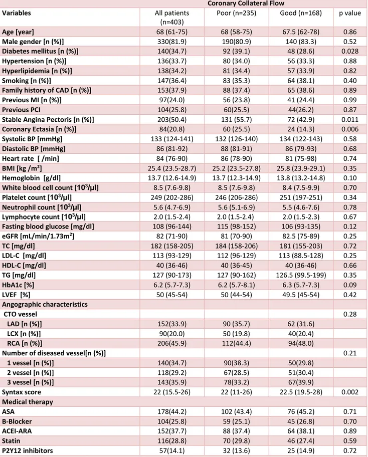
Table 1. Baseline demographic, clinical, laboratory and angiographic characteristic of all CTO patients, patients with poor CCF and patients with good CCF
Coronary Collateral Flow
Variables All patients
(n=403)
Poor (n=235) Good (n=168) p value
Age [year] 68 (61-75) 68 (58-75) 67.5 (62-78) 0.86 Male gender [n (%)] 330(81.9) 190(80.9) 140 (83.3) 0.52 Diabetes mellitus [n (%)] 140(34.7) 92 (39.1) 48 (28.6) 0.028 Hypertension [n (%)] 136(33.7) 80 (34.0) 56 (33.3) 0.88 Hyperlipidemia [n (%)] 138(34.2) 81 (34.4) 57 (33.9) 0.82 Smoking [n (%)] 147(36.4) 83 (35.3) 64 (38.1) 0.40
Family history of CAD [n (%)] 153(37.9) 88 (37.4) 65 (38.6) 0.89
Previous MI [n (%)] 97(24.0) 56 (23.8) 41 (24.4) 0.99
Previous PCI 104(25.8) 60(25.5) 44(26.2) 0.87
Stable Angina Pectoris [n (%)] 203(50.4) 131 (55.7) 72 (42.9) 0.011
Coronary Ectasia [n (%)] 84(20.8) 60 (25.5) 24 (14.3) 0.006
Systolic BP [mmHg] 133 (124-141) 132 (126-140) 134 (122-143) 0.58
Diastolic BP [mmHg] 86 (81-92) 88 (81-91) 86 (79-93) 0.68
Heart rate [ /min] 84 (76-90) 86 (78-90) 81 (75-98) 0.74
BMI [kg /m2] 25.4 (23.5-28.7) 25.2 (23.5-27.8) 25.8 (23.9-29.1) 0.35
Hemoglobin [g/dl] 13.7 (12.6-14.9) 13.7 (12.3-14.9) 13.8 (13.2-14.8) 0.10
White blood cell count [103/µl] 8.5 (7.6-9.8) 8.5 (7.6-9.8) 8.4 (7.5-9.9) 0.70
Platelet count [103/µl] 249 (202-286) 246 (206-286) 251 (197-251) 0.34
Neutrophil count [103/µl] 5.6 (4.7-6.9) 5.6 (5.1-6.9) 5.5 (4.6-7.6) 0.78
Lymphocyte count [103/µl] 2.0 (1.5-2.4) 2.0 (1.5-2.4) 2.0 (1.5-2.3) 0.67
Fasting blood glucose [mg/dl] 108 (96-144) 115 (98-152) 106 (93-135) 0.12
eGFR [mL/min/1.73m2] 82 (71-90) 81 (70-90) 82.5 (75-89) 0.25 TC [mg/dl] 182 (158-205) 184 (158-206) 181 (155-203) 0.72 LDL-C [mg/dl] 113 (93-129) 112 (96-129) 113 (88.5-128) 0.25 HDL-C [mg/dl] 40 (36-46) 40 (36-45) 40 (36-46) 0.66 TG [mg/dl] 127 (90-173) 127 (90-162) 126.5 (99.5-199) 0.35 HbA1c [%] 6.2 (5.7-7.3) 6.2 (5.7-8.1) 6.3 (5.7-7.3) 0.09 LVEF [%] 50 (45-54) 50 (44-54) 49.5 (45-54) 0.42 Angographic characteristics CTO vessel 0.28 LAD [n (%)] 152(33.9) 90 (35.7) 62 (31.6) LCX [n (%)] 90(20.0) 50 (19.8) 40(20.4) RCA [n (%)] 206(45.9) 112(44.4) 94(48.0)
Number of diseased vessel[n (%)] 0.21
1 vessel [n (%)] 140(34.7) 90(38.3) 50(29.8) 2 vessel [n (%)] 118(29.2) 67(28.5) 51(30.4) 3 vessel [n (%)] 143(35.9) 78(33.2) 67(39.9) Syntax score 22 (15.5-26) 22 (11-26) 22.5 (19.5-28) 0.002 Medical therapy ASA 178(44.2) 102 (43.4) 76 (45.2) 0.71 B-Blocker 104(25.8) 59 (25.1) 45 (26.8) 0.70 ACEI-ARA 152(37.7) 88 (37.4) 64 (38.1) 0.89 Statin 116(28.8) 70 (29.8) 46 (27.4) 0.59 P2Y12 inhibitors 57(14.1) 32 (13.6) 25 (14.9) 0.72
Data are expressed median (interquartile range) or count (percentage); ACEI-ARA, angiotensin-converting enzyme inhibitor- angiotensin II receptor antagonist; BMI, body mass index; BP, blood pressure; CAD, coronary artery disease; CTO, chronic total occlusion; eGFR, estimated glomerular filtration rate; HbA1c, hemoglobin A1c; HDL, high density lipoprotein cholesterol; LAD, left anterior descending artery; LCX, left circumflex artery; LDL, low density lipoprotein cholesterol; LVEF, left ventricular ejection fraction; MI, myocardial infarction; RCA, right coronary artery; SYNTAX, SYNergy between Percutaneous Coronary Intervention with TAXus and cardiac surgery; TC, total cholesterol; TG, ttrigyliserid; TIMI, thrombolysis in myocardial infarction
214 The relationship between 9 candidate predictors and good CCF were examined in a model using both
traditional univariable and multivariable logistic regression analyses (Table 2).
Table 2. Traditional univariable and multivariable logistic regression analysis for good CCF
Variable Univariable Multivariable
Unadjusted OR 95 % CI P value Adjusted OR 95% CI p value Age [year] 1.00 0.99-1.02 0.45 Male gender [n (%)] 1.18 0.70-1.99 0.52 Diabetes mellitus [n (%)] 0.62 0.40-0.95 0.032
Stable Angina Pectoris [n (%)]
0.59 0.89-0.40 0.011 0.37 0.22-0.62 0.002
Coronary Artery Ectasia 0.48 0.81- 0.28 0.012 0.51 0.26-0.98 0.046
Hemoglobin [g/dl] 1.07 0.95-1.20 0.25
HbA1c [%] 0.98 0.96-0.99 0.52
Fasting blood glucose [mg/dl]
0.97 0.91-0.99 0.62
SYNTAX score 1.13 1.05-1.41 0.010 1.31 1.11-1.56 0.001
CCF, coronary collateral flow; CTO, chronic total occlusion; SYNTAX, SYNergy between Percutaneous Coronary Intervention with TAXus and cardiac surgery.
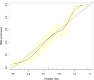
In multivariable logistic regression, stable angina pectoris [OR: 0.37(0.22-0.62), p=0.002], CEA [OR:0.51(0.26-0.98), p=0.046] and SYNTAX score [OR: 1.31(1.11-1.56), p=0.001], were found to be associated with presence of good CCF. As it is shown in Figure 1 the model’s calibration function estimate was slightly nonlinear, with the corrected calibration showing good agreement with the apparent calibration.
Figure 1. Receiver operating characteristic (ROC) curves to discriminate good CCC from poor CCC for the multivariable logistic regression model
There a was good discrimination with a c-index of 0.803 in our apparent model (Figure 2).
Figure 2. Bootstrap overfitting-corrected calibration curve estimate for development of CCC
215
DISCUSSION
To the best of our knowledge, this is the first study evaluating the potential role of CAE on development of CCC in patients with CTO. In our study showed that the presence of CAE in patients with CTO was associated with poorly developed CCC. We also found that SYNTAX score and stable angina were associated with CCC.
It has been shown that coronary collateral vessels develop in two different ways as angiogenesis and arteriogenesis. Angiogenesis is the formation of new vessels from preexisting blood vessels. (22) Arteriogenesis is formation of recently occured vessels or rudimentary colaterals to functional vessels (23). Most of studies have evaluated clinical and other parameters associated with good CCC development. In these studies, the strongest relationship among these parameter is shown in CAD especially in CTOs. In coronary artery disease increased wall stress, cytokines and oxidative stress cause collateral circulation. Coronary collateral development; reduces infarct area and angina in CAD, maintains LV ejection fraction, reduces aneurysmal enlargement (24-26). However, it is known that good CCC development was not observed in all CTO patients.
Although many parameters related to the development of poor CCC have been investigated in the literature, but contradictory results have been obtained. It has not known exactly which parameters reduce collateral growth with which mechanisms yet (27). In particular diabetes mellitus is the only parameter that has consistently shown an association with poor CCC (12). It has been shown that in diabetic patients endothelial dysfunction, impaired endothelial vasodilator response to cytokines, and inadequate neovascularization and coronary collateral development in response to ischemia (13). However, the presence of a process in which a structural deterioration of the vascular endothelium plays an important role can be mentioned for poor CCC.
The pathogenesis of CAE is not completely understood, it is caused by arterial media damage, increased vascular stress, arterial wall thinning and progressive vascular dilatation (28). Although the effect of CAE on coronary circulation has not been
fully elucidated, increased inflammatory markers, cytokines and oxidative stress are thought to impair coronary blood flow (29- 30). The presence of CAE disrupts the physiology of the coronary circulation by converting the laminar flow in the coronary circulation to turbulent flow. Slow or turbulent flow in the enlarged vessel may cause thrombosis in the ectatic segment or embolism in the distal coronary artery. (31-32) In one study, exercise test was positive in 17 of 33 patients with CAE without non-occlusive CAD and concluded that CAE caused ischemia. (33) Güleç et al. (34) showed that epicardial and microvascular perfusion was impaired in patients with ectasia. In another study in which the coronary flow rate was measured, the flow rate was significantly reduced in the aneurysm and was normal in the adjacent normal segment (35). In another studies , it was suggested that this may be due to microvascular dysfunction and / or ischemia in CAE patients with left ventricular diastolic dysfunction detected by tissue doppler imaging. (36-37).
The above-mentioned studies suggest that common pathophysiological mechanisms may play a role in the CAE and coronary collateral development at the vessel level. We aimed to investigate that new vessel formation, such as collateral vessel development may be affected in the presence of a condition such as CAE that causes deterioration of vascular structure and function. In our stduy, we found that the presence of CAE was associated with poor coronary collateral development in CTO patients. Po-Chao Hsu et al. also found that CAE was associated with poor CCC in CAD patients with %70 or more stenosis (38). The findings of this study are consistent with our study. But it is clear that there is a need for in-vitro and in-vivo studies with different designs to reveal this relationship.
CONCLUSION
Our study was the first to investigate relationship between CAE and CCC in patients with CTO. In both univariable and multivariable statistical analysis, we found that the presence of CAE was associated with poor coronary collateral development.
216
Study limitations
This study included only patients who were able to visit at hospital course , and who could be undergone coronary angiography which revealed CTO in at least one of the major coronary artery ; therefore, there could be a selection bias in this study and it is not clear whether identical conclusions can be drawn for all patients with CTO . Angiographically detected collateral flow provides only an estimate of existing absolute collateral flow since only collaterals 100 μm or more in diameter can be identified. Collateral flow can also be evaluated with methods such as myocardial contrast echocardiography, cardiac nuclear imaging, and pressure-derived collateral flow index with better quantification but indirectly. However their routine uses are not feasible in daily clinical practice.
Acknowledgements: None Funding: None
Conflict of Interest: None
REFERENCES
1. Bache RJ, Schwartz JS. Myocardial blood flow during exercise after gradual coronary occlusion in the dog. Am J Physiol 1983, 245: H131-H138.
2. Rentrop KP, Cohen M, Blanke H, Phillips RA. Changes in collateral channelfilling immediately after controlled coronary artery occlusion by anangioplasty balloon in human subjects. J Am Coll Cardiol 1985, 5:587–92.
3. Wustmann K, Zbinden S, Windecker S, Meier B, Seiler C. Is there functional collateralflow during vascular occlusion in angiographically normal coronary arteries? Circulation 2003;107:2213–20.
4. Werner GS, Ferrari M, Betge S, Gastmann O, Richartz BM, Figulla HR. Collateral function in chronic total coronary occlusions is related toregional myocardial function and duration of occlusion. Circulation 2001, 104:2784–90.
5. Fefer P, Knudtson ML, Cheema AN, Galbraith PD, Osherov AB, Yalonetsky S et al. Current perspectives on coronary chronic total occlusions: The Canadian multicenter chronic total occlusions registry. J AmColl Cardiol 2012;59:991–7.
6. Van der Hoeven NW, Teunissen PF, Werner GS, Delewi R, Schirmer SH, Traupe T et al. Clinical parameters associated with collateral development in patients with chronic total coronary occlusion. Heart 2013 Aug; 99(15):1100–5.
7. Lee JH, Kim CY, Kim N, Jang SY, Bae MH, Yang DH et al. Coronary collateral function and clinical outcome betwwen patients with acute and chronic total occlusion. JACC Cardiovasc Interv 2017 Mar 27;10(6):585–93.
8. Azhar G, Gao W, Liu L, et al. Ischemia-reperfusion in the adult mouse heart influence of age. Exp Gerontol 1999;34:699–714.
9. Kurotobi T, Sato H, Kinjo K, et al. Reduced collateral circulation to the infarct-related artery in elderly patients with acute myocardial infarction. J Am Coll Cardiol 2004;44:28–34.
10. van Royen N, Hoefer I, Buschmann I, et al. Effects of local MCP-1 protein therapy on the development of the collateral circulation and atherosclerosis in Watanabe hyperlipidemic rabbits. Cardiovasc Res 2003;57:178–85.
11. Boodhwani M, Nakai Y, Mieno S, et al. Hypercholesterolemia impairs the myocardial angiogenic response in a swine model of chronic ischemia: role of endostatin and oxidative stress. Ann Thorac Surg 2006;81:634–41.
12. Abaci A, Oguzhan A, Kahraman S, et al. Effect of diabetes mellitus on formation of coronary collateral vessels. Circulation 1999;99:2239–42.
13. Di Carli MF, Janisse J, Grunberger G, et al. Role of chronic hyperglycemia in the pathogenesis of coronary microvascular dysfunction in diabetes. J Am Coll Cardiol 2003;41:1387–93.
14. Dincer I, Ongun A, Turhan S, et al. Association between the dosage and duration of statin treatment with coronary collateral development. Coron Artery Dis 2006;17:561–5.
15. Kyriakides ZS, Kolettis T, Antoniadis A, et al. Beta-adrenergic blockade decreases coronary collateral blood flow in patients with coronary artery disease. Cardiovasc Drugs Ther 1998;12:551–9
16. Pohl T, Seiler C, Billinger M, et al. Frequency distribution of collateral flow and factors
217 influencing collateral channel development. Functional collateral channel measurement in 450 patients with coronary artery disease. J Am Coll Cardiol 2001;38:1872–8.
17. Falsetti HL, Carroll RJ. Coronary artery aneurysm. Chest 1976; 69:630-36.
18. Befeler B, Aranda JM, Embi A, et al. Coronary artery aneurysms: Study of their etiology, clinical course and effect on left ventricular function and prognosis. Am J Med 1977;62:597-607.
19. Boztosun B, Günes Y, Kırmal C. Koroner arter ektazisi. Türk Kardiyol Dern Ars 2005;33:356-359.
20. Swaye PS, Fisher LD, Litwin P, et al. Aneurysmal coronary artery disease. Circulation 1983;67:134-138.
21. Sudhir K, Ports TA, Amidon TM , et al. Circulation. 1995 Mar 1;91(5):1375-80.
22. Schultz A, Lavie L, Hochberg I, Beyar R, Stone T, Skorecki K, et al. Interindividual heterogeneity in the hypoxic regulation of VEGF: significance for the development of the coronary artery collateral circulation. Circulation 1999;100:547-52.
23. Schaper W. Tangential wall stress as a molding force in the development of collateral vessels in the canine heart. Experientia 1967;23:595-6.
24. Cohen M, Rentrop KP (1986) Limitation of myocardial ischemia by collateralcirculation during sudden controlled coronary artery oclusion in human subjects: a prospective study. Circulation 74(3): 469–76.
25. Meier P, Gloekler S, Zbinden R, Beckh S, de Marchi SF, et al. (2007) Beneficial effect of recruitable collaterals: a 10-year follow-up study in patients with stable
coronary artery disease undergoing quantitative collateral measurements. Circulation 116(9): 975– 83.
26. Regieli JJ, Jukema JW, Nathoe HM, Zwinderman AH, Ng S, et al. (2009) Coronary collaterals improve prognosis in patients with ischemic heart disease. Int J Cardiol 132(2): 257–62
27. 0. Çelik T, Çelik M, İyisoy A, Işık E. The effect of plasma asymetric dimethylarginine (ADMA) level and L-arginine/ADMA ratio on the development of coronary collaterals. [Article in Turkish-Letter] Türk Kardiyol Dern Arş 2008;36:429-30.
28. Rodbars S, Ikeda K, Montes M (1967) An analysis of mechanisms of poststenotic dilatation. Angiology 18: 349–353.
29. Li JJ, Nie SP, Qian XW, Zeng HS, Zhang CY (2009) Chronic inflammatory status in patients with coronary artery ectasia. Cytokine 46(1): 61–4.
30. Sezen Y, Bas M, Polat M, Yildiz A, Buyukhatipoglu H, et al. (2010) The relationship between oxidative stress and coronary artery ectasia. Cardiol J 17(5): 488–94.
31. Mavrogeni S (2010) Coronary artery ectasia: from diagnosis to treatment. Hellenic J Cardiol 51(2): 158–63.
32. Bal ET, Plokker T, Van den Berg E, et al: Predictability and prognosis of PTCA induced coronary artery aneurysm. Catheterization and Cardiovascular Diagnosis, 22:85,1991.
33. Sayin T, Doven O, Berkalp B, Akyurek O, Gulec S, Oral D. Exercise-induced myocardial ischemia in patients with coronary artery ectasia without obstructive coronary artery disease. Int J Cardiol 2001;78:143-9.
34.Gulec S, Atmaca Y, Kilickap M, Akyurek O, Aras O, Oral D. Angiographic assessment of myocardial perfusion in patients with isolated coronary artery ectasia.Am J Cardiol 2003;91:996-9. 35. Hamaoka K, Onouchi Z, Kamiya Y, Sakata K. Evaluation of coronary flow velocity dynamics and flow reserve in patients with Kawasaki disease by means of a Doppler guide wire. J Am Coll Cardiol 1998;31:833-40.
36. Saglam M, Barutcu I, Karakaya O, Esen AM, Akgun T, Karavelioglu Y, et al. Assessment of left ventricular functions in patients with isolated coronary artery ectasia by conventional and tissue Doppler imaging. Angiology 2008;59:306-11.
218 37. Tuzun N, Tanriverdi H, Evrengul H, Kuru DS, Ergene AO. Aortic elastic properties in patients with coronary artery ectasia. Circ J 2007;71:506-10 38. Po-Chao Hsu, Ho-Ming Su, Hsiang-Chun Lee et al Coronary Collateral Circulation in Patients of Coronary Ectasia with Significant Coronary Artery Disease PLoS One. 2014 Jan27;9(1):e87001.